Functional Residual Capacity — Explained by Medical Evidence, Not Myths

Functional residual capacity is the air left in the lungs after a normal exhalation. It helps prevent the lungs from fully collapsing between breaths and supports steady oxygen and carbon dioxide exchange.
Key Takeaways
- Functional residual capacity is the air left in the lungs after a normal exhalation.
- It helps prevent the lungs from fully collapsing between breaths and supports steady oxygen and carbon dioxide exchange.
- Functional residual capacity can be higher or lower depending on body position, age, weight, lung elasticity, and certain medical conditions.
- Doctors assess it as part of pulmonary function testing when evaluating shortness of breath or suspected lung disease.
- Changes in functional residual capacity may be seen in conditions such as chronic obstructive pulmonary disease, asthma, obesity, or restrictive lung disorders.
Functional residual capacity is the amount of air that remains in the lungs after a relaxed, normal breath out. It is not a disease itself, but an important lung measurement that helps doctors understand breathing mechanics, gas exchange, and whether a lung or airway problem may be present.
Overview: what functional residual capacity means
Functional residual capacity is the volume of air left in the lungs after a person breathes out normally and without force. In simple terms, it is the lungs’ resting air level between regular breaths. This remaining air is important because it helps the lungs stay partly expanded and allows oxygen and carbon dioxide exchange to continue even between inhaling and exhaling.
Medical evidence shows that functional residual capacity is a basic concept in respiratory physiology, not a myth or a vague wellness term. It is one of several lung volumes used to understand how the chest wall and lungs work together. Doctors do not usually focus on this number alone; they interpret it alongside symptoms, examination findings, imaging, and other pulmonary function measurements.
Functional residual capacity is made up of two parts: expiratory reserve volume, which is the extra air a person could breathe out after a normal exhale, and residual volume, which is the air that always remains in the lungs and cannot be exhaled completely. Because some air always stays behind, the lungs do not empty fully with each breath.
This measurement can be especially useful when a clinician is trying to distinguish between obstructive and restrictive breathing patterns. It may be discussed during the evaluation of chronic obstructive pulmonary disease, asthma, obesity-related breathing difficulty, or other conditions that affect lung expansion and airway function.
Why this lung volume matters in everyday breathing
Functional residual capacity serves several practical purposes in normal breathing. First, it acts like a reservoir of air, helping smooth out changes in oxygen and carbon dioxide levels between breaths. Without this reserve, gas exchange would fluctuate more sharply every time a person inhaled or exhaled.
Second, it helps keep the small airways and air sacs open. When functional residual capacity is too low, some parts of the lungs may close earlier during exhalation, which can reduce oxygen transfer and increase the effort needed to breathe. When it is too high, the lungs may become overly inflated, making the breathing muscles work less efficiently.
This balance depends on the natural recoil of the lungs pulling inward and the chest wall tending to move outward. Functional residual capacity is the point where these opposite forces are in equilibrium during quiet breathing. That is why it is often described as the resting position of the respiratory system.
In clinical care, understanding this balance helps explain why breathlessness may worsen when lying flat, after surgery, during pregnancy, with excess weight, or in advanced lung disease. It also helps guide decisions about breathing exercises, oxygen support, and in some cases pulmonary rehabilitation.
What can change functional residual capacity

Functional residual capacity is not fixed. It changes with age, body shape, posture, and health status. In many people, it is lower when lying down than when standing, because the abdominal contents push the diaphragm upward and reduce the space available for the lungs to rest expanded.
Body weight can also affect it. In obesity, especially when fat is concentrated around the abdomen and chest, the lungs may not expand as freely at rest, which can lower functional residual capacity. Pregnancy can cause a similar effect as the growing uterus elevates the diaphragm. After abdominal or chest surgery, pain and shallow breathing may also temporarily reduce this lung volume.
Some lung conditions increase functional residual capacity. This often happens in obstructive disorders where air becomes trapped in the lungs, leading to hyperinflation. By contrast, some restrictive conditions can reduce it because the lungs or chest wall cannot expand normally. Examples include scarring of lung tissue, neuromuscular weakness, and certain chest wall disorders.
Smoking, chronic airway inflammation, reduced physical conditioning, and underlying heart or lung disease may all influence breathing mechanics over time. A change in functional residual capacity does not by itself identify one diagnosis, but it can provide an important clue when placed in the full clinical picture.
Symptoms and conditions associated with abnormal results
Functional residual capacity itself does not cause symptoms in the way a disease does. Instead, symptoms arise from the underlying issue affecting lung function. A person may notice shortness of breath, chest tightness, wheezing, reduced exercise tolerance, fatigue during activity, or the feeling that breathing takes more effort than usual.
When functional residual capacity is increased because of air trapping, the chest may feel overly expanded and exhalation may become prolonged. This pattern can occur in obstructive lung conditions, including asthma and chronic obstructive lung disease. In these situations, the lungs may hold onto too much air, making it harder to take a deep, comfortable next breath.
When functional residual capacity is reduced, smaller airways may close earlier and oxygenation may become less efficient, particularly during sleep, after surgery, or when lying flat. This may contribute to shallow breathing, low stamina, or discomfort with exertion. Restrictive disorders, obesity-related breathing limitations, and some neuromuscular conditions can produce this pattern.
Doctors use these findings as part of a broader workup, especially if a person has ongoing respiratory symptoms, repeated infections, chronic cough, or abnormal oxygen levels. In some cases, more detailed evaluation may include imaging or specialized assessment in a chest diseases evaluation program.
How doctors measure functional residual capacity
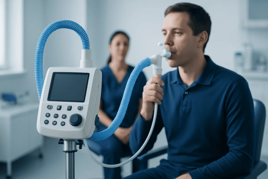
Functional residual capacity cannot be measured accurately with basic spirometry alone, because spirometry does not directly measure the residual volume left in the lungs after maximum exhalation. Instead, clinicians use specific pulmonary function methods such as body plethysmography, gas dilution techniques, or nitrogen washout testing.
Body plethysmography is commonly used in pulmonary laboratories because it can provide detailed information about lung volumes, including trapped air. During the test, the person sits in a clear booth and breathes through a mouthpiece while pressure changes are measured. Gas dilution tests work differently, using inhaled gases to estimate lung volume.
The result is interpreted in relation to age, sex, height, and other predicted values. Doctors also look at associated measurements such as total lung capacity, residual volume, forced expiratory volume, and forced vital capacity. This helps them decide whether the breathing pattern looks obstructive, restrictive, mixed, or within normal limits.
If needed, testing may be combined with chest imaging, oxygen assessment, exercise testing, or specialist review. Depending on symptoms, a clinician may also recommend bronchoscopy or other investigations to look for airway disease, persistent infection, or structural problems.
Treatment depends on the underlying cause
There is no single treatment aimed only at functional residual capacity. Care is directed at the condition that is changing lung mechanics. For example, if the problem is airway narrowing and air trapping, treatment may focus on bronchodilator medicines, inhaled anti-inflammatory therapy, smoking cessation, and exercise-based respiratory support. If the issue is restrictive lung function, treatment depends on whether the cause is obesity, scarring, muscle weakness, pain, or another disorder.
Breathing exercises, posture changes, early mobilization after surgery, and supervised rehabilitation can all help improve breathing efficiency in selected patients. In hospital settings, clinicians may use techniques that support deeper breaths and reduce the chance of small airway closure, especially after operations or prolonged bed rest.
When obesity contributes to low lung volumes, gradual weight management may improve chest wall movement and respiratory comfort. If a medication, occupational exposure, or smoking habit is worsening lung function, addressing those factors is also an important part of care. Long-term management usually includes regular follow-up and monitoring if a chronic lung condition is diagnosed.
For people with complex respiratory symptoms, multidisciplinary assessment can be useful. Near the end of the care pathway, it may be helpful to know that Acibadem International’s multidisciplinary specialists and JCI-accredited hospitals diagnose and treat respiratory conditions for international patients when further evaluation is needed.
Prevention and self-care for healthier lung function
Not every change in functional residual capacity can be prevented, but many supportive habits can protect lung health. Avoiding smoking and secondhand smoke is one of the most important steps. Staying physically active, within a doctor’s guidance, helps maintain breathing muscle strength, chest mobility, and overall endurance.
Good control of chronic conditions such as asthma, allergies, reflux, and sleep-related breathing problems can also reduce day-to-day respiratory strain. Vaccination, hand hygiene, and timely treatment of respiratory infections may lower the risk of complications that affect lung function.
People recovering from surgery or illness may benefit from deep breathing exercises, walking as advised, and careful pain control so they can breathe more fully. Maintaining a healthy weight can also improve breathing mechanics, especially in those who feel short of breath when lying down or during mild exertion.
- Do not ignore persistent cough, wheezing, or breathlessness.
- Use prescribed inhalers correctly and consistently if a doctor recommends them.
- Follow up on abnormal lung tests rather than relying on internet myths or isolated numbers.
- Seek professional advice before starting supplements or devices marketed for “lung detox” or “boosting capacity.”
When to seek medical care
A person should seek medical care if shortness of breath is new, persistent, or getting worse. Medical review is also important for ongoing wheezing, chronic cough, chest discomfort with breathing, reduced exercise tolerance, or repeated chest infections. These symptoms do not always mean a serious illness, but they do deserve proper assessment.
Urgent medical attention is needed for severe difficulty breathing, blue lips, confusion, fainting, sudden chest pain, or an inability to speak in full sentences because of breathlessness. These may be signs of a medical emergency and should not be managed at home.
If pulmonary function testing shows an abnormal functional residual capacity, the next step is usually a discussion with a qualified doctor rather than self-diagnosis. Results need to be interpreted in context, because the same pattern can have different explanations depending on symptoms, history, and physical findings.
Many people feel reassured after a structured evaluation. Clear diagnosis and treatment planning can often improve symptoms and help protect long-term lung health.
Frequently asked questions
What is functional residual capacity in simple terms?
Functional residual capacity is the amount of air left in the lungs after a normal, relaxed exhalation. It is the lungs’ resting volume between everyday breaths and helps keep gas exchange steady.
Is functional residual capacity the same as residual volume?
No. Residual volume is the air that remains in the lungs even after a person exhales as much as possible, while functional residual capacity is the air left after a normal exhale. Functional residual capacity includes residual volume plus expiratory reserve volume.
Can functional residual capacity be too high?
Yes. It can be increased when air gets trapped in the lungs, which may happen in obstructive airway diseases. When this occurs, breathing can feel less efficient and exhalation may become more difficult.
Can functional residual capacity be too low?
Yes. A low functional residual capacity can happen when the lungs or chest wall cannot stay as expanded at rest, such as with obesity, lying flat, some restrictive lung conditions, or after surgery. This may contribute to airway closure and reduced oxygen transfer.
How is functional residual capacity measured?
It is measured using specialized pulmonary function tests such as body plethysmography or gas dilution methods. Basic spirometry alone cannot directly measure it because spirometry does not capture the air that always remains in the lungs.
Does an abnormal result mean lung disease?
Not always. An abnormal result is a clue that may reflect posture, body habitus, temporary illness, or an underlying respiratory condition, but it does not diagnose a disease by itself. A doctor interprets it together with symptoms, examination, and other test results.
This article is for general information only and is not a substitute for professional medical advice. Please consult a qualified doctor about your individual situation.
Persistent digestive symptoms? Get evaluated in Turkey
JCI-accredited · board-certified surgeons · reply within 24h
Add us as a Preferred Source to see more of our trusted health content across Google Search, AI Overviews and Discover.

More from the Health Library

How to loosen a bowel blockage at home? A Doctor-Reviewed Answer

Ozempic Vagina — Explained by Medical Evidence, Not Myths

Guarana: An Evidence-Based Guide for Patients

Health Services — Explained by Medical Evidence, Not Myths

Timed Get Up and Go: A Complete Medical Overview