Gallbladder Cancer: Risk Factors, Imaging Findings, and Treatment Planning

Gallbladder cancer is rare, and many early cases cause few or no specific symptoms. Long-term gallbladder inflammation, gallstones, certain bile duct conditions, and larger gallbladder polyps can increase risk.
Key Takeaways
- Gallbladder cancer is rare, and many early cases cause few or no specific symptoms.
- Long-term gallbladder inflammation, gallstones, certain bile duct conditions, and larger gallbladder polyps can increase risk.
- Ultrasound, CT, MRI/MRCP, and sometimes PET or endoscopic tests help define the tumor, bile ducts, lymph nodes, and possible spread.
- Surgery offers the best chance of long-term control when the cancer is found early and can be completely removed.
- When surgery is not possible, chemotherapy, immunotherapy, radiation, bile duct drainage, and symptom-focused care may help control disease and improve comfort.
- A specialist evaluation is important for unexplained jaundice, persistent right upper abdominal pain, or suspicious gallbladder imaging findings.
Gallbladder cancer is an uncommon cancer of the biliary tract that is often found during imaging or after gallbladder surgery. Careful imaging, staging, and multidisciplinary treatment planning help doctors decide whether surgery, systemic therapy, radiation, or supportive procedures are most appropriate.
Overview
Gallbladder cancer is a cancer that begins in the gallbladder, a small organ under the liver that stores bile. Bile helps the body digest fats and flows from the liver and gallbladder through the bile ducts into the small intestine. Because the gallbladder is close to the liver, bile ducts, stomach, and intestine, treatment planning often requires careful mapping of nearby structures.
Most gallbladder cancers are adenocarcinomas, meaning they start in gland-forming cells of the gallbladder lining. The disease is uncommon, and early gallbladder cancer may not cause clear symptoms. Some cases are discovered unexpectedly after surgery to remove the gallbladder for gallstones or inflammation.
The outlook and treatment options depend mainly on how deeply the cancer has grown into the gallbladder wall, whether it has reached the liver or nearby organs, whether lymph nodes are involved, and whether it has spread to distant sites. Imaging and pathology reports are therefore central to treatment planning. A coordinated team may include hepatobiliary surgeons, medical oncologists, radiation oncologists, gastroenterologists, radiologists, pathologists, and supportive care specialists.
Symptoms and Common Presentation
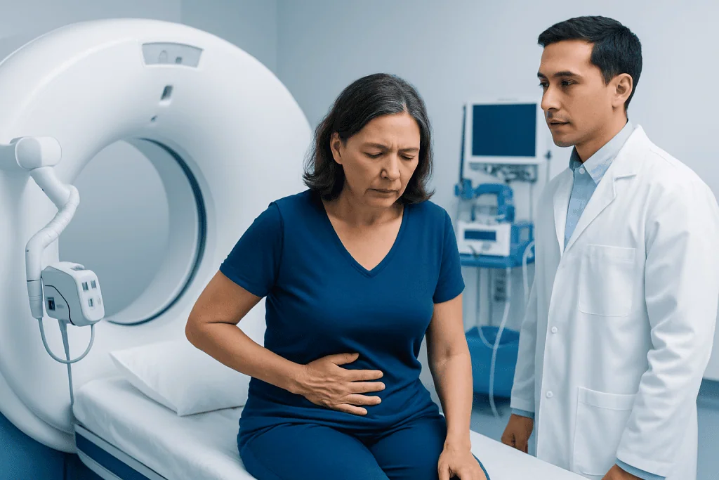
Gallbladder cancer can be difficult to recognize early because symptoms may resemble more common gallbladder problems, such as gallstones or cholecystitis. Some people have no symptoms until imaging is performed for another reason or until the gallbladder is removed and examined by a pathologist.
Possible symptoms include persistent pain or discomfort in the right upper abdomen, nausea, reduced appetite, unintentional weight loss, bloating, fever, or fatigue. If the tumor blocks the bile ducts, jaundice may occur. Jaundice can cause yellowing of the skin or eyes, dark urine, pale stools, and itching.
These symptoms do not necessarily mean cancer is present. Gallstones, infections, hepatitis, and other digestive conditions are much more common causes. However, symptoms that are persistent, worsening, or associated with jaundice should be assessed by a qualified doctor because timely diagnosis can guide appropriate care.
Risk Factors and Who May Be at Higher Risk

A risk factor increases the chance of developing a disease, but it does not mean the disease will occur. Many people with gallstones never develop gallbladder cancer, and some people diagnosed with gallbladder cancer have no obvious risk factor. Risk is usually related to long-term irritation or inflammation of the gallbladder lining.
Recognized risk factors include long-standing gallstones, chronic gallbladder inflammation, a calcified or porcelain gallbladder, and certain gallbladder polyps, especially larger or growing polyps. Risk may also be higher in people with primary sclerosing cholangitis, choledochal cysts, abnormal pancreaticobiliary duct junctions, chronic Salmonella carriage, obesity, older age, and a family history of gallbladder cancer. The condition is more common in some geographic regions and ethnic groups, suggesting that genetics, environment, diet, and gallstone patterns may all play a role.
For patients with gallbladder polyps or other gallbladder abnormalities, doctors consider size, growth over time, symptoms, age, and associated conditions when recommending follow-up or surgery. Routine screening is not generally used for the average-risk population, but targeted monitoring may be appropriate for selected high-risk individuals.
Imaging Findings and Diagnostic Tests
Imaging is essential because it helps doctors distinguish gallbladder cancer from benign gallbladder disease and shows whether the tumor may be removable. Abdominal ultrasound is often the first test. It may show a mass filling or replacing the gallbladder, irregular or asymmetric gallbladder wall thickening, a polypoid lesion, gallstones, or dilated bile ducts.
Contrast-enhanced CT is commonly used to assess the gallbladder, liver, nearby lymph nodes, blood vessels, peritoneum, and distant organs. Findings that may raise concern include direct invasion into the liver, loss of the normal tissue plane between the gallbladder and liver, enlarged regional lymph nodes, or signs of spread in the abdomen. MRI with MRCP can provide detailed views of the bile ducts and liver and may be especially useful when bile duct involvement or obstruction is suspected.
Additional tests are chosen based on the clinical situation. Endoscopic ultrasound may help evaluate the gallbladder wall, bile ducts, and nearby lymph nodes. ERCP may be used when a blocked bile duct needs drainage or sampling. PET/CT can be considered in selected patients to look for disease outside the planned surgical field. Blood tests may include liver function tests and tumor markers such as CA 19-9 or CEA, but these markers cannot diagnose gallbladder cancer on their own.
A tissue diagnosis may be obtained before chemotherapy or radiation, especially if surgery is not planned first. In some potentially resectable cases, doctors may avoid needle biopsy of the main gallbladder mass if it could affect the surgical approach. If cancer is found after routine gallbladder removal, the pathology report becomes a key document for staging and deciding whether additional surgery or treatment is needed.
Staging and Treatment Planning
Staging describes how far the cancer has grown or spread. Doctors evaluate the tumor depth, involvement of the liver or other nearby organs, lymph node status, and distant spread. This information helps determine whether treatment is planned with curative intent, focused on disease control, or primarily aimed at relieving symptoms and maintaining quality of life.
Resectability is a central question. A tumor may be considered resectable if surgeons believe it can be removed completely with clear margins while preserving enough healthy liver and essential structures. A tumor may be unresectable if it has extensive involvement of major blood vessels, widespread lymph node disease, peritoneal spread, or distant metastases. Sometimes diagnostic laparoscopy is performed before major surgery to look for small areas of spread that may not be visible on scans.
Important planning details include the exact location of the tumor, whether the bile ducts are involved, whether jaundice needs to be treated before surgery or chemotherapy, the patient’s liver function, general fitness, nutrition, other medical conditions, and personal preferences. Because gallbladder cancer is uncommon, review by a team experienced in hepatobiliary cancers is particularly valuable.
Treatment Options
Surgery is the main treatment when gallbladder cancer is found early and can be fully removed. For very early cancer limited to the innermost layer of the gallbladder, simple cholecystectomy may be sufficient in some cases. For cancers that have grown deeper into the gallbladder wall, an extended cholecystectomy is often considered. This may include removal of part of the adjacent liver and nearby lymph nodes. Bile duct removal is not automatic, but it may be needed if the duct is involved or if clear margins require it.
If cancer is discovered after a previous gallbladder operation, doctors review the original pathology report and imaging carefully. Depending on tumor depth, margin status, and whether lymph nodes were assessed, a second operation may be recommended to remove additional liver tissue, lymph nodes, or port-site tissue in selected situations. Not every patient needs further surgery, and decisions are individualized.
Systemic therapy treats cancer cells throughout the body. Chemotherapy may be recommended after surgery to reduce the risk of recurrence, or it may be used as the main treatment when the cancer is advanced. Some treatment plans for advanced biliary tract cancers may include immunotherapy in combination with chemotherapy, depending on patient factors, tumor characteristics, and local guidelines. Molecular testing may identify uncommon targets, such as specific gene alterations, that can guide targeted therapy in selected patients.
Radiation therapy may be used in selected cases, often with chemotherapy, especially when there is concern about microscopic disease near surgical margins or local control around the bile ducts. Supportive procedures are also important. If bile flow is blocked, endoscopic or percutaneous stent placement or drainage can reduce jaundice and itching and may make other treatments safer. Acibadem International’s multidisciplinary specialists and JCI-accredited hospitals diagnose and treat gallbladder cancer for international patients using coordinated surgical, oncology, imaging, and supportive care pathways.
Prevention, Self-care, and When to See a Doctor
There is no guaranteed way to prevent gallbladder cancer. However, maintaining a healthy weight, staying physically active, and following a balanced eating pattern may support overall digestive and liver health. People with known gallstones should discuss symptoms and management with a doctor, but gallstones alone do not mean cancer will develop.
Patients with gallbladder polyps, porcelain gallbladder, choledochal cysts, primary sclerosing cholangitis, or recurrent biliary symptoms may need individualized follow-up. Keeping imaging appointments and sharing prior scan reports can help doctors detect changes over time. After treatment for gallbladder cancer, follow-up visits usually include symptom review, physical examination, blood tests, and imaging at intervals determined by the care team.
Medical advice should be sought promptly for yellowing of the skin or eyes, dark urine with pale stools, persistent right upper abdominal pain, unexplained weight loss, ongoing fever with abdominal symptoms, or a new gallbladder mass or suspicious polyp on imaging. Urgent care may be needed if severe abdominal pain, high fever, confusion, or signs of infection occur. Patients should not delay evaluation, but they can also be reassured that many gallbladder symptoms are caused by non-cancerous conditions that can be treated.
Frequently asked questions
Is gallbladder cancer the same as bile duct cancer?
No. Gallbladder cancer starts in the gallbladder, while bile duct cancer starts in the tubes that carry bile. They are both biliary tract cancers and may share some imaging tests and treatments, but staging and surgery can differ.
Can gallstones turn into gallbladder cancer?
Most people with gallstones never develop gallbladder cancer. Long-standing gallstones and chronic inflammation are risk factors, but the overall risk for an individual with gallstones is usually low. A doctor can advise whether symptoms or imaging findings suggest the need for gallbladder removal.
What imaging test is best for gallbladder cancer?
There is no single best test for every patient. Ultrasound often detects the first abnormality, while contrast-enhanced CT and MRI/MRCP are commonly used for staging and surgical planning. Endoscopic ultrasound, ERCP, PET/CT, or diagnostic laparoscopy may be added in selected cases.
Does every gallbladder polyp need surgery?
No. Many small gallbladder polyps are benign and can be monitored. Surgery may be considered when a polyp is large, growing, causing symptoms, or present with other risk factors such as primary sclerosing cholangitis. Decisions should be based on specialist assessment and imaging follow-up.
What happens if gallbladder cancer is found after gallbladder removal?
This is called incidental gallbladder cancer. Doctors review the pathology report to see how deeply the tumor invaded, whether margins were clear, and whether other risk features were present. Additional imaging and, in some cases, further surgery or oncology treatment may be recommended.
Can gallbladder cancer be treated without surgery?
Yes, when surgery is not possible or not appropriate, treatment may include chemotherapy, immunotherapy, targeted therapy in selected cases, radiation, and procedures to relieve bile duct blockage. These treatments aim to control the cancer, reduce symptoms, and support quality of life. The best plan depends on staging, overall health, and patient preferences.
References
- National Cancer Institute
- American Cancer Society
- European Society for Medical Oncology
- National Comprehensive Cancer Network
- World Health Organization
This article is for general information only and is not a substitute for professional medical advice. Please consult a qualified doctor about your individual situation.
Bladder Cancer in Turkey — costs, top hospitals & a free quote
JCI-accredited · board-certified surgeons · reply within 24h
Add us as a Preferred Source to see more of our trusted health content across Google Search, AI Overviews and Discover.

Medical Oncology Department
Medical treatment of cancer with chemotherapy, immunotherapy and targeted therapies under a multidisciplinary tumor board.
60 specialists in this unit