Gallbladder Ultrasonography — Explained by Medical Evidence, Not Myths

Gallbladder ultrasonography uses sound waves, not radiation, to create images of the gallbladder and nearby bile ducts. It is commonly used to detect gallstones, signs of cholecystitis, gallbladder polyps, and bile duct widening.
Key Takeaways
- Gallbladder ultrasonography uses sound waves, not radiation, to create images of the gallbladder and nearby bile ducts.
- It is commonly used to detect gallstones, signs of cholecystitis, gallbladder polyps, and bile duct widening.
- The test is usually painless and often works best after fasting for several hours.
- Normal results do not always explain every type of abdominal pain, so other tests may sometimes be needed.
- Urgent medical review is important for severe pain, fever, jaundice, or persistent vomiting.
Gallbladder ultrasonography is a safe, noninvasive ultrasound test used to look for gallstones, gallbladder inflammation, sludge, and other bile-related problems. It is often the first imaging test doctors choose because it is quick, widely available, and does not use radiation.
Overview: what gallbladder ultrasonography is and why it is used
Gallbladder ultrasonography is an imaging test that uses high-frequency sound waves to create pictures of the gallbladder and nearby structures. In practical terms, it helps doctors see whether the gallbladder contains stones, thickened walls, fluid around it, or other changes that may explain symptoms. Because it does not involve radiation and is usually quick to perform, it is often the first test ordered when gallbladder disease is suspected.
The gallbladder stores bile, a fluid made by the liver that helps digest fats. When bile flow is disrupted or solid particles form into stones, pain and digestive symptoms can occur. Ultrasound is especially useful for seeing gallstones and for checking whether the gallbladder looks inflamed, as can happen in cholecystitis.
This test is guided by medical evidence rather than myths. It does not “cleanse” the gallbladder, it does not prove that every abdominal symptom is gallbladder-related, and it is not a treatment. Its role is diagnostic: it gives doctors visual information that can support or rule out common causes of right upper abdominal pain.
What the test can show
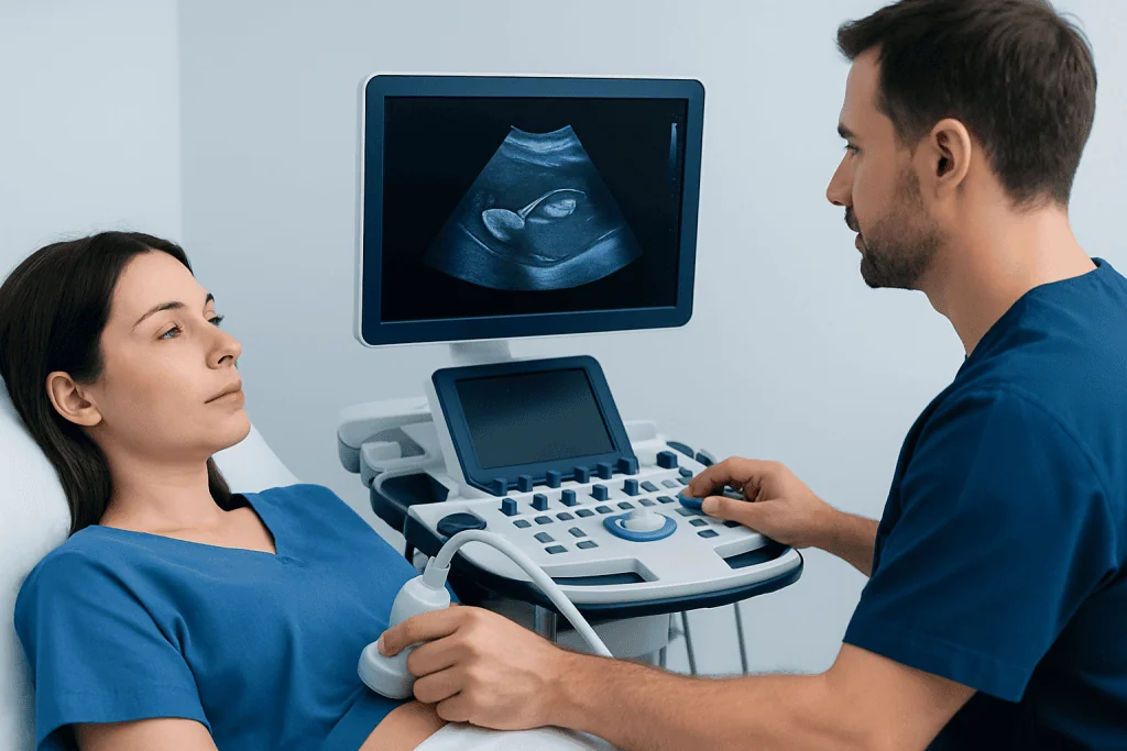
Gallbladder ultrasonography can identify several common problems. The best-known finding is gallstones, which appear as bright structures that may cast a shadow on the image. The scan can also detect sludge, which is a thick material in bile that may contribute to symptoms even when fully formed stones are not seen.
Another important use is evaluating for inflammation. Signs that may suggest acute or chronic gallbladder disease include a thickened gallbladder wall, enlargement of the gallbladder, tenderness during the scan, or fluid around the organ. Doctors may combine the ultrasound findings with symptoms, blood tests, and physical examination to decide whether the picture fits infection, blockage, or another cause.
Ultrasound may also show:
- Gallbladder polyps
- Bile duct widening, which can suggest blockage
- Features that raise concern for infection or complications
- Less commonly, masses or structural abnormalities
Even so, no single test answers every question. Small stones in the bile ducts, very early inflammation, or pain caused by another digestive problem may not be fully explained by a routine gallbladder ultrasound. In those situations, a doctor may recommend other imaging or follow-up testing.
When doctors recommend gallbladder ultrasonography
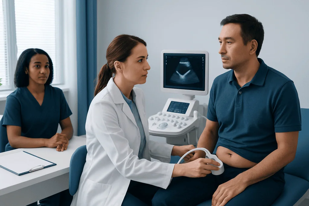
Doctors often order this test when a person has symptoms that fit a biliary pattern. This commonly means pain in the upper right side of the abdomen or in the upper middle abdomen, especially after eating fatty meals. Some people also feel nausea, bloating, pain that spreads to the back or right shoulder, or tenderness under the ribs on the right side.
Gallbladder ultrasonography may also be used when blood tests suggest a liver or bile duct problem, or when a person has unexplained jaundice. In emergency care, it can help quickly assess whether pain may be related to gallstones or inflammation. In outpatient settings, it is frequently used to investigate recurring digestive complaints that have not improved.
Doctors may recommend it in situations such as:
- Suspected gallstones
- Possible acute gallbladder inflammation
- Jaundice or abnormal liver function tests
- Follow-up of known gallbladder polyps or sludge
- Evaluation of upper abdominal pain during pregnancy, when avoiding radiation is especially helpful
It is important to remember that abdominal pain has many possible causes. Stomach ulcers, pancreatitis, liver disease, reflux, and intestinal conditions can sometimes mimic gallbladder pain. Ultrasound helps narrow down the possibilities, but the final diagnosis depends on the full clinical picture.
How to prepare and what happens during the scan
Preparation is usually simple. Many hospitals and imaging centers ask the patient to fast for several hours before the test, often around 6 to 8 hours unless told otherwise. Fasting helps the gallbladder stay full of bile, making it easier to see clearly. Small sips of water for essential medicines may still be allowed, but instructions can vary, so the imaging team’s advice should be followed.
During the examination, the patient lies on an examination bed while gel is placed on the skin over the upper abdomen. A handheld probe called a transducer is moved across the area to create images. The sonographer may ask the patient to change position or hold a breath briefly so the gallbladder can be seen from different angles.
The test is usually painless, though there may be mild pressure when the probe passes over a tender area. Most scans are completed within a short visit, often in less than 30 minutes. Because no sedation is generally needed, people can usually return to normal activities right away unless their doctor has given other instructions.
A radiologist interprets the images and prepares a report for the referring doctor. The doctor then explains what the findings mean in the context of symptoms, examination, and any blood test results. This step matters, because a finding such as a small gallbladder polyp or incidental sludge may not always require treatment.
Understanding the results and their limits
A normal gallbladder ultrasound generally means the gallbladder looks structurally normal and there is no clear sign of stones, wall thickening, or major bile duct enlargement. This can be reassuring, but it does not always mean the symptoms are unrelated to the biliary system. Some people have functional gallbladder problems or small duct stones that are not obvious on standard ultrasound.
An abnormal result may show gallstones, sludge, inflammation, a distended gallbladder, a thickened wall, or dilated bile ducts. These findings help doctors decide on the next step. In some cases, treatment focuses on symptom control and monitoring; in others, more testing or surgery may be the best option.
Further evaluation may include blood tests, repeat ultrasound, CT, MRI, or a specialized bile duct study. When doctors need a more detailed view of the bile ducts and pancreas, MRI may be considered as part of the workup. If symptoms are severe or the diagnosis remains uncertain, treatment decisions are based on both imaging and the person’s clinical condition.
Ultrasound accuracy can also be affected by body habitus, bowel gas, recent eating, or the exact location of a stone. For that reason, patients should not assume that one scan gives a complete answer in every case. A qualified doctor is the best person to explain whether the result is conclusive or whether additional evaluation is needed.
Treatment options after gallbladder ultrasonography
Treatment depends on what the scan shows and whether symptoms are present. If gallstones are seen but cause no symptoms, a doctor may advise watchful waiting rather than immediate treatment. If the stones are causing repeated pain, infection, or complications, surgery to remove the gallbladder may be recommended.
The most common definitive treatment for troublesome gallbladder disease is gallbladder surgery, also called cholecystectomy. This may be discussed when there are repeated attacks of biliary pain, confirmed inflammation, or complications such as blockage. Many people live normally without a gallbladder because bile can still flow from the liver into the intestine.
If the concern is not the gallbladder itself but a possible stone blocking the common bile duct, doctors may consider procedures aimed at the bile ducts. In selected cases, ERCP can be used to diagnose and treat certain bile duct obstructions. Antibiotics, pain relief, fluids, or hospital observation may also be needed when infection or acute inflammation is suspected.
When the ultrasound shows polyps or uncertain findings, the plan may involve follow-up imaging rather than immediate intervention. Care is individualized. Near the end of the evaluation process, some patients may be referred to centers such as Acibadem International, where multidisciplinary specialists in JCI-accredited hospitals diagnose and treat gallbladder and biliary conditions for international patients.
Prevention, self-care, and when to seek medical care
Gallbladder ultrasonography itself is a diagnostic test, so prevention focuses on reducing the risk of gallbladder disease and responding appropriately to symptoms. A balanced eating pattern, maintaining a healthy weight, regular physical activity, and avoiding rapid weight loss may help lower the likelihood of forming some gallstones. People with ongoing digestive symptoms should avoid self-diagnosing and should discuss concerns with a doctor.
Self-care can support comfort while waiting for medical review, but it should not replace assessment if symptoms are significant. Some people notice fewer symptoms when they avoid very heavy or high-fat meals until the cause of the pain is clarified. Staying hydrated and keeping a record of when symptoms occur may also help the doctor identify patterns.
Medical care should be sought promptly if there is severe or persistent pain in the upper abdomen, fever, chills, yellowing of the eyes or skin, repeated vomiting, dark urine, pale stools, fainting, or symptoms that rapidly worsen. These can point to complications such as infection, bile duct blockage, or pancreatitis and should not be ignored.
If pain is mild but keeps returning, or if there is nausea after meals, bloating, or unexplained tenderness in the upper right abdomen, a non-urgent appointment is still worthwhile. Early evaluation can help confirm whether gallbladder ultrasonography is appropriate and whether symptoms may instead be related to another digestive condition.
Frequently asked questions
Is gallbladder ultrasonography the same as an abdominal ultrasound?
Gallbladder ultrasonography is often performed as part of an abdominal ultrasound, but it focuses specifically on the gallbladder and nearby bile ducts. A general abdominal ultrasound may look more broadly at the liver, pancreas, kidneys, spleen, and other organs as well.
Can gallbladder ultrasonography detect gallstones accurately?
It is one of the best first-line tests for gallstones in the gallbladder and is widely used for that reason. However, very small stones or stones located in the bile ducts may sometimes need additional testing to confirm.
Do patients need to fast before a gallbladder ultrasound?
In many cases, yes. Fasting for several hours helps the gallbladder fill with bile, which usually improves image quality and makes abnormalities easier to see. The exact instructions should come from the imaging center or doctor.
Is gallbladder ultrasonography painful or risky?
The test is generally safe, noninvasive, and does not use radiation. Most people feel only slight pressure from the probe, although a tender gallbladder area may be uncomfortable for a moment during the scan.
What if the ultrasound is normal but symptoms continue?
A normal result can be reassuring, but it does not explain every cause of abdominal pain. If symptoms continue, a doctor may consider other digestive causes or order additional tests such as blood work, MRI, CT, or other imaging.
Can gallbladder ultrasonography diagnose cancer?
Ultrasound can sometimes detect a suspicious mass or abnormal thickening, but it usually cannot confirm cancer on its own. If there is concern, doctors typically recommend further imaging and specialist evaluation.
This article is for general information only and is not a substitute for professional medical advice. Please consult a qualified doctor about your individual situation.
Explore treatments in Turkey — costs, top hospitals & a free quote
JCI-accredited · board-certified surgeons · reply within 24h
Add us as a Preferred Source to see more of our trusted health content across Google Search, AI Overviews and Discover.

More from the Health Library

Ginger Shots: What Patients Need to Know

Cryptogenic Stroke: An Evidence-Based Guide for Patients

Mite Bites on Humans: A Complete Medical Overview

Retinopathy: Symptoms, Causes, and Treatment — Complete Patient Guide

It Band: A Complete Medical Overview

Acupuncture for Weight Loss — Explained by Medical Evidence, Not Myths
Gastroenterology Specialists at Acibadem

Prof. Dr. Erkin Öztaş
Gastroenterology
Prof. Dr. Ethem Tankurt
Gastroenterology
Prof. Dr. Fatih Oğuz Önder
Gastroenterology
Dr. Fatma Seçil Kırdök
Gastroenterology