

Heterogeneous Myometrium Treatment: How It Works, Results and What to Expect

A heterogeneous myometrium means the uterine muscle looks uneven in texture on imaging. It is commonly associated with adenomyosis, but imaging findings must be interpreted alongside symptoms and examination results.
Key Takeaways
- A heterogeneous myometrium means the uterine muscle looks uneven in texture on imaging.
- It is commonly associated with adenomyosis, but imaging findings must be interpreted alongside symptoms and examination results.
- People without troublesome symptoms may only need follow-up rather than active treatment.
- Medical treatment can reduce heavy bleeding and pelvic pain, while procedures may help when symptoms persist.
- Fertility wishes are an important part of choosing a treatment plan.
Heterogeneous myometrium treatment is not one standard procedure because the term describes an ultrasound or MRI appearance rather than a diagnosis. Treatment is guided by the underlying cause, such as adenomyosis or fibroids, along with symptoms, age, reproductive plans and personal preferences.
Overview: what heterogeneous myometrium treatment involves
Heterogeneous myometrium treatment aims to address the reason the muscular wall of the uterus appears uneven on an ultrasound or magnetic resonance imaging (MRI) scan. The myometrium is the thick middle layer of the uterus. Normally, it has a relatively uniform appearance; when it is described as heterogeneous, different areas of the muscle appear to have different texture or density.
This is an imaging description, not a disease by itself. A clinician considers the scan together with menstrual symptoms, pelvic examination findings, medical history and, when appropriate, further imaging. Adenomyosis is a frequent explanation, although fibroids, prior uterine surgery, inflammation or normal variation can sometimes contribute to this appearance.
For people who have no concerning symptoms, no treatment may be necessary. When heavy menstrual bleeding, painful periods, pelvic pressure, anemia or fertility concerns are present, treatment can range from symptom-relieving medication to minimally invasive procedures or surgery. The best approach is individualized rather than based on the scan wording alone.
What does it mean when a doctor says my uterus has heterogeneous myometrium?
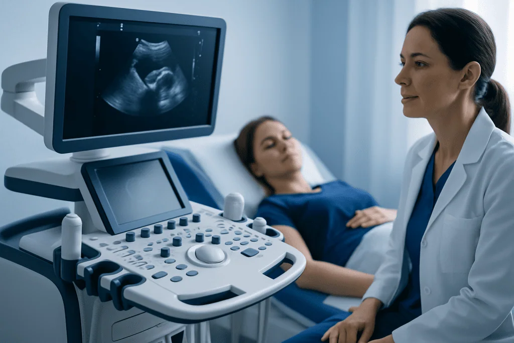
When a doctor says the uterus has a heterogeneous myometrium, they are reporting that the uterine muscle does not look completely uniform on imaging. This can be a diffuse change throughout the uterine wall or a more localized area. It does not automatically mean cancer, and it does not by itself establish a specific diagnosis.
One common cause is adenomyosis, a condition in which tissue similar to the uterine lining grows into the uterine muscle. This may make the uterus look enlarged, irregular or uneven on ultrasound or MRI. Fibroids can also alter the appearance of the myometrium, especially if there are several small fibroids or a fibroid within the uterine wall.
Transvaginal ultrasound is often the first imaging test. MRI may be useful when the diagnosis remains uncertain, when symptoms are significant, or when detailed planning is needed before a procedure. A radiology report should be discussed with a gynecologist, who can explain what the finding means in the individual clinical context.
Should I worry about heterogeneous myometrium?

Heterogeneous myometrium is usually not a reason to panic, but it is appropriate to discuss it with a gynecologist. Its importance depends on the likely cause and on whether the person has symptoms such as unusually heavy bleeding, worsening period pain, pain during sex, pelvic pressure or difficulty becoming pregnant.
Many uterine conditions associated with this finding are benign. However, persistent heavy bleeding can lead to iron-deficiency anemia, and ongoing pelvic pain can affect daily life. Evaluation is especially important when bleeding occurs after menopause, between periods, after sex, or together with unexplained weight loss, fever or severe pain.
A doctor may review the imaging, assess the size and shape of the uterus, check for anemia with blood tests, and consider additional imaging or evaluation of the uterine lining when clinically indicated. This process helps distinguish adenomyosis from fibroids and other possible explanations while excluding conditions that require different care.
Causes, symptoms and who may need assessment
Adenomyosis is often considered when heterogeneous myometrium is seen alongside heavy, painful periods and a tender or enlarged uterus. The condition can occur during reproductive years and is often identified in people in their 30s and 40s, although it can affect younger individuals as well. It may occur together with fibroids, endometriosis or both.
Symptoms vary considerably. Some people have no symptoms, while others experience heavy or prolonged menstrual bleeding, cramping that becomes more severe over time, chronic pelvic pain, bloating, pressure, pain during intercourse or fatigue related to anemia. Symptoms may overlap with other gynecologic conditions, which is why imaging alone is not enough to determine treatment.
Factors associated with adenomyosis may include prior pregnancy and prior procedures involving the uterus, but it is not always possible to identify a clear cause. A person’s reproductive goals are also important. Someone seeking pregnancy may need a different approach from someone who does not wish to preserve fertility or who has completed childbearing.
- Heavy bleeding may require testing for anemia and discussion of bleeding control.
- Persistent pain should be assessed for adenomyosis, endometriosis, fibroids and non-gynecologic causes.
- Difficulty conceiving deserves fertility-focused evaluation rather than assuming the scan finding is the sole explanation.
What is the treatment for heterogeneous myometrium?
Treatment for heterogeneous myometrium depends on the underlying diagnosis. If adenomyosis is suspected and symptoms are mild, observation with periodic review may be appropriate. Pain-relieving anti-inflammatory medicines may help menstrual cramps for some people, while hormonal treatment can reduce bleeding and suppress the hormonal stimulation that contributes to adenomyosis symptoms.
Hormonal options may include combined hormonal contraception, progestin-only treatment, or a levonorgestrel-releasing intrauterine system. The suitability of each option depends on medical history, bleeding pattern, other medications and pregnancy plans. A clinician may also recommend iron treatment if blood tests show iron deficiency or anemia caused by heavy bleeding.
If symptoms remain severe despite medication, or if there are coexisting fibroids or other structural concerns, procedural options may be discussed. Depending on the diagnosis and fertility goals, these may include uterine fibroid embolization for appropriately selected fibroid-related symptoms, focused treatment of a specific lesion, or surgery. Hysterectomy removes the uterus and is the definitive treatment for adenomyosis in people who do not wish to become pregnant in the future, but it is generally considered only after a careful discussion of alternatives.
The expected result is usually improvement in bleeding, pain and quality of life rather than a guaranteed cure from every option. Treatment should be reviewed over time, since symptoms, imaging findings and reproductive priorities can change.
How procedures work: candidacy, steps and recovery
There is no single procedure performed simply because a scan says “heterogeneous myometrium.” A procedure is considered when a defined condition is causing substantial symptoms, when medication has not provided adequate relief, or when a person prefers a procedural approach after informed discussion. Before treatment, the gynecology team confirms the likely diagnosis, reviews imaging, checks blood counts when bleeding is heavy, and discusses fertility wishes.
For a procedure such as uterine artery embolization, an interventional radiologist accesses an artery through a small puncture, commonly in the wrist or groin, and guides a catheter using imaging. Tiny particles are delivered to reduce blood flow to targeted fibroids. This approach is primarily used for fibroids and may not be suitable for all forms of adenomyosis. Recovery often includes pelvic cramping and fatigue for several days, with a gradual return to normal activities as advised by the treating team.
When hysterectomy is chosen, the surgical method may be vaginal, laparoscopic, robotic-assisted or open abdominal, depending on the uterus, other findings and surgical history. Recovery time differs by approach: minimally invasive surgery generally involves a shorter recovery than open surgery. Risks of procedures can include bleeding, infection, anesthesia-related complications, blood clots, injury to nearby organs and the possibility that symptoms may not improve as expected with uterus-preserving options.
Benefits and risks should be considered in relation to symptom severity and future pregnancy plans. A multidisciplinary assessment may include gynecology, radiology, fertility specialists and pain-management professionals when needed. Acibadem International’s multidisciplinary specialists and JCI-accredited hospitals support diagnosis and treatment planning for international patients.
Does adenomyosis cause a big tummy?
Adenomyosis can sometimes make the uterus larger, which may contribute to lower-abdominal fullness or bloating. However, a noticeably enlarged abdomen is not specific to adenomyosis and may have many possible causes, including fibroids, bowel conditions, weight changes, pregnancy, ovarian cysts or fluid accumulation. It should not be assumed to be caused by adenomyosis without medical assessment.
Fibroids are particularly likely to enlarge the uterus and produce pressure or a visible abdominal increase when they are large or numerous. Because adenomyosis and fibroids can occur together, imaging is useful for clarifying whether one or both may be contributing. Related information about uterine fibroids may be helpful when fibroids are mentioned in an imaging report.
New, rapidly worsening or persistent abdominal enlargement should be reviewed by a clinician, especially if it occurs with pain, feeling full quickly, changes in bowel or bladder habits, unexplained weight change, or abnormal bleeding. Prompt assessment helps identify the cause and determine whether treatment is needed.
When to seek medical care
A routine gynecology appointment is appropriate after an imaging report notes heterogeneous myometrium, particularly if menstrual symptoms are affecting work, sleep, exercise, sexual wellbeing or quality of life. Bringing the imaging report, a record of cycle patterns and a list of medicines can help make the consultation more useful.
Medical care should be sought promptly for very heavy bleeding, dizziness or fainting, severe sudden pelvic pain, fever with pelvic symptoms, or possible pregnancy with pain or bleeding. Emergency care may be needed if bleeding is soaking through pads or tampons very quickly, severe weakness develops, or pain is intense and worsening.
People trying to conceive should speak with a gynecologist or fertility specialist if pregnancy has not occurred as expected, particularly when there are also painful or heavy periods. Treatment decisions for suspected adenomyosis require careful balancing of symptom control with reproductive plans, and individualized advice from a qualified clinician is essential.
Frequently asked questions
Can heterogeneous myometrium go away on its own?
The imaging appearance may remain stable, change over time, or improve after menopause if it is related to adenomyosis. Whether symptoms improve without treatment varies from person to person. Follow-up should be guided by symptoms and the clinician’s interpretation of the imaging findings.
Is heterogeneous myometrium the same as adenomyosis?
No. Heterogeneous myometrium is a descriptive imaging finding, whereas adenomyosis is a specific condition that may cause that appearance. A gynecologist uses symptoms, examination and imaging features to decide whether adenomyosis is likely.
Can heterogeneous myometrium affect fertility?
It may be associated with a condition such as adenomyosis, which can be relevant in some people experiencing infertility or pregnancy complications. However, many factors influence fertility, and the scan finding alone cannot predict an individual’s chance of pregnancy. A fertility assessment can identify other possible contributing factors.
Will I need surgery for heterogeneous myometrium?
Most people do not need surgery based on this finding alone. Surgery is usually considered for severe, persistent symptoms, coexisting conditions such as significant fibroids, or when other treatments have not been effective. Fertility goals strongly influence which options are appropriate.
What tests may be needed after this ultrasound finding?
A clinician may review the ultrasound images, perform a pelvic examination and request blood tests if heavy bleeding raises concern about anemia. MRI may be recommended when the diagnosis is uncertain or further detail is needed for treatment planning. Additional tests depend on age, symptoms and bleeding pattern.
Can hormonal treatment help adenomyosis symptoms?
Hormonal treatments can reduce heavy bleeding and pain for many people with suspected or confirmed adenomyosis. Options are chosen according to medical history, side effects, contraception needs and plans for pregnancy. A clinician can explain expected benefits and limitations before treatment begins.
References
- American College of Obstetricians and Gynecologists
- National Institute for Health and Care Excellence
- Mayo Clinic
- Radiological Society of North America
- World Health Organization
This article is for general information only and is not a substitute for professional medical advice. Please consult a qualified doctor about your individual situation.
Explore treatments in Turkey — costs, top hospitals & a free quote
JCI-accredited · board-certified surgeons · reply within 24h
Add us as a Preferred Source to see more of our trusted health content across Google Search, AI Overviews and Discover.

More from the Health Library



Related Specialists

Dt. Bengi Kaya Mert
Dentistry
Prof. Dr. İkbal Esen Aydingöz
Dermatology
Dr. Ertuğrul Yaltı
General Surgery
Dr. Safiye Sayilir
Physical Medicine & Rehabilitation