Hip Labral Tear: Groin Pain, MRI Diagnosis, and Arthroscopy Decisions

A hip labral tear commonly causes deep groin pain, pain with twisting, clicking, catching, or a feeling that the hip is not moving smoothly. Femoroacetabular impingement, hip dysplasia, trauma, repetitive sports movements, and early joint wear can increase the risk of labral injury.
Key Takeaways
- A hip labral tear commonly causes deep groin pain, pain with twisting, clicking, catching, or a feeling that the hip is not moving smoothly.
- Femoroacetabular impingement, hip dysplasia, trauma, repetitive sports movements, and early joint wear can increase the risk of labral injury.
- MRI or MR arthrogram can help confirm a labral tear, but imaging findings must be interpreted together with symptoms and a physical examination.
- Many people improve with non-surgical care, including activity modification, physiotherapy, pain control, and treatment of movement patterns.
- Hip arthroscopy may be considered when pain and mechanical symptoms continue despite appropriate conservative treatment, especially if correctable bony impingement is present.
- Early medical assessment is helpful if groin pain persists, limits walking or sport, or is associated with locking, instability, or loss of hip motion.
A hip labral tear is an injury to the ring of cartilage that helps stabilize the hip joint and often causes deep groin pain, catching, clicking, or stiffness. Diagnosis may require careful examination and advanced imaging, and treatment decisions range from rehabilitation to hip arthroscopy depending on symptoms, anatomy, and daily activity needs.
Overview
A hip labral tear is a tear in the labrum, the strong ring of fibrocartilage that surrounds the rim of the hip socket. The labrum helps deepen the socket, supports joint stability, and contributes to smooth movement between the ball of the thigh bone and the pelvis. When it is irritated or torn, a person may feel pain deep in the groin, a catching sensation, clicking, stiffness, or discomfort with sitting, squatting, running, or turning.
Not every labral tear seen on imaging causes symptoms. Some people have labral changes on MRI without pain, especially as they get older or if they have changes in the shape of the hip joint. For this reason, doctors do not diagnose a hip labral tear from a scan alone. The diagnosis is strongest when the person’s symptoms, physical examination, and imaging findings all fit together.
Hip labral tears are often discussed together with femoroacetabular impingement, also called FAI. In FAI, the ball and socket do not clear each other smoothly during certain movements, and repeated contact can injure the labrum and nearby cartilage. Treatment is individualized and may include education, physiotherapy, medicines, injections, or arthroscopic surgery when symptoms remain limiting.
Symptoms of a Hip Labral Tear
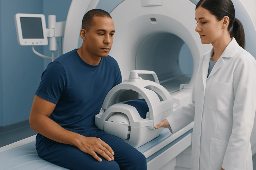
The most typical symptom of a hip labral tear is deep pain in the front of the hip or groin. Some people describe it as a sharp pinch when the hip bends or rotates, while others feel a dull ache after activity or prolonged sitting. Pain may also be felt along the outside of the hip, in the buttock, or down the front of the thigh, although pain below the knee is less typical for an isolated labral problem.
Mechanical symptoms are common but not always present. These can include clicking, catching, locking, popping, or a sensation that the hip briefly sticks during movement. A person may notice discomfort when getting in and out of a car, climbing stairs, rising from a low chair, pivoting during sport, or bringing the knee toward the chest.
Symptoms can overlap with other hip and pelvis conditions, such as muscle or tendon strain, iliopsoas irritation, athletic pubalgia, sacroiliac joint pain, lumbar spine problems, hip osteoarthritis, or stress injury. Important symptom patterns to share with a doctor include when the pain started, whether there was a specific injury, what movements trigger it, and whether the hip feels unstable or blocked.
- Common symptoms include groin pain, hip stiffness, clicking, catching, and pain with twisting.
- Symptoms often worsen with deep hip flexion, prolonged sitting, running, squatting, or pivoting.
- Some labral tears are found incidentally and may not be the main cause of pain.
Causes and Risk Factors
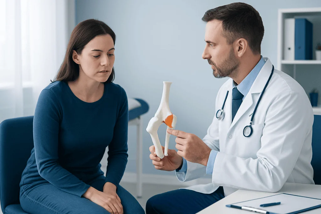
Hip labral tears can occur after a single injury, but they more often develop from repeated stress on the hip joint. Sports and activities involving twisting, kicking, deep squatting, rapid changes of direction, or repetitive hip flexion can place extra load on the labrum. Examples include football, soccer, hockey, ballet, martial arts, distance running, and certain gym-based movements, although a labral tear can also occur in people who are not athletes.
Hip shape is an important factor. In cam-type femoroacetabular impingement, extra bone on the femoral head-neck area can press against the socket during hip flexion and rotation. In pincer-type impingement, extra coverage or positioning of the socket can pinch the labrum. Hip dysplasia, where the socket is shallow or undercovers the ball, can also overload the labrum as it works harder to stabilize the joint.
Other contributors include previous hip trauma, ligament laxity, differences in pelvic or spine mechanics, and early degenerative changes in the joint. As cartilage wear progresses, labral tears may occur together with osteoarthritis. This distinction matters because treatment options and expected recovery can differ when there is more advanced joint damage.
A risk factor does not mean a person will definitely develop a symptomatic tear. Many people with bony variations remain active without pain. The goal of evaluation is to understand whether the labrum is the main pain source, what underlying mechanics may be contributing, and which treatment approach is most appropriate.
Diagnosis and MRI Findings
Diagnosis begins with a medical history and physical examination. The doctor may assess walking pattern, hip range of motion, strength, flexibility, posture, and pain with specific movements. Tests that bring the hip into flexion and rotation may reproduce groin pain, but no single examination test proves a labral tear. The examination also helps identify other possible causes, including lumbar spine, tendon, pelvic, or nerve-related pain.
Plain X-rays are often used first to evaluate hip shape, joint space, dysplasia, signs of impingement, previous injury, or arthritis. X-rays do not show the labrum directly, but they provide important information about the structure of the hip. This is essential because a labral tear is frequently related to the shape of the ball and socket rather than being an isolated soft-tissue injury.
MRI can show the labrum, cartilage, bone marrow, muscles, and surrounding soft tissues. In some cases, an MR arthrogram is recommended. This is a type of MRI performed after contrast fluid is placed into the hip joint to outline the labrum more clearly. MR arthrogram may be especially helpful when the diagnosis remains uncertain or when surgical planning is being considered.
Imaging results should be interpreted carefully. A report may mention a labral tear, fraying, degeneration, cysts, cartilage changes, or impingement morphology, but the scan must be matched to the patient’s pain pattern and examination. Sometimes a diagnostic injection with local anesthetic into the hip joint is used to help confirm whether the hip joint itself is the main source of symptoms.
Treatment Options Without Surgery
Many people with a hip labral tear begin with non-surgical treatment, particularly when symptoms are mild to moderate, there is no true locking, and daily activities are manageable. The first step is often activity modification. This may mean temporarily reducing deep squats, repetitive pivoting, long runs, high-impact exercise, or positions that cause pinching, while maintaining safe general fitness.
Physiotherapy is central to conservative care. A structured program may work on hip and core strength, gluteal control, pelvic stability, gentle mobility, gait mechanics, and gradual return to sport or work tasks. The aim is not simply to stretch the hip, because aggressive stretching into painful impingement positions can worsen symptoms. Instead, rehabilitation focuses on improving control and reducing avoidable stress on the irritated labrum.
Pain relief may include short-term use of anti-inflammatory or analgesic medicines when appropriate, always considering personal medical history and a doctor’s advice. Ice or heat may help some patients. If pain remains difficult to localize, an image-guided hip injection may be used for diagnostic and therapeutic purposes. Corticosteroid or other injection options should be discussed individually, including potential benefits, limitations, and risks.
Improvement can take time. A person’s response depends on the size and type of tear, hip shape, cartilage health, activity demands, and consistency with rehabilitation. If symptoms improve and the hip becomes more functional, surgery may not be needed. If pain, catching, and limitations continue despite a well-guided program, a specialist may discuss arthroscopy.
When Hip Arthroscopy Is Considered
Hip arthroscopy is a minimally invasive surgical technique performed through small incisions using a camera and specialized instruments. It may be considered when a symptomatic hip labral tear continues to limit life despite appropriate non-surgical care, especially when imaging and examination support the labrum as the pain source. The decision is not based only on the presence of a tear on MRI; it also depends on symptoms, function, hip anatomy, cartilage condition, age, activity goals, and overall health.
During arthroscopy, the surgeon may repair the labrum, reconstruct it in selected cases, or remove unstable damaged tissue when repair is not suitable. If femoroacetabular impingement is contributing, the surgeon may reshape the relevant bone to improve clearance between the ball and socket. Treating the underlying impingement can be important because repairing the labrum without addressing a mechanical cause may not solve the problem.
Arthroscopy is generally less invasive than open hip surgery, but it is still an operation and requires careful consideration. Possible risks include infection, bleeding, blood clots, nerve irritation, persistent pain, stiffness, or the need for further procedures. Outcomes may be less predictable when there is significant osteoarthritis or advanced cartilage loss, so the joint condition is an important part of preoperative counseling.
Recovery usually includes protected weight bearing for a period of time, physiotherapy, gradual strengthening, and a staged return to work or sport. Timelines vary according to the procedure performed and the individual’s healing. Patients considering surgery should have a clear discussion about goals, alternatives, rehabilitation commitment, and realistic expectations.
Prevention, Self-care, and When to See a Doctor
Not all hip labral tears can be prevented, especially when hip shape or a sudden injury is involved. However, good movement habits may reduce unnecessary joint stress. Helpful measures include building hip and core strength, increasing training load gradually, using proper technique during squats and sport-specific movements, allowing recovery between high-impact sessions, and avoiding repeated positions that create a sharp groin pinch.
People with recurring hip discomfort should pay attention to early warning signs rather than pushing through persistent pain. Modifying activity for a short period, cross-training with lower-impact exercise, and working with a physiotherapist can help prevent symptoms from becoming more limiting. Maintaining a healthy body weight and treating related back, pelvic, or knee mechanics may also support hip function.
A doctor should be consulted if groin or hip pain lasts more than a few weeks, interferes with walking or sleep, follows a significant injury, or is accompanied by locking, giving way, fever, unexplained weight loss, or inability to bear weight. Urgent medical assessment is needed after major trauma or if the person cannot stand or move the hip normally.
For international patients who need assessment, imaging, rehabilitation planning, or surgical consultation, Acibadem International’s multidisciplinary specialists and JCI-accredited hospitals diagnose and treat hip labral tears as part of comprehensive orthopedic care. Any treatment decision should be made after an individualized examination and discussion with a qualified healthcare professional.
Frequently asked questions
What does a hip labral tear feel like?
A hip labral tear often feels like deep pain in the groin or front of the hip, especially with twisting, squatting, running, or prolonged sitting. Some people notice clicking, catching, locking, or a sense that the hip is not moving smoothly. Symptoms vary, and similar pain can come from tendons, the spine, or other hip joint problems.
Can a hip labral tear heal on its own?
The torn labral tissue may not fully regrow in the same way as some other tissues, but symptoms can still improve significantly without surgery. Physiotherapy, activity modification, and pain control can reduce irritation and improve hip mechanics. The goal is to restore comfortable function, not necessarily to make the MRI appear normal.
Is MRI enough to diagnose a hip labral tear?
MRI or MR arthrogram can provide important evidence, but diagnosis should not rely on imaging alone. Some labral tears found on scans do not cause symptoms. A doctor combines the scan with the patient’s history, physical examination, X-rays, and sometimes a diagnostic injection.
What is the difference between MRI and MR arthrogram for the hip?
A standard MRI uses magnetic imaging to show the hip joint and surrounding tissues. An MR arthrogram involves placing contrast fluid into the joint before the MRI, which can outline small labral tears more clearly in selected cases. The best choice depends on symptoms, prior imaging, and whether surgery is being considered.
When is hip arthroscopy recommended for a labral tear?
Hip arthroscopy may be recommended when pain and mechanical symptoms continue despite a well-planned period of non-surgical treatment. It is more likely to be considered when the tear matches the symptoms and there is a treatable structural problem, such as femoroacetabular impingement. Significant arthritis or cartilage loss can change the expected benefit and should be discussed carefully.
How long does recovery take after hip arthroscopy?
Recovery varies depending on whether the labrum is repaired, bone is reshaped, cartilage is treated, and the person’s overall condition. Most patients need a staged rehabilitation program with early protection, gradual range of motion, strengthening, and progressive return to activity. The surgeon and physiotherapist provide an individualized timeline.
Can someone exercise with a hip labral tear?
Many people can stay active, but they may need to avoid movements that create sharp groin pain, deep pinching, or repeated catching. Lower-impact activities and guided strengthening are often better tolerated during recovery. A physiotherapist or sports medicine doctor can help adapt exercise safely to the person’s symptoms and goals.
References
- American Academy of Orthopaedic Surgeons
- British Hip Society
- International Society for Hip Arthroscopy
- Radiological Society of North America
- National Institute for Health and Care Excellence
This article is for general information only and is not a substitute for professional medical advice. Please consult a qualified doctor about your individual situation.
Arthroscopy in Turkey — costs, top hospitals & a free quote
JCI-accredited · board-certified surgeons · reply within 24h
Add us as a Preferred Source to see more of our trusted health content across Google Search, AI Overviews and Discover.

Orthopedics & Spine
Care for bones, joints and the musculoskeletal system, including joint replacement, sports injuries and trauma.
125 specialists in this unit