

Image-Guided Biopsy: How Tissue Samples Are Taken Safely
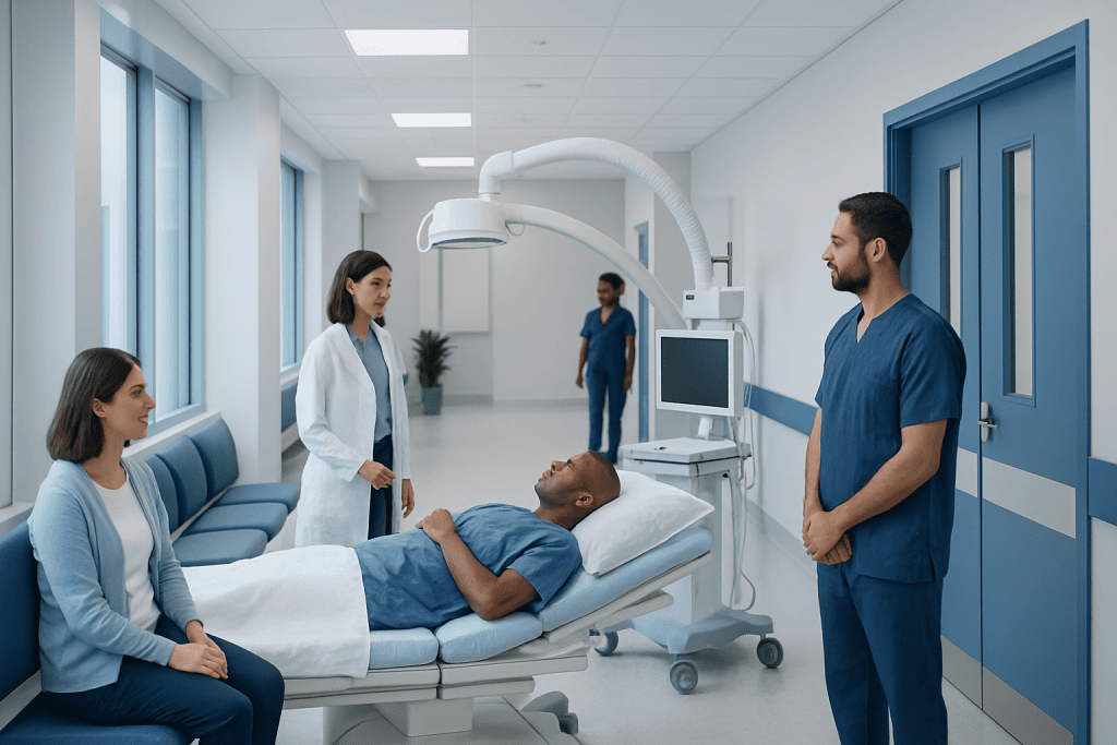
Image-guided biopsy uses ultrasound, CT, MRI, mammography, or fluoroscopy to guide a needle accurately to the area being examined. Most procedures are performed with local anesthesia and do not require a large incision or a hospital stay.
Key Takeaways
- Image-guided biopsy uses ultrasound, CT, MRI, mammography, or fluoroscopy to guide a needle accurately to the area being examined.
- Most procedures are performed with local anesthesia and do not require a large incision or a hospital stay.
- The tissue sample is examined by a pathologist, and results help doctors confirm a diagnosis and plan the most appropriate care.
- Preparation may include blood tests, reviewing medications such as blood thinners, and fasting if sedation is planned.
- Mild soreness or bruising is common after biopsy, while serious complications are uncommon when the procedure is carefully planned and monitored.
An image-guided biopsy is a minimally invasive procedure that uses imaging technology to help doctors collect a small tissue sample from a precise area of the body. It is commonly used to diagnose or rule out conditions such as cancer, infection, inflammation, and other abnormal tissue changes.
Overview
An image-guided biopsy is a medical procedure used to remove a small sample of tissue, cells, or fluid from the body for laboratory examination. The key feature is that the doctor uses imaging, such as ultrasound, computed tomography, magnetic resonance imaging, mammography, or fluoroscopy, to see the target area and guide the biopsy needle with precision.
This approach is often recommended when an abnormal area is found during a scan, physical examination, or screening test. The sample is usually examined by a pathologist, a specialist who studies cells and tissues under a microscope. The findings can help determine whether an abnormality is benign, cancerous, infectious, inflammatory, or related to another condition.
Compared with open surgical biopsy, many image-guided biopsies are less invasive. They typically involve a small skin puncture rather than a large incision, and many patients go home the same day. The exact experience depends on the part of the body being examined, the imaging method used, the type of needle, and the person’s overall health.
Why an Image-Guided Biopsy Is Performed
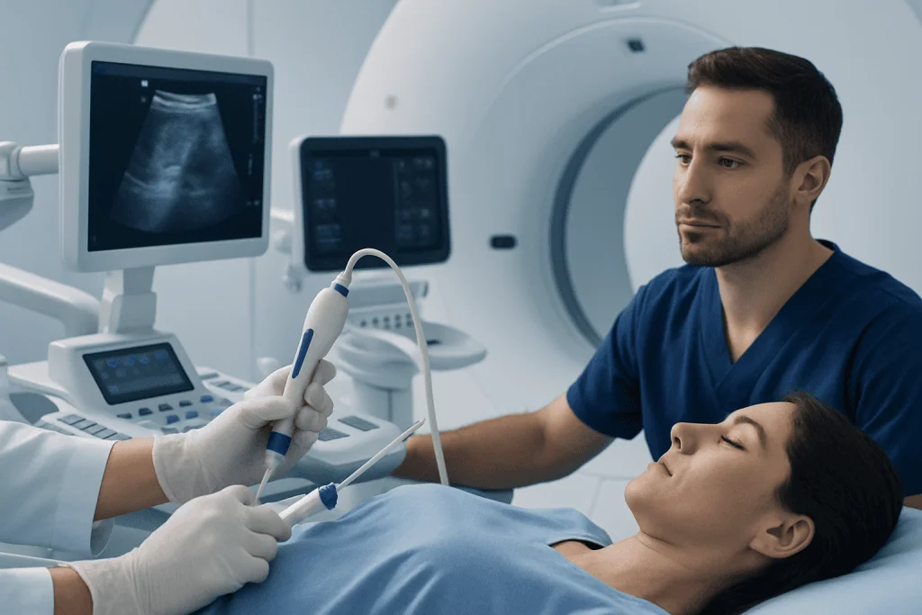
Doctors request an image-guided biopsy when imaging tests show an area that needs a more definite explanation. Scans can describe the size, shape, and location of an abnormality, but a tissue sample is often needed to confirm what it is. This is especially important when treatment decisions depend on the exact diagnosis.
Image-guided biopsy may be used for many organs and tissues, including the breast, thyroid, liver, lung, kidney, prostate, lymph nodes, bone, soft tissues, and abdominal or pelvic masses. It can also be used to sample fluid collections or suspected infection when identifying the cause may guide treatment.
Common reasons for the procedure include:
- Clarifying whether a lump, nodule, or mass is benign or malignant.
- Confirming the type of cancer before surgery, chemotherapy, radiotherapy, or targeted treatment.
- Checking for infection, inflammation, or autoimmune-related tissue changes.
- Investigating abnormal blood tests, such as liver or kidney changes, when imaging suggests a target area.
- Reassessing disease that has changed over time or has not responded as expected to treatment.
Types of Image Guidance and Biopsy Techniques
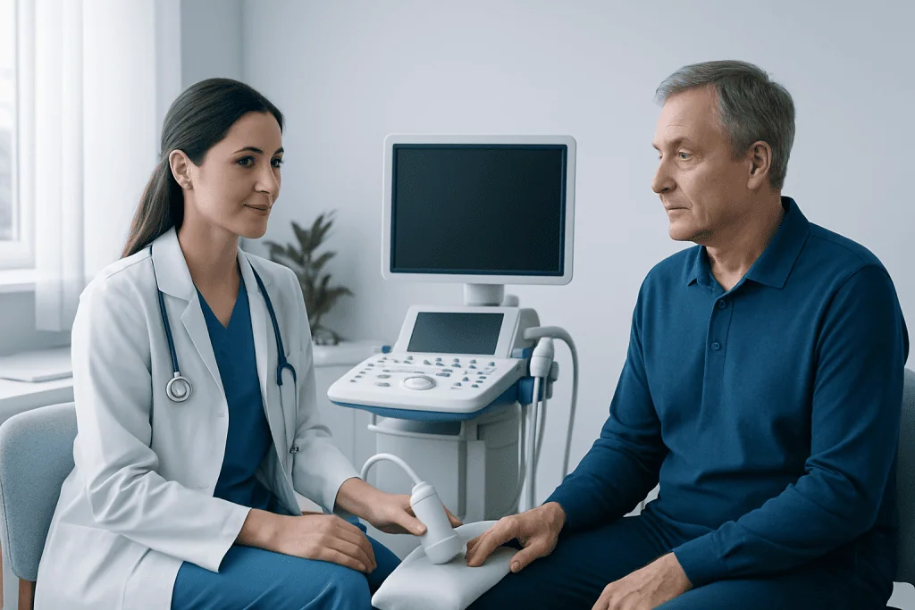
The imaging method is chosen according to where the abnormality is located and which method shows it most clearly. Ultrasound is often used for areas near the surface, such as the thyroid, breast, lymph nodes, or some liver lesions. CT is commonly used for deeper areas in the chest, abdomen, pelvis, bone, or lung. MRI can be helpful when the target is best seen on MRI, such as certain breast, prostate, or soft-tissue findings. Mammography or stereotactic guidance is frequently used for some breast abnormalities, especially tiny calcifications.
Several biopsy techniques may be used. Fine needle aspiration uses a thin needle to collect cells or fluid. Core needle biopsy uses a slightly larger needle to remove a small cylinder of tissue, which often provides more information about tissue structure. Vacuum-assisted biopsy may collect multiple samples through one small skin opening, especially in breast procedures. In selected cases, a marker clip may be placed to identify the sampled area for future imaging or surgery.
The choice of technique depends on the suspected condition, the size and location of the target, nearby blood vessels or organs, and how much tissue is needed for diagnosis. The doctor aims to collect enough material for accurate testing while keeping the procedure as safe and comfortable as possible.
How to Prepare for the Procedure
Preparation begins with a review of the patient’s medical history, allergies, previous imaging, and current medications. It is important to tell the medical team about blood thinners, antiplatelet medicines, supplements that may affect bleeding, pregnancy or possible pregnancy, implanted medical devices, and any previous reaction to anesthesia or contrast agents. Patients should not stop prescribed medicines unless a doctor specifically advises them to do so.
Some patients need blood tests before biopsy, especially if the procedure involves an organ with a higher bleeding risk or if there is a history of bleeding problems. If sedation is planned, fasting may be required for a certain period before the appointment. For many biopsies performed with local anesthesia only, fasting may not be necessary, but instructions vary by procedure and facility.
Patients are usually asked to bring previous scans or reports if they were performed elsewhere. Wearing comfortable clothing and arranging for someone to drive home may be recommended, particularly if sedation is used or if the biopsy is in a location that may require longer observation afterward.
What Happens During an Image-Guided Biopsy
Before the biopsy, the team confirms the patient’s identity, the planned biopsy site, relevant medical details, and consent. The patient is positioned so the target area can be seen clearly with the imaging equipment. The skin is cleaned with antiseptic solution, and sterile drapes are placed. Local anesthetic is injected to numb the skin and deeper tissues. Patients may feel pressure, pushing, or brief discomfort, but sharp pain should be reported so more local anesthetic can be considered.
The doctor then advances the biopsy needle while watching its position on the imaging screen. In some procedures, the needle may be repositioned slightly to collect samples from the best part of the target. Several small samples are often taken to improve diagnostic accuracy. Patients may be asked to hold their breath briefly during lung, liver, or upper abdominal biopsies to keep the target still.
After the samples are collected, pressure is applied to the site to reduce bleeding, and a small dressing is placed. The tissue is sent to the laboratory for processing and microscopic examination. Some samples may also undergo special stains, cultures, molecular tests, or genetic marker testing, depending on the clinical question. Results may take several days or longer if advanced testing is required.
Safety, Risks, and Recovery
Image-guided biopsy is generally considered safe and is performed by trained specialists who plan the needle path carefully. Imaging helps avoid important structures such as major blood vessels, nerves, and organs whenever possible. Even so, every medical procedure has potential risks, and these depend on the biopsy site and the patient’s individual health factors.
Common after-effects include mild soreness, bruising, or a small amount of bleeding at the puncture site. These usually improve with simple care and rest as advised by the medical team. Less common risks include infection, more significant bleeding, an inadequate sample that requires repeat biopsy, or injury to nearby structures. Lung biopsy can sometimes cause air to leak around the lung, known as pneumothorax, which is why observation and a follow-up chest X-ray may be used in selected patients.
After the procedure, patients may be monitored for a short time or longer depending on the biopsy type. Discharge instructions usually include how to care for the dressing, when normal activities can resume, and which activities to avoid temporarily, such as heavy lifting or strenuous exercise. Patients should follow their specific instructions because recovery recommendations differ for breast, liver, lung, kidney, bone, and other biopsies.
Results, Next Steps, and When to See a Doctor
The biopsy report describes what the pathologist found in the sample. A benign result may still require follow-up imaging if the doctor wants to make sure the sampled tissue matches the imaging appearance. A malignant or suspicious result usually leads to further discussion with the relevant specialist team, such as oncology, surgery, radiology, pathology, or organ-specific specialists. Sometimes the sample is inconclusive, meaning the result does not fully explain the abnormality, and repeat biopsy or another diagnostic approach may be recommended.
Patients should contact their healthcare provider promptly if they develop increasing pain, swelling, persistent bleeding, fever, redness or warmth at the biopsy site, dizziness, shortness of breath, chest pain, or any symptom specifically listed in their discharge instructions. These symptoms do not always mean a serious problem, but they should be assessed so the patient can receive timely guidance.
At Acibadem International, multidisciplinary specialists in JCI-accredited hospitals diagnose and treat conditions that may require image-guided biopsy for international patients. Patients considering the procedure should discuss the reason for biopsy, the expected benefits, possible alternatives, and follow-up plan with a qualified doctor who knows their medical history.
Frequently asked questions
Is an image-guided biopsy painful?
Most patients feel pressure or brief discomfort rather than significant pain because local anesthetic is used to numb the area. Some soreness or bruising afterward is common and usually improves within a few days. Patients should tell the doctor during the procedure if they feel sharp pain.
How long does an image-guided biopsy take?
The needle sampling itself may take only a short time, but preparation, positioning, imaging, and observation afterward add to the appointment length. Many procedures are completed as outpatient visits. More complex biopsies, or those requiring sedation or longer monitoring, may take longer.
Will a biopsy spread cancer if the lump is malignant?
This is a common concern, but modern needle biopsy techniques are designed to minimize risk, and biopsy is routinely used to diagnose cancer safely. The benefits of confirming the diagnosis and choosing the right treatment usually outweigh this very low theoretical risk. Patients should discuss any specific concern with their treating physician.
What is the difference between fine needle aspiration and core needle biopsy?
Fine needle aspiration uses a very thin needle to collect cells or fluid, while core needle biopsy removes a small piece of tissue. Core biopsy often gives more information about tissue architecture and tumor features. The best method depends on the body area, the suspected diagnosis, and what tests the laboratory needs to perform.
When will the biopsy results be ready?
Some results may be available within a few days, but timing varies by laboratory and by the type of tests needed. Special stains, cultures, molecular testing, or second review can take longer. The healthcare team should explain how and when results will be shared.
Can patients return to normal activities after biopsy?
Many patients return to light activities soon after the procedure, especially after a simple superficial biopsy. The doctor may advise avoiding heavy lifting, vigorous exercise, or certain medications for a short time depending on the biopsy site and bleeding risk. Patients should follow the written discharge instructions provided by their care team.
References
- American College of Radiology
- Radiological Society of North America
- National Cancer Institute
- Mayo Clinic
- Society of Interventional Radiology
This article is for general information only and is not a substitute for professional medical advice. Please consult a qualified doctor about your individual situation.
Oncology care in Turkey — second opinion and treatment plan
JCI-accredited · board-certified surgeons · reply within 24h
Add us as a Preferred Source to see more of our trusted health content across Google Search, AI Overviews and Discover.

More from the Health Library



Related Specialists

Prof. Dr. Ercan Karaarslan
Radiology
Prof. Dr. Bora Peynircioğlu
Interventional Radiology
Prof. Dr. Koray Güven
Interventional Radiology
Assoc. Prof. Dr. Deniz Can Aliş
Radiology