

Interventional Neuroradiology: How Minimally Invasive Brain Vessel Procedures Work

Interventional neuroradiology treats blood vessel problems in the brain, spine, head, and neck using catheters and live imaging. Common procedures include cerebral angiography, aneurysm coiling, embolization, stenting, and mechanical thrombectomy for some strokes.
Key Takeaways
- Interventional neuroradiology treats blood vessel problems in the brain, spine, head, and neck using catheters and live imaging.
- Common procedures include cerebral angiography, aneurysm coiling, embolization, stenting, and mechanical thrombectomy for some strokes.
- Many treatments are performed through a small puncture in the wrist or groin rather than open surgery.
- Recovery is often faster than with traditional surgery, but each procedure still has risks and requires specialist evaluation.
- Treatment choice depends on the exact condition, symptoms, anatomy, and the patient’s overall health.
Interventional neuroradiology is a medical specialty that uses imaging guidance and thin catheters to diagnose and treat disorders of the brain, spine, head, and neck blood vessels. These procedures can often offer an effective alternative to open surgery, with smaller incisions and shorter recovery times for selected patients.
Overview
Interventional neuroradiology is a highly specialized field that combines advanced imaging with minimally invasive treatment techniques. Instead of making a large surgical opening, the specialist guides very thin tubes called catheters through the blood vessels, usually starting from the wrist or groin, to reach a problem area in the brain, spine, head, or neck.
Using live X-ray imaging and contrast dye, the doctor can see blood flow in real time and deliver treatment directly where it is needed. This approach is often called endovascular treatment because it works from inside the blood vessels. For many patients, it can reduce tissue disruption, shorten hospital stay, and support a quicker return to daily activities compared with some open operations.
Interventional neuroradiology may be used for both diagnosis and treatment. A diagnostic test such as cerebral angiography can map the blood vessels in great detail. Therapeutic procedures can then be used to close an abnormal vessel, open a blocked artery, repair an aneurysm, or reduce blood supply to a tumor before surgery.
These procedures are typically performed by a multidisciplinary team that may include interventional neuroradiologists, neurologists, neurosurgeons, anesthesiologists, and stroke specialists. Treatment planning is individualized, because the safest and most effective approach depends on the patient’s symptoms, imaging findings, and overall health.
What conditions can it treat?
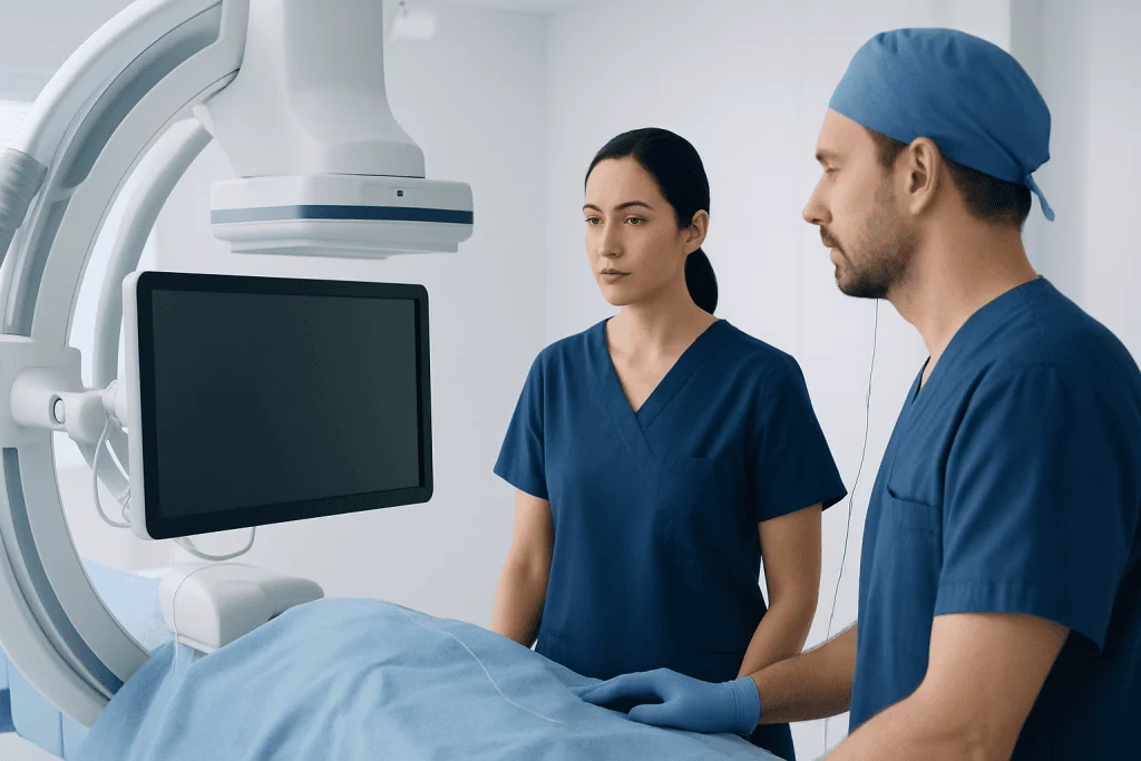
Interventional neuroradiology is used for a range of vascular and related conditions affecting the nervous system. One of the best-known examples is the treatment of a brain aneurysm, where coils, stents, or flow-diverting devices may help prevent rupture or rebleeding. It is also used to treat arteriovenous malformations and fistulas, which are abnormal connections between arteries and veins.
Another major area is stroke care. In selected patients with a large vessel blockage, an urgent endovascular procedure may remove the clot and restore blood flow to brain tissue. This is called mechanical thrombectomy, and it is now an important part of modern treatment for certain ischemic strokes.
Interventional neuroradiology may also help manage narrowing of blood vessels, bleeding, some spinal vascular disorders, and conditions that cause pulsating noise in the ear or neurological symptoms related to abnormal circulation. In some cases, it is used before surgery to reduce blood flow to highly vascular tumors, making later treatment safer and technically easier.
Commonly treated conditions and procedures include:
- Brain aneurysms
- Arteriovenous malformations and dural arteriovenous fistulas
- Acute ischemic stroke due to large vessel occlusion
- Carotid or intracranial artery narrowing in selected cases
- Bleeding from certain vascular abnormalities
- Preoperative tumor embolization
- Spinal vascular malformations
How minimally invasive brain vessel procedures work

Most interventional neuroradiology procedures follow the same basic principle. After the skin is cleaned and numbed, the doctor makes a very small puncture in an artery, often in the wrist or groin. A short tube is placed into the vessel, and thin catheters and wires are guided through the body under continuous imaging until they reach the target area.
Contrast dye is injected so the blood vessels appear clearly on X-ray. This step helps the team understand the exact shape of the vessel problem and safely place treatment devices. Depending on the condition, the doctor may use coils to fill an aneurysm, a stent to support a vessel wall, a clot retriever to remove a blockage, or embolic material to close an abnormal vessel.
A key diagnostic procedure is cerebral angiography, which provides a detailed map of the brain’s circulation. In some patients, angiography is all that is needed to confirm a diagnosis. In others, it is performed immediately before treatment so the team can move directly from diagnosis to therapy in one session.
Many procedures are performed under local anesthesia with sedation, while others require general anesthesia. The decision depends on the urgency of the situation, the complexity of the case, and how still the patient needs to remain. Throughout the procedure, the team closely monitors blood pressure, heart rhythm, oxygen levels, and neurological status.
Diagnosis and pre-procedure evaluation
Before recommending an interventional neuroradiology procedure, doctors usually begin with a detailed medical history, physical examination, and neurological assessment. They also review any symptoms such as sudden severe headache, weakness, speech difficulty, visual changes, dizziness, or pulsating sounds in the ear. The goal is to understand whether symptoms match a vascular problem and how urgent treatment may be.
Imaging plays a central role in diagnosis. Depending on the situation, a patient may have CT, MRI, CT angiography, MR angiography, ultrasound, or formal angiography. Each test provides different information about blood flow, vessel structure, bleeding, stroke, or surrounding brain tissue.
Doctors also check for factors that affect procedural safety, including kidney function, allergies to contrast dye, blood-thinning medications, diabetes, pregnancy, and previous bleeding or clotting problems. Blood tests may be needed before treatment. Patients are commonly advised not to eat or drink for a period before the procedure, and medication instructions are tailored individually.
Because these treatments are highly specialized, decision-making often involves a team discussion. The patient and family are usually informed about the diagnosis, the expected benefits, possible alternatives such as observation or open surgery, and the potential risks. This shared approach helps patients understand why a minimally invasive treatment may or may not be the best option.
Common treatment options and what recovery is like
Treatment is chosen according to the exact condition. For aneurysms, options may include aneurysm coiling or other endovascular techniques that redirect or reduce blood flow into the weak area. For abnormal vessel connections, embolization may be used to block the problematic channels. For narrowed arteries, angioplasty or stenting may sometimes be considered in carefully selected patients.
In acute stroke, rapid treatment is essential. If imaging shows a suitable blockage, mechanical thrombectomy may be performed to physically remove the clot. In other situations, a person may need medication, surgery, or monitoring rather than an endovascular procedure. The best approach depends on time from symptom onset, imaging results, and the location of the problem.
After the procedure, most patients spend several hours in a recovery area where nurses monitor the puncture site, blood pressure, and neurological condition. Some patients go home the same day after a diagnostic test, while others stay overnight or longer, especially after treatment for stroke, bleeding, or a complex vascular abnormality. Temporary instructions may include avoiding heavy lifting, drinking fluids if advised, and watching for swelling or bleeding at the access site.
Recovery varies by procedure and overall health. Many people appreciate the small incision and relatively short recovery period, but follow-up remains important. Repeat imaging may be needed to confirm that the vessel has healed, the aneurysm remains sealed, or the abnormal connection has stayed closed.
Benefits, risks, and possible complications
The main benefit of interventional neuroradiology is that it can treat delicate problems deep inside the body without the larger incisions of open surgery. In the right patient, this may mean less postoperative pain, a shorter hospital stay, a lower burden on surrounding tissues, and a faster return to normal routines. It can also make treatment possible for people who may not be good candidates for conventional surgery.
However, minimally invasive does not mean risk-free. As with any procedure involving blood vessels and contrast dye, there can be complications. These may include bruising or bleeding at the access site, infection, allergic reaction to contrast, kidney strain, blood vessel injury, clot formation, stroke, or recurrence of the treated problem over time.
The level of risk depends on the condition being treated, the blood vessel anatomy, the devices used, and the patient’s age and medical history. Some procedures are performed in emergency situations where the underlying condition itself already carries significant risk. For that reason, specialists carefully balance the expected benefit of treatment against the potential complications.
Patients are encouraged to ask practical questions before treatment, such as what the procedure aims to achieve, what recovery will involve, whether follow-up imaging will be needed, and what warning signs to watch for at home. Clear communication can reduce uncertainty and help patients feel more prepared.
Prevention, follow-up, and when to seek medical care
Not every condition treated by interventional neuroradiology can be prevented, but vascular health can often be supported through everyday habits. Managing blood pressure, avoiding smoking, controlling diabetes, staying physically active, and following treatment for high cholesterol may lower the risk of some blood vessel diseases. People with a family history of aneurysm or stroke may benefit from discussing screening or risk assessment with their doctor when appropriate.
Follow-up care is an important part of successful treatment. Some patients need repeat scans after aneurysm repair or embolization to ensure that the treated area remains stable. Others may need medications such as antiplatelet therapy after certain stent procedures, always under a doctor’s supervision. Attending scheduled visits helps the team detect changes early and adjust care if needed.
Urgent medical attention is needed for possible stroke symptoms, such as sudden facial drooping, arm weakness, speech difficulty, vision loss, severe imbalance, or sudden confusion. Emergency care is also important for a sudden explosive headache, loss of consciousness, new seizures, or significant bleeding from a recent catheter site. These symptoms do not always mean a serious complication, but they should be assessed promptly.
For people seeking evaluation or treatment across borders, Acibadem International’s multidisciplinary specialists and JCI-accredited hospitals diagnose and treat complex neurovascular conditions for international patients. A specialist consultation can help determine whether interventional neuroradiology, surgery, medication, or observation is the most appropriate next step.
Frequently asked questions
Is interventional neuroradiology the same as brain surgery?
No. Interventional neuroradiology usually treats problems from inside the blood vessels using catheters guided by imaging, rather than through a large surgical opening. In some cases it is an alternative to open surgery, while in others it is used alongside neurosurgical care.
Are these procedures painful?
Many patients feel little pain during the procedure because the access site is numbed and sedation or anesthesia may be used. Afterward, there may be mild soreness or bruising where the catheter was inserted. The care team gives instructions on how to stay comfortable during recovery.
How long does recovery usually take?
Recovery depends on the type of procedure and the condition being treated. A simple diagnostic angiogram may allow a quick return home, while treatment for stroke or a complex vascular problem may require a hospital stay and closer follow-up. The doctor will explain activity limits and when normal routines can resume.
Who is a good candidate for interventional neuroradiology?
Good candidates are people whose imaging and symptoms show a condition that can be safely reached and treated through the blood vessels. Suitability depends on the exact diagnosis, vessel anatomy, age, medical history, and whether the situation is urgent. A specialist review is needed before a decision is made.
What is the difference between angiography and treatment?
Angiography is mainly a diagnostic test that creates a detailed picture of the blood vessels using contrast dye and X-ray imaging. Treatment uses similar catheter techniques, but the doctor also places devices or materials to repair, open, or block a vessel. Sometimes both happen during the same session.
Can interventional neuroradiology help in an emergency stroke?
Yes, in some patients with a large artery blocked by a clot, an urgent endovascular procedure can remove the clot and improve blood flow. This is time-sensitive, so emergency evaluation is essential. Not every stroke is treated this way, and imaging helps determine the best option.
References
- World Stroke Organization
- American Heart Association
- Society of NeuroInterventional Surgery
- Radiological Society of North America
- National Institute of Neurological Disorders and Stroke
This article is for general information only and is not a substitute for professional medical advice. Please consult a qualified doctor about your individual situation.
Interventional Neuroradiology in Turkey — costs, top hospitals & a free quote
JCI-accredited · board-certified surgeons · reply within 24h
Add us as a Preferred Source to see more of our trusted health content across Google Search, AI Overviews and Discover.
Check your numbers in seconds
BMI, calories, due date, blood pressure and 30+ more clinical calculators — free, instant, doctor-reviewed ranges.

Robotic Surgery
Robot-assisted procedures across urology, gynecology and general surgery for greater precision and faster recovery.
More from the Health Library



Related Specialists

Dt. Ayşe Nur Gümüş
Oral & Dental Health
Prof. Dr. Ahmet Cem Batukan
Gynecology & Obstetrics
Prof. Dr. Mert Erkan
General Surgery
Dr. Abdullah Etöz
Aesthetic Plastic & Reconstructive Surgery