Interventional Neuroradiology: Minimally Invasive Treatment for Brain Blood Vessels

Interventional neuroradiology uses advanced imaging and very thin catheters to treat selected brain and spinal blood vessel conditions from inside the blood vessels. Commonly treated conditions include ischemic stroke, brain aneurysms, arteriovenous malformations, dural fistulas, and certain vascular tumors.
Key Takeaways
- Interventional neuroradiology uses advanced imaging and very thin catheters to treat selected brain and spinal blood vessel conditions from inside the blood vessels.
- Commonly treated conditions include ischemic stroke, brain aneurysms, arteriovenous malformations, dural fistulas, and certain vascular tumors.
- Procedures may include mechanical thrombectomy, aneurysm coiling, flow diversion, stenting, and embolization, depending on the diagnosis.
- Treatment decisions are individualized by a multidisciplinary team, often involving neuroradiologists, neurosurgeons, neurologists, anesthesiologists, and intensive care specialists.
- Urgent medical care is important for stroke-like symptoms, sudden severe headache, seizures, or new neurological changes.
Interventional neuroradiology is a specialized field that treats blood vessel conditions of the brain, head, neck, and spine using image-guided, minimally invasive techniques. Many procedures are performed through a small puncture in the groin or wrist, allowing doctors to reach delicate vessels without open surgery.
Overview
Interventional neuroradiology, also called endovascular neurosurgery or neurointervention, is a medical specialty focused on diagnosing and treating problems in the blood vessels of the brain, head, neck, and spine. Instead of opening the skull or spine, specialists use real-time imaging to guide tiny tubes called catheters through the blood vessels to the area that needs treatment.
Most procedures begin with a small puncture, usually in an artery in the groin or wrist. Through this access point, the doctor navigates catheters and microcatheters to the target vessel using X-ray guidance, contrast dye, and high-resolution angiography. This approach can be especially useful in areas where open surgery may be more complex because the blood vessels are small, deep, or close to important brain structures.
Interventional neuroradiology is used in both emergency and planned care. In emergencies, it can restore blood flow during certain types of stroke. In planned treatment, it can help secure aneurysms, close abnormal vessel connections, or reduce bleeding risk before surgery. The goal is always to choose the safest and most effective option for the individual patient.
Conditions Treated
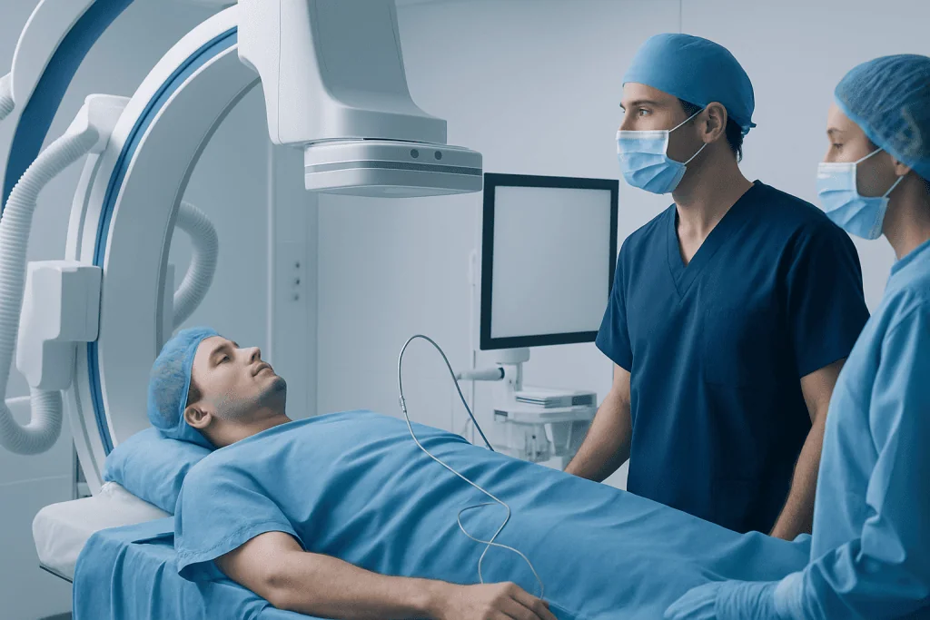
Interventional neuroradiology is most often associated with diseases of the arteries and veins that supply the brain and spinal cord. These conditions vary widely: some cause sudden symptoms, while others are discovered during imaging for headaches, dizziness, trauma, or unrelated health concerns.
Common conditions treated by neurointerventional specialists include:
- Ischemic stroke: a blockage in a brain artery that may be treated with mechanical thrombectomy in selected patients.
- Brain aneurysm: a weakened, bulging area in a blood vessel that may be treated with coiling, stent-assisted coiling, or flow diversion.
- Arteriovenous malformation (AVM): an abnormal tangle of arteries and veins that can sometimes be treated with embolization.
- Dural arteriovenous fistula: an abnormal connection between arteries and veins in the coverings of the brain or spine.
- Carotid or intracranial artery narrowing: selected cases may be managed with angioplasty or stenting.
- Vascular tumors or severe nosebleeds: embolization may reduce blood supply to a tumor or bleeding site.
Not every vascular condition requires a procedure. Some aneurysms, malformations, or vessel narrowings may be monitored with imaging and risk-factor management. The decision depends on the size, location, symptoms, overall health, and the balance of benefits and risks.
Symptoms and Warning Signs

Many brain blood vessel conditions do not cause symptoms until they affect blood flow, bleed, press on nearby structures, or irritate the brain. Symptoms can also overlap with other neurological conditions, so medical evaluation is important rather than self-diagnosis.
Possible symptoms include sudden weakness or numbness on one side of the body, facial drooping, speech difficulty, vision loss, severe dizziness, imbalance, confusion, or a new seizure. A sudden, unusually severe headache may occur with bleeding around the brain and should be assessed urgently. Some aneurysms or vascular malformations may cause persistent headaches, double vision, ringing in the ears that pulses with the heartbeat, or gradual neurological changes.
Stroke-like symptoms should be treated as a medical emergency because time-sensitive treatments may be possible. Even if symptoms improve, a transient ischemic attack, sometimes called a mini-stroke, can be a warning sign that further evaluation is needed. Prompt care allows doctors to identify the cause and discuss treatment or prevention strategies.
Diagnosis and Treatment Planning
Diagnosis usually begins with a medical history, neurological examination, and imaging. Doctors may ask when symptoms started, whether they were sudden or gradual, and whether there are risk factors such as high blood pressure, smoking, previous stroke, clotting disorders, or a family history of aneurysm.
Imaging tests may include computed tomography (CT), magnetic resonance imaging (MRI), CT angiography, MR angiography, and catheter angiography. Catheter angiography, also known as digital subtraction angiography, provides detailed views of blood flow and vessel anatomy. It may be used when the diagnosis is uncertain or when a procedure is being considered.
Treatment planning is typically multidisciplinary. A neurointerventionalist may work with neurologists, neurosurgeons, radiologists, anesthesiologists, intensive care physicians, and rehabilitation specialists. This team approach helps determine whether a minimally invasive procedure, open surgery, medication, observation, or a combination of treatments is most appropriate.
Minimally Invasive Treatment Options
The exact procedure depends on the condition being treated. In mechanical thrombectomy for selected ischemic strokes, a catheter is guided to the blocked artery and a device is used to remove or capture the clot. Restoring blood flow as early as possible can help protect threatened brain tissue, although eligibility depends on imaging findings, timing, and the patient’s overall condition.
For brain aneurysms, endovascular treatment may involve placing soft platinum coils inside the aneurysm to promote clotting and reduce blood flow into the bulge. Some aneurysms need stent-assisted coiling, where a small mesh tube helps keep coils in place. Flow diverters are another type of stent designed to redirect blood away from certain aneurysms so the vessel can gradually heal.
For AVMs, dural fistulas, and some tumors, embolization may be used. During embolization, the doctor delivers special materials, such as medical glue, particles, or coils, to block abnormal blood flow. Embolization may be a stand-alone treatment or may be performed before surgery or radiosurgery to make further treatment safer and more controlled.
In selected cases of narrowed arteries, angioplasty or stenting may be considered to improve blood flow. These treatments are not suitable for every patient and are carefully weighed against medication-based management. The best option is determined after reviewing symptoms, vessel anatomy, stroke risk, and procedural risk.
Benefits, Risks, and Recovery
A major benefit of interventional neuroradiology is that it can treat complex vascular conditions through a small access point rather than a large incision. This may reduce tissue disruption and allow faster early recovery compared with some open surgical approaches. However, the procedure still involves important blood vessels and the brain, so careful planning and monitoring are essential.
Potential risks vary by procedure and condition. They may include bleeding or bruising at the access site, contrast dye reaction, kidney strain in susceptible patients, vessel injury, blood clots, stroke, infection, or incomplete treatment that requires follow-up care. Doctors explain the individual risk profile before planned procedures and take precautions such as imaging checks, blood tests, medication review, and close monitoring during and after treatment.
Recovery depends on the reason for treatment. A patient treated for an unruptured aneurysm may spend a short time in hospital for observation, while a patient treated for a major stroke or bleeding may need intensive care and rehabilitation. Follow-up imaging is often recommended because some treated aneurysms or malformations need long-term surveillance.
Preparation and Self-Care
Before a planned neurointerventional procedure, patients are usually given instructions about eating and drinking, regular medications, blood thinners, allergies, and previous reactions to contrast dye. It is important to tell the medical team about kidney disease, pregnancy, bleeding disorders, implanted devices, and all prescription or non-prescription medicines.
After the procedure, the care team monitors the puncture site, blood pressure, neurological status, and comfort level. Patients may be asked to lie still for a period of time, especially if the groin artery was used. Drinking fluids may be encouraged when appropriate to help clear contrast dye, but instructions depend on the patient’s medical condition.
Long-term self-care focuses on protecting blood vessels and reducing future risk. This may include managing blood pressure, diabetes, and cholesterol; stopping smoking; limiting alcohol; taking prescribed medicines consistently; and attending follow-up imaging appointments. Patients should avoid changing antiplatelet or anticoagulant medicines without speaking to their doctor, especially after stent or flow-diverter procedures.
When to See a Doctor
Immediate medical attention is needed for sudden weakness, numbness, facial drooping, speech trouble, vision changes, severe imbalance, sudden confusion, new seizure, or the worst headache a person has ever experienced. These symptoms may have several causes, but they require urgent assessment because some treatments are time-sensitive.
A non-urgent medical appointment is also appropriate for persistent headaches with neurological symptoms, pulsating noises in the ear, double vision, unexplained fainting, or an incidental finding such as an aneurysm or vascular malformation on imaging. A specialist can explain what the finding means and whether monitoring or treatment is recommended.
For international patients, Acibadem International’s multidisciplinary specialists and JCI-accredited hospitals evaluate and treat neurovascular conditions, including cases that may be suitable for interventional neuroradiology. Patients should bring previous imaging, reports, medication lists, and discharge summaries to help the medical team provide a well-informed opinion.
Frequently asked questions
Is interventional neuroradiology the same as brain surgery?
It is related to neurosurgical care but uses a different approach. Instead of making an open incision in the skull or spine, the specialist works through the blood vessels using catheters and imaging guidance. Some patients still need open surgery, and some benefit from a combination of approaches.
Is a patient awake during an interventional neuroradiology procedure?
It depends on the procedure, the patient’s condition, and the hospital protocol. Some procedures are done with local anesthesia and sedation, while others require general anesthesia. The anesthesia team discusses the safest option before treatment whenever possible.
How long does recovery take after a neurointerventional procedure?
Recovery varies widely. Some planned procedures require only short hospital observation, while emergency stroke or bleeding cases may require intensive care and rehabilitation. The doctor will give individualized instructions about activity, medications, wound care, and follow-up imaging.
Can all brain aneurysms be treated with coiling or a flow diverter?
No. The best treatment depends on the aneurysm’s size, shape, location, whether it has ruptured, and the patient’s overall health. Some aneurysms are monitored, some are treated endovascularly, and others may be better suited to microsurgical clipping.
What is mechanical thrombectomy for stroke?
Mechanical thrombectomy is an endovascular procedure used in selected ischemic strokes caused by a large artery blockage. A specialist guides a catheter to the clot and uses a device to remove it or restore blood flow. Eligibility depends on timing, brain imaging, symptoms, and medical factors.
Will follow-up imaging be needed after treatment?
Often, yes. Follow-up imaging helps confirm that an aneurysm, fistula, or malformation remains closed or stable and that blood vessels are healing as expected. The timing and type of imaging are individualized according to the condition and procedure performed.
References
- American Heart Association and American Stroke Association
- Society of NeuroInterventional Surgery
- European Society of Minimally Invasive Neurological Therapy
- National Institute of Neurological Disorders and Stroke
- Radiological Society of North America
This article is for general information only and is not a substitute for professional medical advice. Please consult a qualified doctor about your individual situation.
Interventional Neuroradiology in Turkey — costs, top hospitals & a free quote
JCI-accredited · board-certified surgeons · reply within 24h
Add us as a Preferred Source to see more of our trusted health content across Google Search, AI Overviews and Discover.
Check your numbers in seconds
BMI, calories, due date, blood pressure and 30+ more clinical calculators — free, instant, doctor-reviewed ranges.

Robotic Surgery
Robot-assisted procedures across urology, gynecology and general surgery for greater precision and faster recovery.