

Interventional Radiology: Minimally Invasive Treatments Guided by Imaging
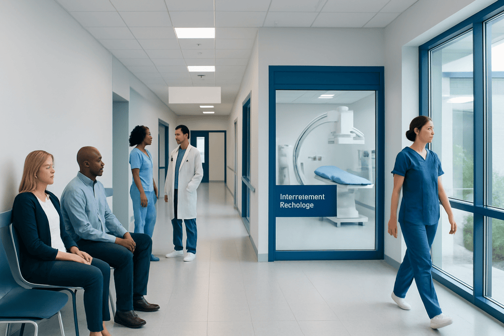
Interventional radiology combines advanced imaging with minimally invasive techniques to treat many vascular and non-vascular conditions. Procedures are commonly guided by ultrasound, fluoroscopy, CT, MRI, or angiography to help doctors work precisely inside the body.
Key Takeaways
- Interventional radiology combines advanced imaging with minimally invasive techniques to treat many vascular and non-vascular conditions.
- Procedures are commonly guided by ultrasound, fluoroscopy, CT, MRI, or angiography to help doctors work precisely inside the body.
- Common treatments include angioplasty, stenting, embolization, drainage procedures, biopsies, and tumor ablation.
- Recovery is often shorter than with open surgery, but each procedure still has benefits, risks, and preparation requirements.
- Patients should discuss alternatives, anesthesia, follow-up, and warning symptoms with a qualified interventional radiologist or treating physician.
Interventional radiology is a medical specialty that uses imaging technologies to guide small instruments through the body for diagnosis and treatment. Many procedures can be performed through a tiny skin puncture, often reducing pain, hospital stay, and recovery time compared with traditional surgery.
Overview
Interventional radiology is a specialty in which doctors use medical imaging to guide diagnosis and treatment from inside the body. Instead of making a large incision, the interventional radiologist usually reaches the treatment area through a small puncture in the skin, often using a catheter, needle, wire, balloon, stent, or other tiny instrument.
Imaging is central to the procedure. Ultrasound, X-ray fluoroscopy, computed tomography, magnetic resonance imaging, and angiography can show blood vessels, organs, tumors, fluid collections, or other targets in real time or near real time. This guidance helps the physician place instruments accurately while avoiding nearby structures as much as possible.
Interventional radiology is used across many fields, including cardiology, oncology, gastroenterology, urology, nephrology, gynecology, trauma care, and pain management. Some procedures are performed to diagnose a problem, such as an image-guided biopsy, while others are therapeutic, such as opening a narrowed blood vessel or blocking abnormal bleeding.
How Image-Guided Procedures Work
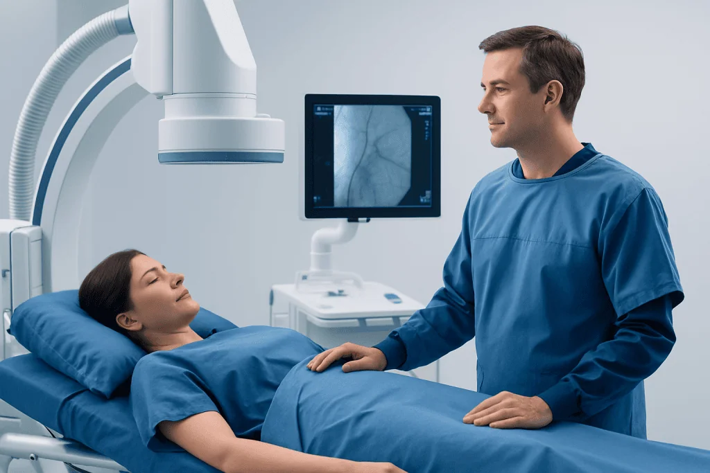
Most interventional radiology procedures begin with careful planning. The physician reviews the patient’s symptoms, medical history, laboratory results, previous imaging, current medications, allergies, and treatment goals. The best imaging method and route into the body are then selected according to the target area and the patient’s overall health.
During the procedure, the skin is cleaned and numbed. Depending on the type and length of the procedure, patients may receive local anesthesia, conscious sedation, or general anesthesia. A small needle puncture is made, and instruments are guided to the treatment site while the physician watches the images on a monitor.
After treatment or tissue sampling is completed, the instruments are removed and the puncture site is covered with a dressing. Many procedures do not require stitches. Patients are monitored for a period of time afterward, especially if sedation was used or if a blood vessel was treated.
Common Interventional Radiology Treatments

Interventional radiology includes a wide range of procedures. Some focus on blood vessels, while others treat organs, tumors, infections, fluid collections, or painful conditions. The exact recommendation depends on the diagnosis, available alternatives, and the patient’s individual risk profile.
Common examples include:
- Angioplasty and stenting: A balloon and sometimes a stent are used to open narrowed or blocked blood vessels.
- Embolization: Tiny particles, coils, plugs, or other materials are used to block blood flow to an abnormal vessel, bleeding site, fibroid, or selected tumor supply.
- Image-guided biopsy: A small tissue sample is taken using imaging guidance to support an accurate diagnosis.
- Drainage procedures: A catheter is placed to drain an abscess, bile, urine, or other fluid collection.
- Tumor ablation: Heat, cold, or other energy is used to destroy selected tumors in organs such as the liver, kidney, lung, or bone.
- Central venous access: Ports, dialysis catheters, or other lines are placed into large veins for treatment or blood access.
Some procedures are planned in advance, while others may be performed urgently, for example to control internal bleeding or restore blood flow. In many hospitals, interventional radiology is part of a multidisciplinary team, meaning treatment decisions are made together with surgeons, oncologists, vascular specialists, gastroenterologists, or other physicians as needed.
Conditions That May Be Treated
Interventional radiology can be used for many conditions because it focuses on reaching a problem precisely without a large incision. In vascular care, it may help manage peripheral artery disease, certain blocked veins, dialysis access problems, deep vein thrombosis in selected cases, varicose veins, aneurysms, and acute bleeding.
In cancer care, interventional radiologists may perform biopsies, place ports for chemotherapy, treat pain related to bone tumors, drain blocked bile ducts, or use ablation and embolization techniques for selected tumors. These treatments may be used alone or alongside surgery, chemotherapy, immunotherapy, radiation therapy, or other cancer treatments.
Other examples include uterine fibroid embolization, prostate artery embolization in selected patients with benign prostate enlargement, treatment of varicocele, kidney or urinary tract drainage, abscess drainage, vertebral augmentation for certain spine fractures, and procedures to relieve blocked bile ducts. Not every patient is a candidate, and careful imaging review is needed to decide whether an interventional approach is appropriate.
Benefits and Possible Risks
A major advantage of interventional radiology is that many treatments can be performed through a small puncture rather than a large incision. This may mean less tissue trauma, less pain, lower need for general anesthesia, shorter hospital stay, and faster return to daily activities. For some patients who are not ideal candidates for open surgery, an image-guided option may provide an alternative.
However, minimally invasive does not mean risk-free. Possible risks vary by procedure and may include bleeding, infection, bruising, contrast reaction, damage to nearby tissues, blood vessel injury, blood clots, pain, or incomplete treatment. Procedures involving radiation imaging are planned to use the lowest practical radiation exposure while still allowing safe guidance.
Patients can help reduce risks by sharing complete medical information before the procedure. This includes pregnancy status or possibility of pregnancy, kidney disease, diabetes, blood-thinning medications, allergies to contrast materials or medicines, prior reactions to anesthesia, implanted devices, and recent infections.
Preparation and What to Expect Afterward
Preparation depends on the procedure. Patients may be asked not to eat or drink for a certain period if sedation or anesthesia is planned. Blood tests may be needed to check clotting and kidney function. Some medications, especially blood thinners or diabetes medicines, may need temporary adjustment under medical supervision.
Patients should arrange transportation if sedation is expected, because driving afterward is usually not advised. They should also follow instructions about clothing, jewelry, implanted devices, prior imaging, and current medication lists. A consent discussion should explain why the procedure is recommended, what alternatives exist, and what symptoms should be reported afterward.
Recovery ranges from a short observation period to an overnight hospital stay or longer, depending on the procedure and the patient’s condition. Mild soreness, bruising, or fatigue can occur. Patients are usually given instructions about wound care, bathing, physical activity, medication use, diet, and follow-up imaging or clinic visits.
Prevention, Self-Care, and Follow-Up
Interventional radiology procedures often treat a specific problem, but long-term health may also depend on controlling the underlying condition. For vascular disease, this may include not smoking, managing blood pressure and cholesterol, controlling diabetes, following a heart-healthy diet, staying physically active when safe, and taking prescribed medications consistently.
For infections, tumors, urinary obstruction, bile duct problems, or chronic pain conditions, follow-up is essential. Some treatments require repeat imaging to confirm that the target area has responded or that a catheter, stent, or drainage tube remains in the correct position. Patients should keep follow-up appointments even if they feel well.
After a procedure, patients should contact their healthcare team if they have increasing pain, fever, persistent bleeding, swelling, new weakness or numbness, shortness of breath, chest pain, severe abdominal pain, or any symptom specifically listed in their discharge instructions. Early communication helps doctors address concerns promptly and safely.
When to See a Doctor
A patient may be referred to an interventional radiologist when imaging shows a condition that could be diagnosed or treated through a minimally invasive approach. Referral can come from a family physician, surgeon, oncologist, cardiologist, nephrologist, gastroenterologist, urologist, gynecologist, or emergency physician.
Patients should ask what the goal of the procedure is, whether it is diagnostic or therapeutic, what imaging will be used, what type of anesthesia is expected, and how success will be measured. It is also reasonable to ask about alternatives, including medication, observation, endoscopic treatment, surgery, or radiation therapy, depending on the condition.
Acibadem International’s multidisciplinary specialists and JCI-accredited hospitals diagnose and treat many conditions using interventional radiology for international patients. As with any procedure, the best treatment plan should be individualized after a qualified medical evaluation and review of imaging results.
Frequently asked questions
Is interventional radiology the same as surgery?
Interventional radiology is different from traditional open surgery because it usually uses a small puncture and imaging guidance rather than a large incision. Some procedures can achieve goals similar to surgery, such as stopping bleeding or opening a blocked vessel. The best option depends on the condition, anatomy, overall health, and available treatments.
Are interventional radiology procedures painful?
Most procedures use local anesthesia to numb the skin, and many also use sedation to help the patient relax. Patients may feel pressure, mild discomfort, or soreness afterward, but severe pain is not expected and should be reported. Pain control plans vary according to the procedure and the patient’s medical needs.
Will a patient need to stay in the hospital?
Some image-guided procedures are done as outpatient treatments, with discharge the same day after monitoring. Others require overnight observation or a longer hospital stay, especially if the condition is urgent, complex, or requires anesthesia. The care team will explain the expected recovery time before the procedure whenever possible.
Does interventional radiology use radiation?
Some procedures use X-ray fluoroscopy or CT, which involve ionizing radiation, while others may use ultrasound or MRI. When radiation is needed, the team plans the procedure to use the lowest practical exposure while maintaining safe and effective guidance. Patients who are pregnant or may be pregnant should inform the medical team before imaging or treatment.
What is embolization?
Embolization is a procedure that intentionally blocks blood flow in a selected vessel. It may be used to control bleeding, reduce blood supply to fibroids, treat certain vascular abnormalities, or support selected tumor treatments. The materials and technique are chosen according to the reason for treatment and the patient’s anatomy.
How should a patient prepare for an image-guided biopsy?
Preparation may include blood tests, review of blood-thinning medicines, fasting if sedation is planned, and bringing prior imaging studies. The doctor will explain where the biopsy will be taken from, how imaging will guide the needle, and how results will be communicated. Patients should follow all medication instructions exactly and ask questions before the procedure.
Can interventional radiology replace chemotherapy or surgery for cancer?
In cancer care, interventional radiology may be used to diagnose cancer, relieve symptoms, treat selected tumors, or support other treatments. It does not automatically replace chemotherapy, surgery, immunotherapy, or radiation therapy. Treatment decisions are usually made by a multidisciplinary team based on cancer type, stage, location, and the patient’s overall health.
References
- Society of Interventional Radiology
- Cardiovascular and Interventional Radiological Society of Europe
- Radiological Society of North America
- American College of Radiology
- National Institute for Health and Care Excellence
This article is for general information only and is not a substitute for professional medical advice. Please consult a qualified doctor about your individual situation.
Minimally Invasive in Turkey — costs, top hospitals & a free quote
JCI-accredited · board-certified surgeons · reply within 24h
Add us as a Preferred Source to see more of our trusted health content across Google Search, AI Overviews and Discover.

Robotic Surgery
Robot-assisted procedures across urology, gynecology and general surgery for greater precision and faster recovery.
More from the Health Library



Related Specialists

Prof. Dr. Ercan Karaarslan
Radiology
Prof. Dr. Bora Peynircioğlu
Interventional Radiology
Prof. Dr. Koray Güven
Interventional Radiology
Assoc. Prof. Dr. Deniz Can Aliş
Radiology