Intraoperative CT Scan: Real-Time Imaging During Surgery Explained
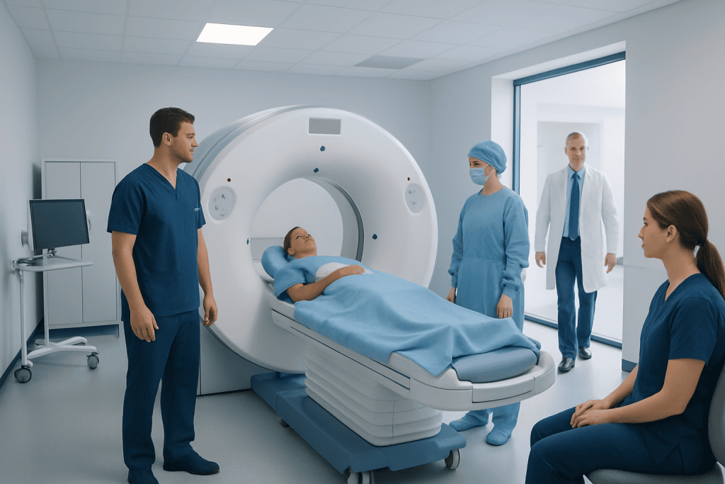
An intraoperative CT scan is done during surgery, not before or after it. It gives detailed cross-sectional images that can guide surgical decisions in real time.
Key Takeaways
- An intraoperative CT scan is done during surgery, not before or after it.
- It gives detailed cross-sectional images that can guide surgical decisions in real time.
- It is often used in neurosurgery, spine surgery, trauma, and some ENT or orthopedic procedures.
- The main benefits are improved accuracy, immediate verification, and fewer surprises after surgery.
- Not every operation needs intraoperative CT; the decision depends on the procedure, anatomy, and clinical goals.
An intraoperative CT scan is a computed tomography scan performed during an operation to give surgeons real-time images while the procedure is still in progress. It can help improve precision, confirm placement of implants or instruments, and support safer decision-making in selected surgeries.
Overview: What Is an Intraoperative CT Scan?
An intraoperative CT scan is a computed tomography scan performed while a patient is still in the operating room. Unlike a standard CT that is done before surgery for planning or after surgery for follow-up, this scan gives the surgical team updated images during the procedure itself. That immediate information can be especially useful when anatomy is complex or when precise placement of implants, screws, or surgical tools is important.
CT technology uses X-rays and computer processing to create detailed cross-sectional images of the body. During surgery, these images can show bones, some soft tissues, surgical hardware, and the relationship between treated areas and nearby structures. The goal is not simply to “take another picture,” but to support safer and more informed surgical decisions while there is still an opportunity to make adjustments.
Intraoperative CT is one form of image-guided surgery. It may be combined with navigation systems, robotics, fluoroscopy, or other advanced technologies that help translate images into surgical guidance. For patients, this usually happens behind the scenes, but it can play a valuable role in improving precision in carefully selected operations.
How It Works in the Operating Room
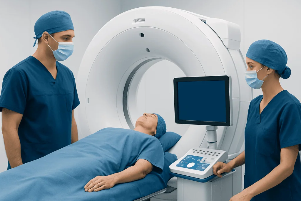
The exact setup depends on the hospital and the type of scanner used. In some operating rooms, a mobile CT scanner moves to the patient when images are needed. In others, the operating table and imaging system are integrated into a specialized hybrid operating room. The patient remains positioned for surgery, and the team takes steps to maintain sterility, monitor vital signs, and protect staff from unnecessary radiation exposure.
When the surgeon requests imaging, the operation is briefly paused. The scan is performed, and the images are reviewed immediately on monitors in the operating room. This allows the team to check whether the anatomy matches the surgical plan, whether a target has been reached, or whether hardware such as screws, cages, or implants is correctly placed.
If needed, the surgeon can revise the plan at once. For example, they may adjust implant position, remove remaining bone fragments, or confirm that decompression has been adequate before closing the incision. This real-time feedback is one of the main reasons intraoperative CT can be helpful in complex procedures.
When Intraoperative CT May Be Used

Intraoperative CT is not used in every surgery. It is most helpful when surgical accuracy must be confirmed in real time or when traditional visual inspection alone may not provide enough information. Common areas of use include spine surgery, neurosurgery, trauma surgery, and selected orthopedic and ear, nose, and throat procedures.
In spine operations, intraoperative CT can help with the placement of pedicle screws and other hardware, especially when anatomy is altered by scoliosis, prior surgery, or injury. In neurosurgery, it may support navigation or help the team evaluate changes during the procedure. Patients exploring advanced brain procedures may also hear about related approaches in neurosurgery and brain tumor surgery, where precise imaging can be important.
Other possible uses include facial trauma repair, sinus or skull base surgery, and some complex orthopedic operations. In institutions with advanced surgical imaging, intraoperative CT may work alongside robotic surgery or navigation systems to improve spatial accuracy. Whether it is appropriate depends on the surgical goal, the area being treated, and the experience of the multidisciplinary team.
- Spine stabilization and deformity correction
- Neurosurgical procedures requiring detailed navigation
- Complex fracture repair or trauma surgery
- Selected ENT, skull base, or sinus operations
- Revision surgery where anatomy has changed
Benefits and Limitations
The main advantage of intraoperative CT is that it provides immediate confirmation. Instead of waiting until after surgery to discover that an implant needs repositioning or that a treatment target was not fully reached, the surgeon can review updated images and respond before the operation ends. This may improve precision and reduce the need for early reoperation in some cases.
Another benefit is better navigation in difficult anatomy. Scar tissue, congenital differences, severe degeneration, fractures, or tumors can make surgery more challenging. Real-time CT images can help the team understand the current anatomy rather than relying only on preoperative scans, which may no longer perfectly reflect the surgical situation once the procedure has begun. This can be relevant in conditions such as brain tumor or scoliosis, where anatomical detail matters.
However, intraoperative CT also has limitations. It involves radiation, although the amount depends on the body area, scanner, and imaging protocol. It may lengthen the procedure slightly, requires specialized equipment and trained staff, and is not necessary for many routine surgeries. CT also shows some tissues better than others, so in certain situations other imaging tools, such as MRI, ultrasound, or fluoroscopy, may be more suitable.
What Patients Can Expect Before, During, and After Surgery
Before surgery, the surgical team explains whether intraoperative CT is planned and why it may be useful. Patients do not usually need special preparation solely for the scan beyond the preparation already required for the operation itself. The consent discussion may include the expected benefits, possible alternatives, and the fact that X-ray-based imaging involves radiation.
During the operation, the patient is under anesthesia or appropriate procedural sedation according to the surgical plan. The scan itself is performed without the patient needing to do anything. Monitoring continues throughout, and the operating room team carefully coordinates positioning, sterility, anesthesia, and image acquisition.
After surgery, recovery is usually determined by the operation rather than by the intraoperative CT scan. The scan does not typically add pain or create a separate recovery process. In many cases, it simply becomes one more tool used during the procedure to support the surgeon’s work. If a patient has questions about radiation exposure or why imaging was necessary, the surgical team can explain how those decisions were made.
Safety, Radiation, and Decision-Making
Radiation exposure is an understandable concern whenever CT is discussed. Intraoperative CT uses ionizing radiation, but teams aim to keep exposure as low as reasonably achievable while still obtaining clinically useful images. The decision to use it is based on whether the expected benefit in surgical guidance and confirmation outweighs the added imaging exposure.
Safety in the operating room also involves careful coordination. The team may step back or use shielding during image acquisition, while the patient remains monitored at all times. Modern systems are designed to fit into surgical workflows and to reduce disruption, though they still require planning, experience, and quality control.
For many patients, the most helpful question is not whether intraoperative CT is “better” in general, but whether it is useful for their specific procedure. The answer depends on factors such as surgical complexity, the need for hardware placement, anatomical variability, prior operations, and whether real-time confirmation could change management during the operation.
Questions to Ask the Surgeon and When to Seek Further Advice
Patients may feel more comfortable when they understand why a technology is being recommended. Useful questions include whether intraoperative CT is expected to change surgical planning, whether it will be used routinely or only if needed, and what alternatives exist. It is also reasonable to ask about radiation, how the images are interpreted, and whether the hospital has dedicated experience with image-guided surgery.
Anyone preparing for surgery should speak with their doctor if they do not understand the purpose of imaging or if they have concerns about risks, prior radiation exposure, implants, or the expected recovery process. A second opinion may be helpful when surgery is complex or when more than one treatment approach is possible. The goal is informed, shared decision-making rather than avoiding technology that may be beneficial.
Near the end of planning, some patients seek care in centers with advanced operating rooms and multidisciplinary teams. Acibadem International’s multidisciplinary specialists and JCI-accredited hospitals diagnose and treat complex conditions for international patients, including procedures where advanced intraoperative imaging may be part of care when clinically appropriate.
Frequently asked questions
Is an intraoperative CT scan the same as a regular CT scan?
It uses the same basic CT imaging principle, but it is performed during an operation rather than before or after it. The main difference is timing: intraoperative CT gives the surgeon updated images while the procedure is still underway.
Why would a surgeon need a CT scan during surgery?
A surgeon may use intraoperative CT to check anatomy, guide instrument placement, confirm implant position, or verify that the intended surgical goal has been achieved. This can be especially helpful in complex procedures where precision is critical.
Does intraoperative CT make surgery safer?
It can improve surgical accuracy and provide immediate confirmation in selected cases, which may support safer decision-making. However, whether it improves safety for an individual patient depends on the type of surgery, the clinical situation, and the experience of the team.
Will the patient feel the scan during surgery?
No. Patients are typically under anesthesia or sedation according to the surgical plan, and the scan itself does not cause a separate sensation. From the patient's perspective, it is usually just one part of the overall operation.
Does intraoperative CT expose the patient to radiation?
Yes. Because CT uses X-rays, there is radiation exposure. The medical team weighs this against the potential benefit of getting precise, real-time information during surgery and uses protocols designed to limit exposure as much as possible.
Is intraoperative CT used in every surgery?
No. Many operations do not need it. It is usually reserved for procedures where real-time imaging may meaningfully improve accuracy, navigation, or confirmation of the final result.
References
- World Health Organization
- Radiological Society of North America
- American College of Radiology
- National Institute of Neurological Disorders and Stroke
- North American Spine Society
This article is for general information only and is not a substitute for professional medical advice. Please consult a qualified doctor about your individual situation.
Explore treatments in Turkey — costs, top hospitals & a free quote
JCI-accredited · board-certified surgeons · reply within 24h
Add us as a Preferred Source to see more of our trusted health content across Google Search, AI Overviews and Discover.

More from the Health Library



Related Specialists

Prof. Dr. Muzaffer Olcay Çizmeli
Interventional Neuroradiology
Prof. Dr. Yaşar Çolak
Gastroenterology
Prof. Dr. Burak Akan
Orthopedic Surgery & Traumatology
Dr. Mustafa Engin Çakmakçı
Otorhinolaryngology