Mediastinum — Explained by Medical Evidence, Not Myths

The mediastinum is the space in the middle of the chest between the lungs. It contains several essential organs and structures, including the heart, trachea, esophagus, thymus, lymph nodes, and major blood vessels.
Key Takeaways
- The mediastinum is the space in the middle of the chest between the lungs.
- It contains several essential organs and structures, including the heart, trachea, esophagus, thymus, lymph nodes, and major blood vessels.
- Mediastinal problems may include infection, inflammation, enlarged lymph nodes, cysts, or tumors.
- Symptoms vary widely and depend on which nearby structure is being pressed on or affected.
- Diagnosis usually relies on imaging such as chest X-ray, CT, MRI, and sometimes biopsy.
- Treatment depends on the underlying cause and may include monitoring, medicines, drainage, surgery, or cancer care.
The mediastinum is the central compartment of the chest, located between the lungs and containing the heart, major blood vessels, trachea, esophagus, thymus, lymph nodes, and important nerves. It is not a disease itself, but problems in this area can cause symptoms such as chest pain, cough, shortness of breath, or difficulty swallowing depending on which structures are affected.
Overview: what the mediastinum is
The mediastinum is the central space inside the chest between the right and left lungs. It is best understood as an anatomical region rather than a single organ. This area contains many vital structures packed closely together, which is why disorders involving the mediastinum can affect breathing, swallowing, circulation, or cause chest discomfort.
Within the mediastinum are the heart, the large blood vessels connected to it, the trachea, the esophagus, lymph nodes, nerves, and the thymus gland. In children, the thymus is relatively larger and plays an important role in immune system development. In adults, it becomes smaller, but it still remains part of the mediastinal anatomy.
Doctors often divide the mediastinum into compartments such as anterior, middle, and posterior regions. This helps narrow down possible causes when a scan shows a mass, swelling, enlarged lymph nodes, or another abnormality. Different conditions tend to arise in different compartments, so location can give important diagnostic clues.
Because the mediastinum holds so many essential structures, a finding in this area should be evaluated carefully. However, not every mediastinal abnormality is dangerous. Some are benign cysts, temporary inflammatory changes, or enlarged lymph nodes related to infection that improve with appropriate care.
What can affect the mediastinum
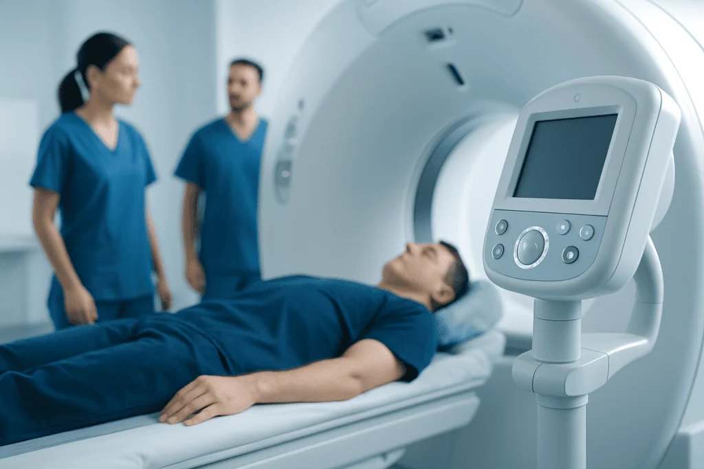
The mediastinum itself is not an illness, but many different conditions can develop in or spread to this area. These include enlarged lymph nodes, thymus disorders, thyroid tissue extending into the chest, cysts, infections, inflammatory conditions, and both benign and malignant tumors. Some abnormalities are found only by chance during imaging done for another reason.
A common clinical question is whether a person has a mediastinal mass, which means an abnormal growth or area seen on imaging. A mediastinal mass may come from the thymus, lymph nodes, nerves, connective tissue, thyroid tissue, or nearby structures. The causes vary greatly by age, symptoms, and the exact location within the chest.
Examples of conditions that may involve this area include lymphoma, thymoma, germ cell tumors, cysts, and enlarged lymph nodes due to infection or inflammatory disease. In some cases, mediastinal involvement is related to lung disorders or to cancers that spread from nearby structures. For readers learning about related chest conditions, it may be helpful to understand lung cancer and how it can affect nearby tissues and lymph nodes.
Because many different disorders can occur in the mediastinum, it is important not to assume that any single symptom or scan result means cancer. A careful, step-by-step medical evaluation is usually needed before drawing conclusions or choosing treatment.
Symptoms and possible warning signs

Mediastinal problems do not always cause symptoms. Small lesions or mildly enlarged lymph nodes may be discovered incidentally on a chest X-ray or CT scan. When symptoms do occur, they usually happen because a growth, infection, or inflammation is pressing on nearby structures or disrupting their normal function.
Possible symptoms can include chest pain or pressure, shortness of breath, cough, hoarseness, wheezing, fever, unexplained weight loss, fatigue, or difficulty swallowing. Some people notice swelling in the face, neck, or upper chest if blood flow through major veins is affected. Others may have recurrent respiratory infections or a persistent cough that does not improve as expected.
The pattern of symptoms may help guide diagnosis. For example, difficulty swallowing may suggest pressure on the esophagus, while hoarseness can occur if a nerve to the voice box is affected. Breathing symptoms can occur when the trachea or main airways are narrowed or irritated. Symptoms such as fever or night sweats may point doctors toward infection, inflammation, or certain cancers, but they are not specific on their own.
Any new or ongoing chest-related symptom deserves attention, especially if it persists, worsens, or is associated with weight loss, breathing difficulty, or swallowing problems. A healthcare professional can decide whether the symptoms are likely to come from the mediastinum or another part of the chest.
Causes and risk factors
The causes of mediastinal abnormalities depend on the person’s age, medical history, and which mediastinal compartment is involved. In children, some masses are more likely to be related to developmental cysts, lymphatic conditions, or certain tumors. In adults, thymic tumors, enlarged lymph nodes, thyroid-related masses, and nerve sheath tumors may be more common depending on the location.
Infections can also affect the mediastinum. These may occur after severe chest infections, after surgery, or in rare cases after injury or a tear in the esophagus. Inflammatory conditions such as sarcoidosis can enlarge mediastinal lymph nodes. Autoimmune disease may also involve structures in this part of the chest, especially the thymus.
Risk factors vary by diagnosis rather than by the mediastinum itself. Smoking, prior cancer, radiation exposure, chronic infection, and some immune-related disorders may increase the likelihood of particular mediastinal conditions. Family history may matter in selected cases, but many mediastinal findings occur without a clear inherited cause.
Importantly, a risk factor does not confirm a diagnosis, and the absence of risk factors does not rule one out. The role of testing is to identify exactly what the abnormality is, whether it is affecting nearby organs, and whether treatment is needed now or simple monitoring is enough.
How doctors diagnose mediastinal conditions
Diagnosis usually begins with a medical history and physical examination, followed by imaging. A chest X-ray may provide the first clue, but cross-sectional imaging is often needed for more detail. MRI and computed tomography can help show the size, location, shape, and relationship of a mediastinal abnormality to surrounding organs and vessels.
Depending on the situation, doctors may also order blood tests, lung function testing, or imaging that evaluates metabolic activity. If enlarged lymph nodes or a suspicious mass is present, a tissue sample may be necessary to make a definite diagnosis. This may be obtained through bronchoscopy, needle biopsy, endoscopic procedures, or surgery, depending on where the lesion sits and what structures are nearby.
In some cases, surgery is part of both diagnosis and treatment. For carefully selected lesions, minimally invasive approaches such as robotic surgery or thoracoscopic techniques may be considered. The best approach depends on the lesion’s size, compartment, and whether doctors suspect a benign cyst, thymic tumor, lymphoma, or another condition.
The most important principle is that imaging alone does not always tell the whole story. A precise diagnosis often requires combining radiology findings, symptoms, laboratory results, and sometimes pathology from a biopsy or operation.
Treatment options and what they depend on
Treatment for a mediastinal condition depends entirely on the cause. Some benign cysts or small incidental findings can be monitored with follow-up imaging if they are not causing symptoms and do not look concerning. Infections may require antibiotics, drainage, or urgent care if severe. Inflammatory conditions may improve with treatment directed at the underlying disorder.
If a mediastinal mass is confirmed or strongly suspected to be a tumor, the treatment plan may include surgery, chemotherapy, radiation therapy, or a combination of these. Thymic tumors and some cysts are often treated surgically. Lymphoma usually requires specialized medical treatment after tissue diagnosis rather than immediate removal. Management is individualized because the same symptom can come from very different diseases.
When cancer treatment is needed, options may include chemotherapy or radiation oncology as part of a broader plan. The aim may be to remove the lesion, shrink it, control spread, relieve pressure on nearby organs, or address symptoms such as pain or airway narrowing.
Care is often multidisciplinary, involving chest physicians, thoracic surgeons, oncologists, radiologists, and pathologists. Near the end of the diagnostic journey, some international patients seek evaluation at centers such as Acibadem International, where multidisciplinary specialists and JCI-accredited hospitals diagnose and treat mediastinal conditions using coordinated care plans.
Prevention, self-care, and follow-up
There is no single way to prevent all mediastinal disorders because they arise from many different causes. However, general chest and lung health measures are still useful. Avoiding smoking, keeping up with routine medical care, and getting prompt treatment for significant infections may reduce the risk of some conditions or complications.
Self-care should focus on supporting overall health and following medical advice rather than attempting to manage a mediastinal problem without evaluation. If a doctor recommends observation, it is important to attend follow-up appointments and repeat imaging on schedule. This helps track whether a lesion remains stable, shrinks, or grows over time.
People who already have a diagnosed mediastinal condition should report any change in breathing, swallowing, voice, fever, or chest discomfort. Even when a finding is thought to be benign, new symptoms can change the urgency of treatment. Rest, hydration, and symptom tracking can be helpful, but they do not replace proper assessment.
Questions during follow-up often include whether a lesion is stable, whether biopsy is needed, and what symptoms should trigger faster review. Asking for clear explanations can help patients feel more confident and informed throughout the process.
When to seek medical care
Medical care should be sought if there is persistent chest pain, unexplained cough, shortness of breath, wheezing, trouble swallowing, hoarseness, or swelling of the face or neck. These symptoms do not always mean a mediastinal disorder, but they should be evaluated, especially if they are new, progressive, or unexplained.
Urgent care is important for severe breathing difficulty, sudden chest pain, fainting, blue lips, or signs of serious infection such as high fever and rapidly worsening illness. Symptoms caused by pressure on the airway or major blood vessels may need prompt hospital assessment.
If imaging has already shown a mediastinal mass or enlarged lymph nodes, follow-up should not be delayed even if symptoms are mild. Early specialist review can help clarify the diagnosis and avoid unnecessary worry.
Patients should seek guidance from a qualified doctor rather than relying on symptoms alone to judge seriousness. Because the mediastinum contains multiple vital structures, professional assessment is the safest way to understand what is happening and what next steps are appropriate.
Frequently asked questions
What is the mediastinum in simple terms?
The mediastinum is the space in the center of the chest between the lungs. It contains important structures such as the heart, major blood vessels, trachea, esophagus, thymus, nerves, and lymph nodes.
Is the mediastinum an organ?
No. The mediastinum is an anatomical compartment, not a single organ. It is a region that holds several organs and other vital structures.
Can a mediastinal mass be benign?
Yes. Some mediastinal masses are benign, including certain cysts, noncancerous tumors, or enlarged lymph nodes related to infection or inflammation. Imaging and sometimes biopsy are needed to determine the exact cause.
What symptoms can a mediastinal problem cause?
Symptoms may include chest pain, cough, shortness of breath, wheezing, hoarseness, or difficulty swallowing. Some people have no symptoms at all, and the problem is discovered only on imaging.
How is a mediastinal abnormality diagnosed?
Doctors usually begin with chest imaging such as X-ray, CT, or MRI. Depending on the findings, further tests may include blood work, bronchoscopy, or biopsy to identify the cause.
Does a finding in the mediastinum always mean cancer?
No. Cancer is only one possible cause of a mediastinal abnormality. Infections, inflammatory conditions, benign cysts, thyroid-related masses, and enlarged lymph nodes can also affect this area.
References
- National Cancer Institute
- American College of Radiology
- American Thoracic Society
- National Heart, Lung, and Blood Institute
- MedlinePlus
This article is for general information only and is not a substitute for professional medical advice. Please consult a qualified doctor about your individual situation.
Explore treatments in Turkey — costs, top hospitals & a free quote
JCI-accredited · board-certified surgeons · reply within 24h
Add us as a Preferred Source to see more of our trusted health content across Google Search, AI Overviews and Discover.