MRI and Brain: What Patients Need to Know
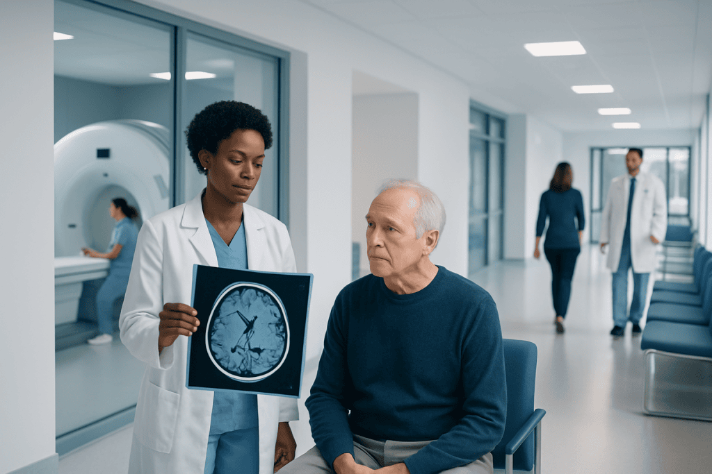
A brain MRI provides detailed images of brain tissue, blood vessels, and nearby structures without radiation. It may be ordered for symptoms, injuries, follow-up care, or to clarify findings from other tests.
Key Takeaways
- A brain MRI provides detailed images of brain tissue, blood vessels, and nearby structures without radiation.
- It may be ordered for symptoms, injuries, follow-up care, or to clarify findings from other tests.
- Some scans use contrast dye to highlight inflammation, tumors, infection, or blood vessel problems.
- MRI is usually painless, but patients should tell the care team about implants, kidney problems, pregnancy, or claustrophobia.
- Urgent medical assessment is important for sudden neurologic symptoms such as weakness, speech changes, or severe new headache.
MRI and brain imaging uses strong magnets and radio waves to create detailed pictures of the brain without using ionizing radiation. It helps doctors investigate symptoms such as headaches, seizures, weakness, vision changes, memory problems, and suspected stroke or tumors.
Overview: What MRI and Brain Imaging Means
MRI and brain imaging refers to magnetic resonance imaging used to examine the brain, surrounding tissues, and sometimes the blood vessels that supply them. A brain MRI uses a strong magnet, radio waves, and a computer to produce very detailed pictures. Unlike X-rays and CT scans, it does not use ionizing radiation.
Doctors use brain MRI when they need more detail than a physical examination alone can provide. It can help explain symptoms such as persistent headaches, dizziness, seizures, numbness, weakness, balance problems, vision changes, hearing changes, memory difficulties, or changes in behavior. It is also commonly used after head injury, during follow-up of known brain conditions, or when another imaging test has raised questions.
Brain MRI is not a diagnosis by itself. Instead, it is a tool that helps a doctor understand what may be happening and decide on the next step. In many cases, the scan is normal, which can be reassuring. In other cases, it may show changes that need monitoring, treatment, or referral to a specialist.
What a Brain MRI Can Show
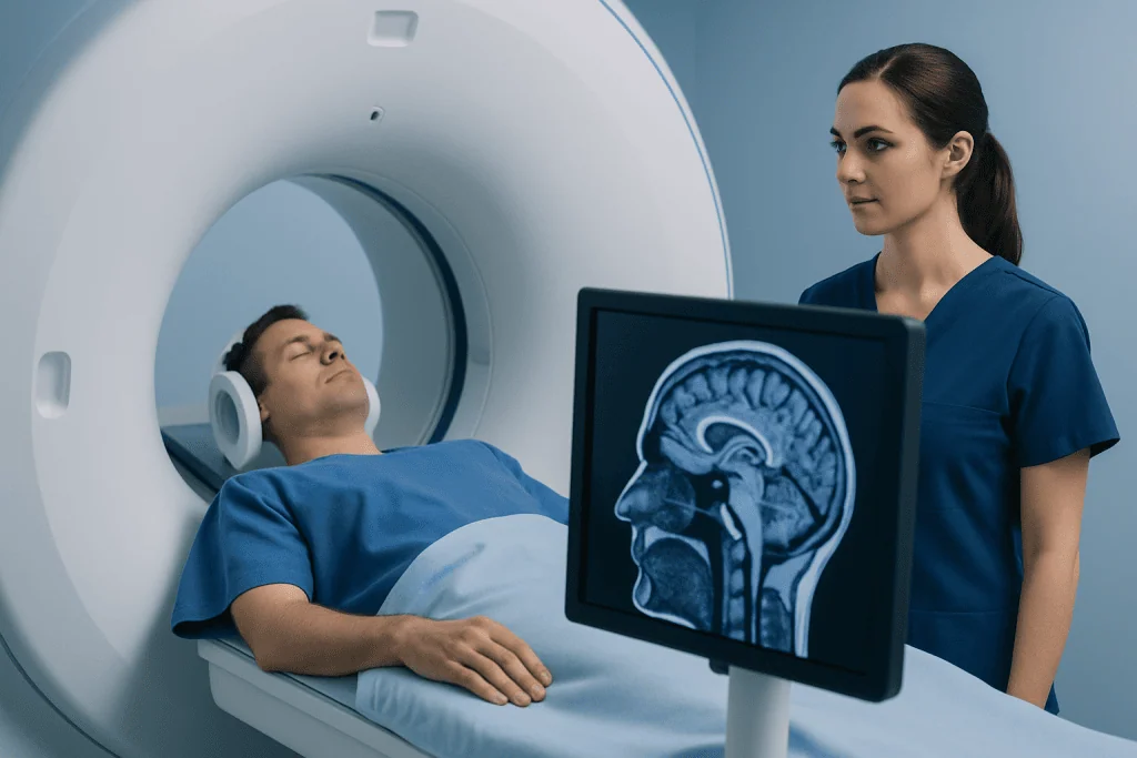
A brain MRI can show the shape and condition of brain tissue in much greater detail than many other imaging tests. It may help identify areas of swelling, bleeding, tissue injury, infection, inflammation, abnormal fluid buildup, or structural differences present from birth. It can also help assess the pituitary gland, optic nerves, inner ear region, and skull base when needed.
Different MRI sequences highlight different features. Some are better for anatomy, while others are more useful for spotting recent stroke, demyelination, inflammation, or subtle tissue changes. If blood vessels need closer evaluation, the doctor may request MR angiography or venography as part of the study.
Conditions that may be evaluated with MRI include suspected stroke, brain tumors, multiple sclerosis, epilepsy-related changes, infection, hydrocephalus, and complications after trauma. Depending on the reason for the scan, MRI may also support the assessment of brain tumor or stroke. The radiologist interprets the images in the context of the patient’s symptoms, history, and other test results.
- Recent or old stroke-related changes
- Brain tumors or other masses
- Inflammation or demyelinating disease
- Bleeding, swelling, or trauma-related injury
- Infection, abscess, or fluid collection
- Abnormalities in blood vessels or blood flow
Why Doctors Order MRI for Brain Symptoms
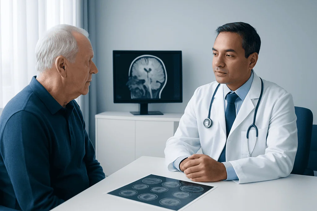
Doctors choose MRI when they need detailed information to evaluate symptoms that could come from the brain or nearby nerves. A scan may be ordered for new seizures, unexplained weakness or numbness, persistent headaches with concerning features, fainting with neurologic symptoms, memory or cognitive changes, or changes in coordination and balance. MRI can also help when symptoms develop gradually and the cause is uncertain.
In emergency situations, CT is often the first imaging test because it is fast and widely available. MRI may follow later to provide more detail, especially for small strokes, inflammation, posterior brain structures, or complex cases. This is one reason patients may have more than one imaging test during the same illness.
MRI is also important in follow-up care. A specialist may repeat scans over time to monitor a known tumor, assess treatment response, track disease activity, or check recovery after surgery. In selected cases, advanced MRI methods may support planning for Gamma Knife radiosurgery or other neurological treatment strategies.
What Happens During a Brain MRI
Before the scan, the imaging team asks about metal implants, previous surgeries, pacemakers, insulin pumps, aneurysm clips, hearing devices, medication patches, and any metal fragments in the body. This safety screening is very important because MRI uses a strong magnet. Patients usually remove jewelry, watches, hairpins, and other metal items before entering the scan room.
During the scan, the patient lies on a table that slides into the MRI machine. A head coil, which helps produce clear images, is placed around the head. The machine makes loud tapping or knocking sounds while pictures are being taken, so ear protection is provided. It is important to remain still because movement can blur the images and may require repeat sequences.
Some people receive a contrast agent, usually gadolinium-based dye, through a vein in the arm. Contrast can make certain abnormalities easier to detect, including tumors, inflammation, or areas where the blood-brain barrier is affected. The test is painless, although some patients may find the enclosed space uncomfortable. If claustrophobia is a concern, it is helpful to mention it in advance so the team can discuss practical options.
Preparing for the Scan and Understanding Safety
Most brain MRI scans require little preparation. In many cases, people can eat, drink, and take their usual medicines unless they are told otherwise. If contrast will be used, the care team may ask about kidney disease, allergies, or previous reactions to contrast. Patients should also mention pregnancy or the possibility of pregnancy, even though MRI itself does not use radiation.
MRI is considered safe for many people, but not every implant is MRI-compatible. The exact model and manufacturer of a device may need to be checked before the test. For patients with kidney problems, the team carefully weighs whether contrast is needed. In children or adults who cannot stay still, sedation may occasionally be considered, but this depends on the clinical situation and local practice.
Many patients worry that a brain MRI means a serious diagnosis is likely. In reality, MRI is often used to rule out important conditions and provide reassurance. The value of the scan is that it can narrow down possibilities and guide what should happen next, whether that is observation, medication, referral, or a procedure such as brain tumor surgery in carefully selected cases.
How Results Are Reported and What They Mean
After the scan, a radiologist reviews the images and prepares a report for the referring doctor. The report usually describes the technique used, whether contrast was given, and any findings in the brain tissue, fluid spaces, blood vessels, and nearby structures. Results are interpreted together with the patient’s symptoms, examination findings, and medical history.
A normal brain MRI means that no structural abnormality was seen on that study, but it does not always explain every symptom. Some conditions affect function more than structure, and some symptoms can come from causes outside the brain. Conversely, an abnormal result does not automatically mean cancer or a dangerous disease. MRI can detect old injuries, small age-related changes, incidental benign findings, and abnormalities that simply need monitoring.
If the MRI shows a possible problem, the next steps may include blood tests, repeat imaging, vascular imaging, lumbar puncture, EEG, or referral to a neurologist or neurosurgeon. In patients with blood vessel disorders, specialists may discuss options such as interventional neuroradiology when that is appropriate. Clear communication with the treating doctor can help patients understand which findings are urgent and which are not.
When to Seek Medical Care
Many brain MRI scans are arranged routinely, but some symptoms need urgent assessment rather than waiting for an outpatient appointment. Immediate medical care is important for sudden weakness, facial drooping, new speech difficulty, sudden confusion, loss of consciousness, a seizure in someone without a known seizure disorder, or a severe sudden headache unlike usual headaches. These symptoms can indicate a time-sensitive emergency.
Medical review is also important for persistent headaches with vomiting, a new neurologic symptom after a head injury, steadily worsening balance problems, unexplained vision loss, or new behavior and memory changes that interfere with daily life. A doctor can decide whether MRI is needed and how quickly it should be arranged.
For planned evaluation, patients should still seek non-emergency care if symptoms continue, recur, or raise concern even when they seem mild. If specialist assessment is required, Acibadem International’s multidisciplinary specialists in JCI-accredited hospitals diagnose and treat neurological conditions for international patients using coordinated imaging and follow-up care.
Frequently asked questions
What does a brain MRI show?
A brain MRI shows detailed pictures of brain tissue, fluid spaces, and nearby structures. It can help identify stroke, tumors, inflammation, infection, bleeding, trauma-related changes, and some blood vessel problems.
Is a brain MRI safe?
For most people, brain MRI is safe because it does not use ionizing radiation. The main safety issue is the strong magnet, so patients must tell the imaging team about implants, devices, metal fragments, pregnancy, or kidney disease.
Why might contrast be needed for a brain MRI?
Contrast can make certain abnormalities stand out more clearly on the images. It may help the doctor assess tumors, inflammation, infection, active lesions, or blood vessel-related changes more accurately.
How long does a brain MRI take?
The exact time varies, but many brain MRI scans take around 20 to 45 minutes. Studies with contrast or extra sequences can take longer.
Can a brain MRI detect stroke?
Yes, MRI can detect many strokes and may be especially useful for small or early ischemic strokes. In emergencies, however, CT is often performed first because it is faster and helps quickly identify bleeding.
What if the MRI is normal but symptoms continue?
A normal MRI can be reassuring, but it does not always explain every symptom. If symptoms persist, the doctor may look for causes outside the brain or order other tests based on the clinical picture.
References
- Radiological Society of North America
- American College of Radiology
- National Institute of Neurological Disorders and Stroke
- National Institutes of Health
- World Health Organization
This article is for general information only and is not a substitute for professional medical advice. Please consult a qualified doctor about your individual situation.
Explore treatments in Turkey — costs, top hospitals & a free quote
JCI-accredited · board-certified surgeons · reply within 24h
Add us as a Preferred Source to see more of our trusted health content across Google Search, AI Overviews and Discover.

More from the Health Library



Related Specialists

Dr. Yelda Başbuğ
Pulmonary Medicine
Prof. Dr. Behice Kurtaran
Infectious Diseases & Clinical Microbiology
Dr. Etem Aytaç Yazar
Orthopedic Surgery & Traumatology
Dr. Öznur İşcan
Ophthalmology