MRI Bone Metastases: Preparation, Procedure and Results
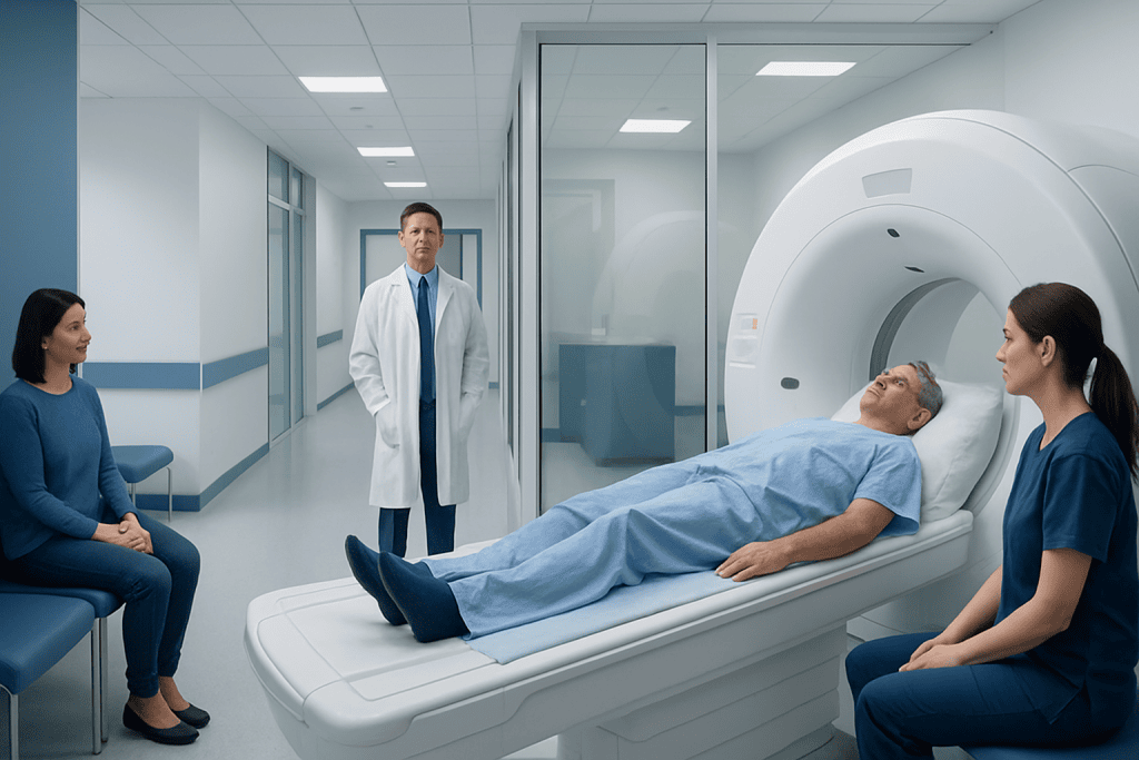
MRI can identify changes in bone marrow that may suggest bone metastases, sometimes before changes are visible on standard X-rays. The examination is painless, usually takes 30 to 60 minutes, and may include contrast dye when clinically appropriate.
Key Takeaways
- MRI can identify changes in bone marrow that may suggest bone metastases, sometimes before changes are visible on standard X-rays.
- The examination is painless, usually takes 30 to 60 minutes, and may include contrast dye when clinically appropriate.
- An MRI result can raise or lower suspicion for metastases, but diagnosis is interpreted in the context of the person’s cancer history and other tests.
- No single imaging test is best for every cancer type, body area or clinical question.
- Bone metastases are often treatable even when they cannot be fully cured, with care focused on cancer control, symptom relief and bone protection.
MRI bone metastases imaging uses magnetic fields and radio waves to create detailed pictures of bone marrow and nearby soft tissues, helping clinicians assess whether cancer may have spread to bone. It does not use ionizing radiation and is often used alongside other scans, blood tests and, when needed, biopsy results.
Overview: What MRI Bone Metastases Imaging Shows
MRI bone metastases imaging is a scan used to look for possible cancer spread to the bones. It is particularly good at showing bone marrow, the soft tissue inside many bones where metastatic deposits can develop. MRI creates detailed images with a strong magnetic field and radio waves rather than X-rays or radiation.
Bone metastases occur when cancer cells from another part of the body travel to bone. They are not the same as a primary bone cancer, which begins in bone itself. Metastases may develop with several cancers, including breast, prostate, lung, kidney and thyroid cancers, although an individual person’s risk depends on the type and stage of cancer.
An MRI may be ordered to investigate persistent focal bone pain, unexplained abnormalities on another scan, possible spinal involvement, or symptoms suggesting pressure on nerves. For mri bone metastases breast cancer assessment, MRI may be used when there is a concern about marrow involvement or when another imaging study needs clarification.
MRI findings are reviewed by a radiologist and then considered with symptoms, examination findings, blood tests and previous imaging. A suspicious area on MRI is important, but it does not always prove metastasis because infection, injury, arthritis-related change and some noncancerous marrow conditions can also alter scan appearances.
How MRI Detects Bone Metastasis
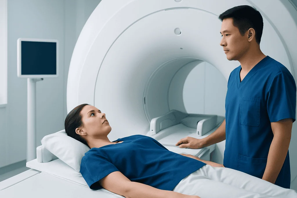
Healthy bone marrow contains varying proportions of fat and blood-forming tissue. A metastatic tumor can replace or change normal marrow, affecting how the area appears on different MRI sequences. Radiologists compare these sequences to evaluate the location, size, pattern and relationship of any abnormality to nearby bone, nerves and soft tissues.
MRI is especially valuable for the spine, pelvis and other regions with substantial marrow. It can help show whether a lesion extends beyond bone or affects the spinal canal. When doctors are concerned about possible spinal cord compression, MRI can be an urgent and highly informative examination.
Some MRI examinations use intravenous contrast, usually a gadolinium-based agent. Contrast can make blood flow and tissue patterns easier to assess, helping radiologists characterize an abnormal area. It is not needed for every patient or every MRI protocol.
Whole-body MRI may be considered in selected circumstances, while a focused MRI may examine a painful area or a specific section of the spine. The choice is made by the treating team based on the known or suspected cancer, symptoms, prior test results and the question the scan needs to answer.
Candidacy, Preparation and Safety Checks

A doctor may recommend MRI when a person has a known cancer and new bone symptoms, when another test is inconclusive, or when detailed evaluation of marrow or the spine is needed. MRI may also be part of staging or follow-up planning. It is not automatically required for every person with cancer, and the appropriate imaging pathway differs between cancer types.
Before the scan, the imaging team asks about implanted medical devices, previous surgery, metal fragments, medication patches and pregnancy. Many modern implants are MRI-compatible under specific conditions, but the team must verify the exact device and follow its safety instructions. People who have worked with metal or had a possible metal eye injury may need additional screening.
Patients should tell the team about kidney disease, prior reactions to contrast agents, claustrophobia or difficulty lying flat. If contrast is planned, kidney function may be reviewed in some situations. Severe reactions to MRI contrast are uncommon, but staff are prepared to manage them.
Preparation instructions vary by facility and by whether contrast or sedation is planned. In many cases, people can eat, drink and take regular medicines as usual. They should bring prior scan reports or images when requested and wear comfortable clothing without metal fasteners, or change into a gown.
MRI Procedure: Step by Step
At the appointment, the patient removes metal objects such as jewelry, watches, hearing aids, credit cards and removable dental items. A radiographer positions the person on a padded table. A receiving device called a coil may be placed around the body area being scanned to improve image quality.
The table moves slowly into the MRI scanner. During image acquisition, the machine makes repeated loud tapping or knocking sounds, so earplugs or headphones are provided. The patient can communicate with staff through an intercom and is usually given a call button. Remaining still is important because movement can blur the images.
If contrast is required, a small intravenous line is placed, often in the arm or hand. Images may be taken before and after the contrast injection. The scan itself is painless, though lying still in a confined space can be uncomfortable for some people. The team can discuss strategies for anxiety or pain before the appointment.
A focused scan may take about 30 to 60 minutes, while whole-body or more complex examinations can take longer. Once imaging is complete, the table moves out of the scanner and the patient can generally leave shortly afterward unless sedation has been used.
Recovery, Benefits and Possible Risks
There is usually no recovery period after a standard MRI. People can return to normal daily activities immediately and typically resume normal eating and drinking. If sedating medication has been given, the person will need a responsible adult to accompany them home and should follow the facility’s advice about driving, work and important decisions.
The main benefit of MRI is its detailed view of marrow, nerves, soft tissues and the extent of a suspected lesion. This information can help the multidisciplinary cancer team decide whether further tests are needed and support planning for systemic therapy, radiation treatment, surgery or measures to protect a weakened bone.
MRI does not expose patients to ionizing radiation. However, it has limitations: it can be lengthy, may be difficult for people with severe claustrophobia, and may not be suitable with certain implanted devices or metal objects. The scanner noise and need to remain still can also be challenging.
Contrast-related side effects are usually mild when they occur, such as a temporary cool sensation or nausea. Serious allergic-type reactions are rare. Gadolinium-based contrast requires particular consideration in people with severe kidney impairment, so the clinical team weighs the expected benefit against individual risks.
Understanding Results and Choosing the Most Accurate Test
Results may be described as normal, indeterminate, suspicious for metastases or consistent with metastatic disease. The report often comments on the number and location of lesions, possible fracture risk, soft-tissue extension and, in spinal imaging, any pressure on the spinal cord or nerve roots. The clinician who ordered the test explains what the findings mean for the individual.
Do bone metastases show on MRI? Yes, bone metastases can often show on MRI because they may alter normal marrow signals and may enhance after contrast. However, no scan is perfect, and appearance can overlap with noncancerous conditions. Additional imaging, follow-up MRI, laboratory tests or a biopsy may be appropriate when the diagnosis remains uncertain.
What is the most accurate test for bone metastases? There is no single most accurate test for every situation. MRI is highly sensitive for marrow and spinal disease; CT is useful for bone structure and fractures; PET/CT can assess many body sites and metabolic activity; and nuclear medicine bone scans can survey much of the skeleton. When confirmation will change treatment and imaging is not definitive, a biopsy may provide the most direct diagnosis.
Care is commonly planned with specialists in medical oncology, radiation oncology, radiology, pathology, orthopedics and pain management as needed. Acibadem International’s multidisciplinary specialists and JCI-accredited hospitals diagnose and treat cancer-related bone involvement for international patients, with investigations and treatment decisions tailored to the clinical situation.
Outlook, Treatment and Supportive Care
Can bone metastasis be cured? Bone metastasis is often considered advanced cancer and may not be curable in many cases. Still, it is frequently treatable. Treatment may slow cancer growth, reduce pain, lower the risk of skeletal complications and help maintain mobility and quality of life.
The treatment plan depends on the original cancer, the number and location of bone lesions, molecular features of the tumor, previous treatment, general health and personal priorities. Options can include systemic anticancer treatments, radiation therapy for painful or high-risk areas, bone-strengthening medicines, surgery or minimally invasive procedures when stabilization is needed.
Supportive care is an important part of treatment at every stage. It may include pain management, physical therapy, advice on safe activity and measures to reduce fall risk. People should not start supplements, exercise programs or complementary treatments for bone health without discussing them with their cancer team, as individual needs vary.
Follow-up imaging is chosen according to the treatment plan and symptoms. Imaging changes do not always immediately reflect treatment response, so clinicians evaluate scans together with pain levels, physical function, blood tests and the broader cancer assessment.
When to Seek Medical Care
People with a current or past cancer should contact their cancer team promptly if they develop new, persistent or worsening bone pain, especially pain that occurs at night or is not explained by an injury. New limitation in movement, increasing tenderness over a bone, or pain with weight-bearing also deserves medical assessment.
Urgent medical attention is needed for sudden severe back or neck pain with weakness, numbness, difficulty walking, loss of bladder or bowel control, or new numbness around the groin or buttocks. These symptoms can indicate pressure on the spinal cord or nerves and require prompt evaluation.
A sudden inability to bear weight, severe pain after minimal trauma, or a visibly deformed limb may indicate a fracture and should be assessed urgently. Even without known cancer, persistent unexplained bone pain should be discussed with a qualified clinician, particularly when it is progressive or associated with other concerning symptoms.
Most new aches and pains are not caused by metastases. Early communication with a healthcare professional helps ensure that symptoms are assessed appropriately and that imaging is used when it is likely to provide useful answers.
Frequently asked questions
How long does it take to get results from a bone scan for cancer?
The scan images are often available to the radiologist soon after the examination, but the final report may take from the same day to several working days depending on the facility and clinical urgency. The doctor who ordered the test will explain the results in the context of the person’s symptoms, cancer history and other investigations. Urgent findings are generally communicated more quickly.
Do bone metastases show on MRI?
Yes. MRI can show many bone metastases by identifying changes in normal bone marrow and nearby soft tissues. It is particularly helpful for assessing the spine and pelvis, but results may need to be compared with CT, PET/CT, a bone scan or biopsy because some noncancerous conditions can look similar.
Can bone metastasis be cured?
Bone metastasis is often not curable, but it can commonly be treated and controlled. Treatment may help slow cancer growth, reduce pain, prevent fractures or spinal complications, and support daily functioning. The outlook varies substantially according to the primary cancer and response to therapy.
What is the most accurate test for bone metastases?
The best test depends on the cancer type and the clinical question. MRI is very effective for bone marrow and spinal disease, while PET/CT, CT and bone scans each provide different useful information. If imaging cannot establish the diagnosis and the result would affect treatment, a biopsy may be needed.
Is MRI better than a bone scan for bone metastases?
MRI and nuclear medicine bone scans answer different questions. MRI gives highly detailed images of marrow, nerves and soft tissues in a targeted area or, in some settings, throughout the body. A bone scan can provide a broad skeletal survey, so doctors may use one test, the other, or both depending on the situation.
Does an MRI for bone metastases require contrast?
Not always. Many MRI protocols can provide important information without contrast, while contrast may improve characterization of a suspicious lesion or show its relationship to surrounding tissues. The radiologist and referring clinician decide whether contrast is appropriate based on the reason for the scan and the person’s medical history.
References
- National Cancer Institute
- American Cancer Society
- RadiologyInfo.org
- European Society for Medical Oncology
- National Health Service
This article is for general information only and is not a substitute for professional medical advice. Please consult a qualified doctor about your individual situation.
Oncology care in Turkey — second opinion and treatment plan
JCI-accredited · board-certified surgeons · reply within 24h
Add us as a Preferred Source to see more of our trusted health content across Google Search, AI Overviews and Discover.