MRI vs CT for Neurological Symptoms: How Doctors Choose
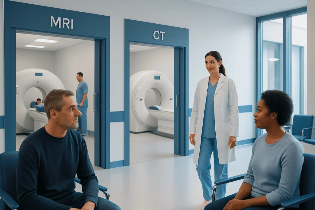
CT is usually the first choice in emergencies because it is fast and widely available. MRI provides more detailed images of the brain, spinal cord, and soft tissues.
Key Takeaways
- CT is usually the first choice in emergencies because it is fast and widely available.
- MRI provides more detailed images of the brain, spinal cord, and soft tissues.
- The best test depends on symptoms, timing, medical history, and safety factors such as metal implants or kidney function.
- Some people may need both CT and MRI because the tests provide complementary information.
- Imaging is only one part of diagnosis and is interpreted together with the neurological exam and other tests.
MRI and CT are both important imaging tests for neurological symptoms, but they answer different clinical questions. Doctors choose between them based on how urgent the situation is, the suspected cause, and which test can provide the safest and most useful information.
Overview: Why imaging matters in neurological symptoms
When a person develops neurological symptoms such as sudden weakness, severe headache, confusion, seizures, vision changes, dizziness, or numbness, doctors often need imaging quickly. Brain and spine imaging can help identify whether symptoms are related to bleeding, stroke, infection, inflammation, tumor, injury, or another structural problem affecting the nervous system.
The two most common imaging tools are computed tomography, called CT, and magnetic resonance imaging, called MRI. Both create pictures of the brain and other parts of the nervous system, but they do so in different ways. A CT scan uses X-rays to produce cross-sectional images, while MRI uses a strong magnetic field and radio waves to create highly detailed images of soft tissues.
Doctors do not usually choose one test because it is simply “better” overall. Instead, they choose the test that best fits the clinical situation. In many urgent situations, CT is preferred first because it is fast. In other cases, MRI is more helpful because it can show subtle changes in brain tissue that CT may miss.
For patients and families, it can be reassuring to know that this decision follows a clear medical logic. The choice is based on what symptom needs to be explained, how quickly an answer is needed, and whether the person has any factors that make one test safer or more appropriate than the other.
How CT and MRI differ
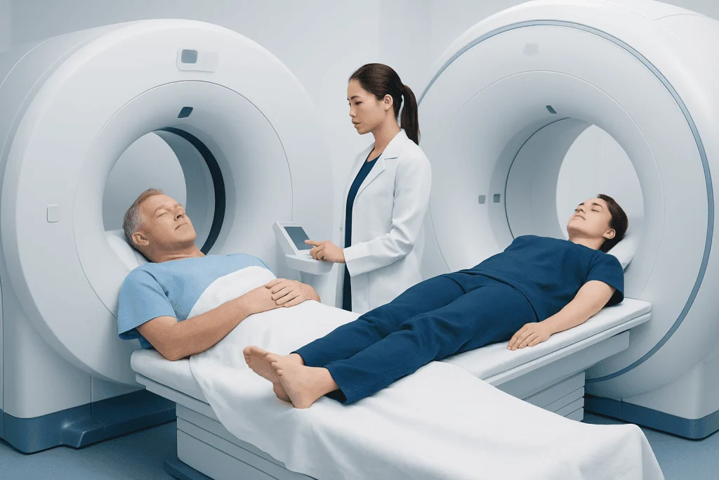
CT is often the quickest imaging option in acute care. The scan itself usually takes only a few minutes, and the machine is commonly available in emergency departments. CT is especially useful for detecting fresh bleeding in or around the brain, major skull injuries, some fractures, and signs that may point to an urgent stroke evaluation.
MRI takes longer, but it provides more detailed information about the brain, spinal cord, nerves, and surrounding soft tissues. It is better at showing small areas of ischemic stroke, demyelination, inflammation, many tumors, and subtle structural abnormalities. MRI can also image the brain in multiple sequences, each highlighting different tissue characteristics.
Another practical difference is patient experience. CT is relatively open and quick, while MRI involves lying still in a narrow scanner for longer and can be noisy. Some people find MRI uncomfortable if they have claustrophobia, difficulty staying still, or significant pain. In certain cases, mild sedation may be considered to help complete the exam safely.
Doctors also think about safety. CT exposes the patient to ionizing radiation, so unnecessary repeat scans are avoided when possible. MRI does not use radiation, but it may not be suitable for people with certain implanted devices, metal fragments, or other MRI-related safety concerns. If contrast dye is needed, kidney function and allergy history may also be reviewed before either test.
When doctors usually choose CT first
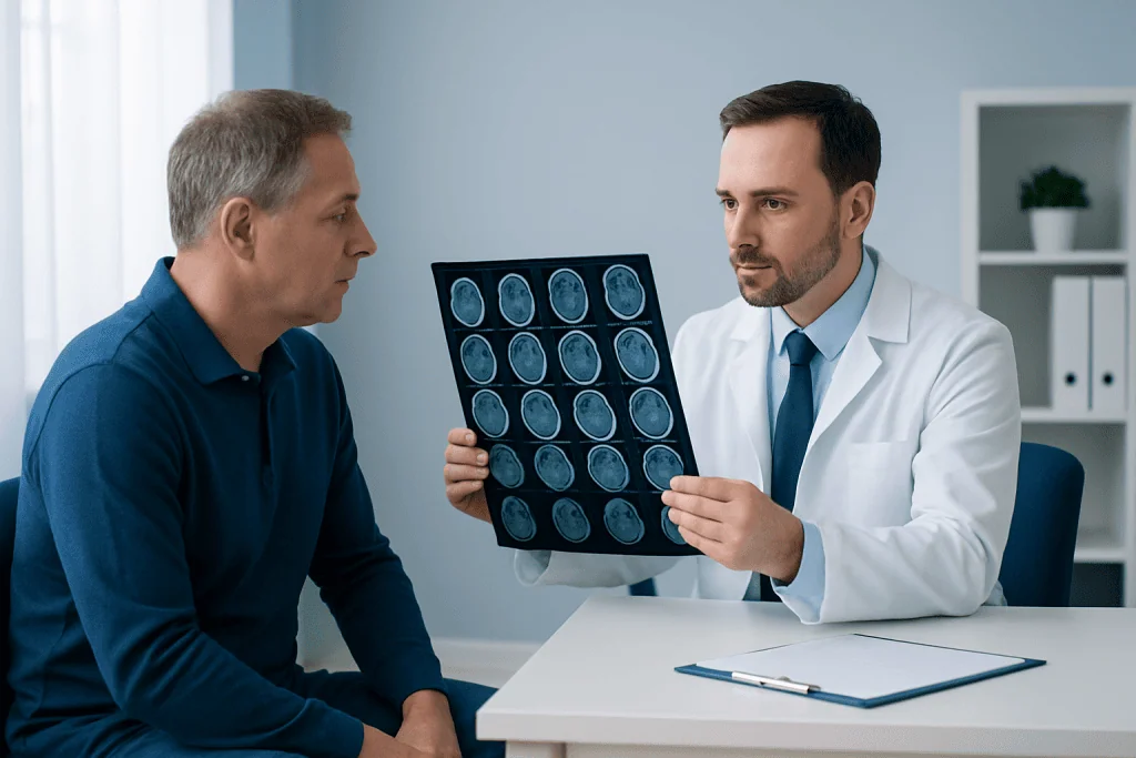
CT is commonly chosen first when symptoms are sudden, severe, or potentially life-threatening. Examples include a suspected stroke, serious head injury, sudden loss of consciousness, severe new headache with concern for bleeding, or rapid neurological decline. In these situations, speed matters, and CT can quickly show whether there is blood in the brain or another emergency that needs immediate treatment.
In stroke care, CT is often the first brain scan because it helps distinguish bleeding from ischemic stroke. This distinction is essential because treatment decisions can differ greatly. A non-contrast head CT is widely used early in stroke evaluation, and some patients may also have CT angiography to assess the brain and neck blood vessels when a blocked artery is suspected.
CT may also be preferred when a person cannot safely undergo MRI, such as those with certain non-compatible implanted devices or metal-related risks. It can be a practical choice if a patient is very unstable, attached to emergency monitoring equipment, or unable to remain still long enough for a longer scan.
Even when CT is the first test, it is not always the last. If CT results are normal but symptoms still suggest a neurological problem, MRI may be recommended afterward to provide more detailed information.
When doctors usually choose MRI
MRI is often chosen when doctors need a closer look at the brain or spinal cord and the situation is stable enough to allow more detailed imaging. It is especially helpful for symptoms that are unexplained, ongoing, recurrent, or subtle. Examples include chronic headaches with red-flag features, unexplained seizures, gradual weakness, balance problems, sensory changes, memory concerns, or possible inflammatory or demyelinating disease such as multiple sclerosis.
MRI is particularly sensitive for early ischemic stroke, especially when symptoms are mild or the affected area is small. It can also better show lesions in the posterior fossa, brainstem, and spinal cord, where CT is less detailed. Doctors may request brain MRI when they need to evaluate brain tissue more closely or look for causes that would not be clearly visible on CT.
If symptoms suggest a problem in the spine rather than the brain, MRI is often the preferred test because it can show discs, nerves, the spinal cord, and surrounding soft tissues. For example, a patient with weakness, numbness, severe neck pain, or bowel or bladder symptoms may need spine MRI to look for compression, inflammation, or another structural cause.
MRI is also commonly used in follow-up care. Once an emergency is ruled out, MRI may help characterize a lesion in more detail, monitor changes over time, or guide treatment planning for conditions such as tumors, inflammatory disorders, or chronic vascular disease.
What doctors consider before choosing a scan
The choice between MRI and CT is based first on the symptom pattern and how quickly the answer is needed. Sudden symptoms that could represent bleeding, major trauma, or an acute vascular event usually push doctors toward CT first. More gradual or complex symptoms may favor MRI because of its stronger soft-tissue detail.
Medical history also matters. Doctors ask about prior strokes, seizures, cancer, infections, migraine history, immune conditions, and recent injuries. They also review any implanted devices, metal fragments, kidney disease, pregnancy status, and prior reactions to contrast material. These details help determine whether contrast is needed and whether one test may be safer than another.
The body area being evaluated is another factor. CT is very useful for bone, acute blood, and urgent vascular assessment. MRI is often preferred for brain tissue, spinal cord disorders, nerve pathways, and subtle inflammatory or demyelinating changes. Sometimes the clinical question is so specific that the order becomes clear right away.
Availability can also influence timing, especially in emergency settings or overnight care. A doctor may begin with the fastest useful test and then arrange a second study if needed. This stepwise approach is common in neurology and does not mean anything was missed; it reflects how different imaging tools complement each other.
Contrast, special scans, and why some people need both
Some CT and MRI exams are done without contrast, while others use contrast material to highlight blood vessels, inflammation, tumors, or areas where the blood-brain barrier is disrupted. A doctor may request MRI with contrast when extra detail is needed to better define an abnormality. The decision depends on the suspected diagnosis and on whether contrast can be given safely.
Specialized scans may also be used for particular neurological questions. For example, CT angiography can help visualize arteries in suspected stroke or aneurysm evaluation, while MRI angiography or venography may be used to study blood vessels without exposing the patient to radiation. Different MRI sequences can also reveal acute stroke, old injury, inflammation, or tiny areas of bleeding.
It is common for a person to have both CT and MRI during the same illness. For example, CT may be used first in the emergency department to rule out bleeding, followed later by MRI to confirm a small stroke, assess inflammation, or clarify a lesion. This does not mean one test failed; it means each test contributed different information.
Radiologists and neurologists interpret these studies together with the neurological examination, blood tests, and sometimes lumbar puncture, EEG, or vascular studies. Imaging is highly valuable, but it works best as part of a complete diagnostic picture.
How patients can prepare and what to expect
For a CT scan, preparation is usually simple. In urgent cases, the test may happen almost immediately. If contrast is planned, staff may ask about allergies, kidney function, and medications. The scan is quick, and many people can return to normal activities soon afterward unless they are being treated for the underlying neurological problem.
For MRI, patients are usually asked to remove metal objects and complete a safety questionnaire about implants, prior surgeries, pacemakers, or possible metal exposure. Remaining still is important for clear images, and the scanner can be loud, so ear protection is commonly provided. If a person is anxious in enclosed spaces, they should tell the care team in advance.
Patients should also share details about pregnancy, kidney disease, and any prior reactions to contrast agents. If a child or someone with severe pain, confusion, or movement difficulty is having MRI, special arrangements may sometimes be needed to help them stay still safely during the scan.
Near the end of the care pathway, patients may benefit from centers where neurologists, neurosurgeons, and neuroradiologists work together. Acibadem International’s multidisciplinary specialists and JCI-accredited hospitals diagnose and treat neurological conditions for international patients, with imaging used as part of coordinated clinical evaluation.
When to seek urgent medical care for neurological symptoms
Some neurological symptoms need urgent medical assessment, whether or not imaging has already been discussed. These include sudden weakness or numbness on one side, trouble speaking, facial drooping, sudden vision loss, a severe and unusual headache, a seizure, fainting, confusion, or difficulty walking that starts suddenly. These symptoms can signal a medical emergency.
Head injury with vomiting, drowsiness, loss of consciousness, worsening headache, or behavior change also requires prompt evaluation. Emergency doctors may decide that CT is the safest first step because it can rapidly check for bleeding or significant injury.
Less sudden symptoms should still be assessed if they are persistent, progressive, or unexplained. Ongoing headaches with neurological changes, repeated episodes of numbness, balance problems, memory decline, or new weakness deserve medical attention even if they are not dramatic at first.
Patients should not try to choose the scan on their own. The safest approach is to seek care promptly and allow a qualified doctor to decide whether CT, MRI, or both are appropriate based on the full clinical picture.
Frequently asked questions
Is MRI better than CT for neurological symptoms?
Not always. CT is often better for urgent situations because it is fast and can quickly detect bleeding or major injury, while MRI is better for detailed views of brain and spinal cord tissues. The best test depends on the symptom, timing, and the clinical question.
Why would a doctor order a CT scan first and then an MRI later?
This is a common and appropriate approach. CT can rapidly rule out emergencies such as bleeding, and MRI can then provide more detailed information if the diagnosis remains unclear or if subtle abnormalities are suspected.
Which scan is used for suspected stroke?
In many emergency settings, CT is the first scan used for suspected stroke because it is fast and helps show whether there is bleeding. MRI may follow in selected cases to detect small or early ischemic strokes or to clarify the affected brain area.
Does MRI expose the body to radiation?
No. MRI does not use ionizing radiation; it uses magnetic fields and radio waves instead. CT does use X-rays, so doctors generally avoid unnecessary repeat CT scans when another suitable option is available.
Can everyone have an MRI?
No, not everyone can. Some implanted devices, metal fragments, or other safety concerns may make MRI unsuitable or require special review before scanning. The imaging team checks these issues carefully before the test.
Is contrast always needed for brain imaging?
No. Many CT and MRI studies are done without contrast, especially in urgent situations such as an initial head CT for suspected bleeding. Contrast is used only when it is likely to add important diagnostic information and when it is safe for the patient.
References
- World Health Organization
- American College of Radiology
- Radiological Society of North America
- National Institute of Neurological Disorders and Stroke
- American Stroke Association
This article is for general information only and is not a substitute for professional medical advice. Please consult a qualified doctor about your individual situation.
Joint and spine care in Turkey — expert assessment & treatment
JCI-accredited · board-certified surgeons · reply within 24h
Add us as a Preferred Source to see more of our trusted health content across Google Search, AI Overviews and Discover.