Navicular Bone: An Evidence-Based Guide for Patients

The navicular bone sits on the inner side of the midfoot and plays a key role in arch support and movement. Pain over the top or inner side of the midfoot may be linked to a navicular stress fracture, accessory navicular, arthritis, or tendon strain.
Key Takeaways
- The navicular bone sits on the inner side of the midfoot and plays a key role in arch support and movement.
- Pain over the top or inner side of the midfoot may be linked to a navicular stress fracture, accessory navicular, arthritis, or tendon strain.
- Diagnosis may involve a physical exam and imaging such as X-ray, CT, or MRI, depending on the suspected problem.
- Treatment ranges from rest, footwear changes, and physical therapy to immobilization or surgery in selected cases.
- Persistent foot pain, swelling, or inability to bear weight should be assessed by a qualified doctor.
The navicular bone is a small but important bone in the midfoot that helps support the arch and transfer force during walking and running. Problems affecting the navicular bone can lead to pain, swelling, and reduced mobility, but timely diagnosis and appropriate care often lead to good recovery.
Overview: What Is the Navicular Bone?
The navicular bone is one of the small bones of the foot, located in the midfoot on the inner side. It connects the ankle-side bones to the bones in the front of the foot and helps maintain the arch. Because it sits at an important junction, the navicular bone absorbs and transfers force with every step.
Patients usually search for the navicular bone because of pain, swelling, tenderness, or a visible bump on the inner foot. The most common concerns include stress fractures, irritation from an accessory navicular, arthritis, overuse injuries, and problems involving nearby tendons such as the posterior tibial tendon. These conditions can feel similar at first, so the cause is not always obvious without medical assessment.
Unlike very general foot pain guides, understanding the navicular bone specifically helps explain why some symptoms worsen during running, jumping, prolonged standing, or wearing certain shoes. This is also why some navicular problems need more careful follow-up than a simple sprain. Early attention can reduce the risk of prolonged pain and delayed healing.
Where the Navicular Bone Is and Why It Matters
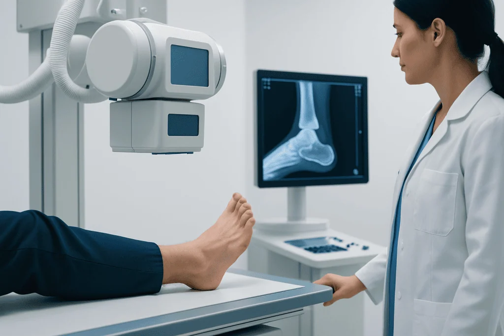
The navicular bone lies in the middle of the foot, just in front of the talus and behind the cuneiform bones. It forms part of the medial longitudinal arch, the structure that helps the foot support body weight and adapt to different surfaces. Its shape and position make it essential for both stability and efficient movement.
Several structures attach to or move around the navicular bone. One of the most important is the posterior tibial tendon, which helps support the arch and control foot motion during walking. If this tendon becomes irritated or weakened, pain may be felt near the navicular area, and the arch can gradually flatten. In some patients, this may overlap with flat feet symptoms.
Some people are also born with an accessory navicular, which is an extra piece of bone or cartilage near the navicular bone. This variant is not rare and may cause no symptoms at all. However, if it becomes irritated by shoes, activity, or tendon pull, it can lead to inner foot pain and swelling.
Symptoms of Navicular Bone Problems
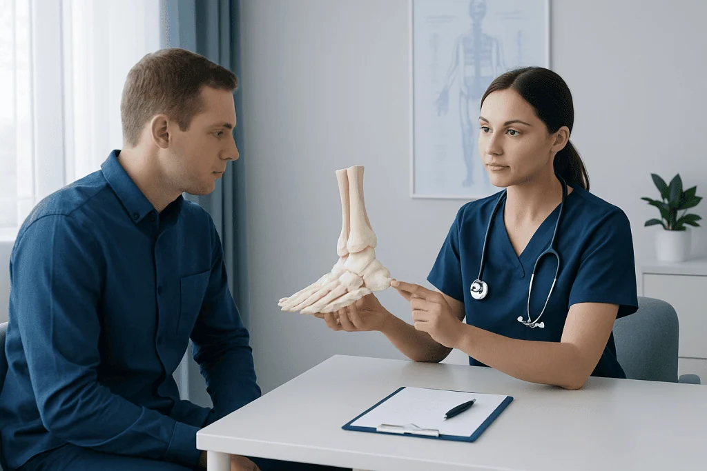
Symptoms vary depending on the underlying cause, but the most common complaint is pain in the midfoot or along the inner side of the foot. The discomfort may start gradually with activity or appear more suddenly after an injury. Many patients describe tenderness when pressing on the top-middle part of the foot or the area over the arch.
Other symptoms can include swelling, bruising, a limp, stiffness, and pain during running, climbing stairs, or pushing off the foot. In stress injuries, pain may improve with rest at first but return when activity resumes. In more advanced cases, walking can become difficult even during routine daily tasks.
An accessory navicular may produce a visible or palpable bump on the inner side of the foot. Shoe pressure can make this area sore, red, or irritated. If tendon strain is also present, the arch may feel tired and the foot may become less stable over time.
- Localized midfoot tenderness
- Pain that worsens with sports or prolonged standing
- Swelling on the inner or top part of the foot
- Difficulty bearing weight or walking normally
- A bony prominence on the inner foot in accessory navicular syndrome
Common Causes and Risk Factors
Navicular bone pain has several possible causes. One important cause is a navicular stress fracture, which develops from repeated loading rather than a single major injury. This is more likely in runners, jumping athletes, dancers, and people whose training volume increases quickly. Hard surfaces, poor footwear, and inadequate recovery can also contribute.
Another common source of symptoms is accessory navicular syndrome. In this condition, an extra navicular-related bone becomes painful because of shoe friction, overuse, or pulling from the posterior tibial tendon. Flat arches or altered foot mechanics can place more strain on the area. Some patients may also have ankle pain or broader foot discomfort at the same time.
Acute injuries such as twisting the foot, direct trauma, or falls can bruise or fracture the navicular bone. Degenerative changes, inflammatory conditions, and nearby tendon disorders may also produce similar pain patterns. Less commonly, reduced bone strength from nutritional factors, low bone density, or certain medical conditions may increase the risk of stress injury.
Risk factors often include a combination of anatomy, activity, and loading patterns rather than one single cause. For this reason, treatment usually works best when it addresses both the painful structure and the factors that triggered it.
How Doctors Diagnose Navicular Bone Conditions
Diagnosis begins with a detailed history and physical examination. A doctor will ask when the pain started, whether it followed increased exercise or trauma, and which movements make it worse. During the exam, they may check for tenderness over the navicular bone, swelling, gait changes, arch shape, and pain related to tendon function.
Imaging is often helpful because different navicular conditions can appear similar. Plain X-rays may show an accessory navicular, some fractures, arthritis, or alignment changes, but they do not detect every stress injury. If symptoms strongly suggest a stress fracture and the X-ray is normal, further imaging may be needed.
MRI can help identify bone stress reactions, tendon problems, and soft tissue inflammation. CT scans can be useful for defining fracture lines and understanding bone healing in more detail. In selected cases, doctors may also assess foot alignment, training habits, or underlying bone health if a stress injury seems likely.
If surgery is being considered or the diagnosis remains uncertain, assessment by an orthopedic or foot-and-ankle specialist may be recommended. In many centers, this evaluation is paired with MRI imaging or CT scanning to guide treatment decisions.
Treatment Options for Navicular Bone Pain and Injury
Treatment depends on the exact diagnosis, the severity of symptoms, and whether the problem is acute or related to overuse. For mild irritation, conservative care may include reducing activity, applying ice, using supportive footwear, and avoiding movements that trigger pain. Short-term immobilization in a boot may help if walking is painful.
Navicular stress fractures usually need a more structured approach because healing can be slow. Rest from impact activity is often essential, and some patients need non-weight-bearing protection for a period recommended by their doctor. Follow-up imaging may be used in certain cases to confirm healing before returning to sport.
Accessory navicular syndrome may improve with shoe modifications, arch support, activity adjustment, and physical therapy. Therapy may focus on foot mechanics, calf flexibility, and strengthening the muscles that support the arch. If symptoms are related to tendon overload, care may overlap with treatment for tendonitis.
Surgery is not needed for most patients, but it may be considered for fractures that do not heal properly, ongoing pain despite conservative treatment, or persistent symptomatic accessory navicular. Depending on the problem, treatment planning may involve orthopedic rehabilitation and, in selected cases, orthopedic surgery evaluation.
Recovery, Prevention, and Self-Care
Recovery time varies widely. A mild soft tissue irritation may settle within weeks, while a stress fracture can require a longer healing period and careful return to activity. Patients often recover best when they do not rush back into running or high-impact exercise before pain has resolved and their doctor confirms it is safe.
Good self-care focuses on reducing repeated strain. Supportive shoes, sport-specific training progression, rest days, and attention to running or walking mechanics can all help. If there is a known foot shape issue, insoles or orthotics may be recommended. Strengthening and flexibility work may also reduce recurrent overload.
Bone health matters as well, especially for people with repeated stress injuries. Adequate nutrition, appropriate calcium and vitamin D intake if advised by a clinician, and addressing menstrual, hormonal, or metabolic concerns may be part of prevention. These issues are particularly relevant for competitive athletes and highly active adults.
Patients with persistent arch pain should avoid self-diagnosing every inner-foot bump as harmless. While many navicular problems are manageable, ongoing symptoms deserve proper evaluation so that a fracture, tendon disorder, or structural issue is not missed.
When to Seek Medical Care
Medical review is important if foot pain lasts more than a few days, returns repeatedly with activity, or interferes with walking. It is especially important to seek care for pain after a fall or twist, a visibly swollen midfoot, or inability to bear weight comfortably. These signs may suggest a fracture or another injury that needs targeted treatment.
Patients should also seek assessment if they notice a growing painful bump on the inner foot, loss of arch support, or pain that keeps returning during sports. Early diagnosis can prevent a minor overload problem from becoming a longer recovery issue. A clinician can also identify whether the pain is actually coming from the navicular bone or a nearby joint, ligament, or tendon.
For international patients who need specialist assessment, Acibadem International’s multidisciplinary teams and JCI-accredited hospitals diagnose and treat foot and ankle conditions using appropriate imaging, rehabilitation, and orthopedic care plans. A qualified doctor can advise on the safest next steps based on symptoms, examination findings, and imaging results.
Frequently asked questions
What does the navicular bone do?
The navicular bone helps connect the back and middle parts of the foot and supports the arch. It also helps the foot transfer force during walking, running, and pushing off the ground.
Where is navicular bone pain usually felt?
Pain is usually felt on the inner side of the midfoot or over the top-middle part of the foot. Some people also notice tenderness when pressing on the area, especially after activity.
Can a navicular stress fracture heal without surgery?
Yes, many navicular stress fractures can heal without surgery if they are diagnosed early and managed properly. Treatment often involves strict activity restriction and sometimes immobilization, but the exact plan depends on the fracture pattern and symptoms.
Is an accessory navicular always a problem?
No, many people have an accessory navicular and never develop symptoms. It becomes a problem only if it is irritated by footwear, overuse, or strain from the posterior tibial tendon.
How is a navicular bone problem diagnosed?
Doctors usually diagnose it through a physical exam and imaging. X-rays may be the first step, but MRI or CT can be more helpful when a stress fracture or complex injury is suspected.
When should someone worry about navicular bone pain?
They should seek medical advice if the pain is persistent, gets worse with walking, or follows an injury. Swelling, limping, or inability to bear weight are also reasons for prompt assessment.
This article is for general information only and is not a substitute for professional medical advice. Please consult a qualified doctor about your individual situation.
Explore treatments in Turkey — costs, top hospitals & a free quote
JCI-accredited · board-certified surgeons · reply within 24h
Add us as a Preferred Source to see more of our trusted health content across Google Search, AI Overviews and Discover.

More from the Health Library

Jet Lag: A Complete Medical Overview

Emotional Abuse: An Evidence-Based Guide for Patients

Bosu Ball Exercises: What Patients Need to Know

Balsamic Reduction: A Complete Medical Overview

Mp Joint: A Complete Medical Overview

Vocal Cord Polyp — Explained by Medical Evidence, Not Myths
Physical Medicine & Rehabilitation Specialists at Acibadem

Dr. Cem Tekin Baran
Orthopedic Surgery & Traumatology
Fzt. Ceren Kandemir Gençsoylu
Physical Medicine & Rehabilitation
Fzt. Ceren Keser
Physical Medicine & Rehabilitation
Dr. Ceyhun Başoğlu
Physical Medicine & Rehabilitation