Neuro Scans Explained: MRI, CT, and Angiography for Brain and Spine Problems
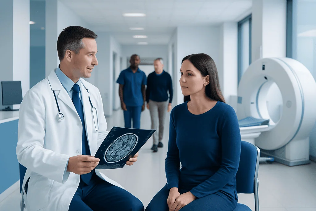
MRI, CT, and angiography are used for different brain and spine questions. MRI shows soft tissues, nerves, discs, and many brain abnormalities in great detail.
Key Takeaways
- MRI, CT, and angiography are used for different brain and spine questions.
- MRI shows soft tissues, nerves, discs, and many brain abnormalities in great detail.
- CT is fast and is often used in emergencies to look for bleeding, fractures, or major swelling.
- Angiography focuses on blood vessels and can help detect aneurysms, narrowing, or blockages.
- The best scan depends on symptoms, medical history, urgency, and safety factors such as pregnancy or metal implants.
Neuroimaging tests such as MRI, CT, and angiography help doctors examine the brain, spine, and blood vessels in detail. Each scan has a different purpose, and understanding how they work can help patients feel more prepared and informed.
Overview: What neuro scans are and why they are used
Neuro scans are imaging tests that help doctors look at the brain, spinal cord, spine, and the blood vessels that supply them. They are commonly used when a person has symptoms such as severe headache, weakness, numbness, seizures, memory changes, dizziness, back pain, neck pain, or signs of stroke. These scans can also be used to monitor a known condition or to plan treatment.
The three most common types discussed in neuroradiology are magnetic resonance imaging (MRI), computed tomography (CT), and angiography. While they may seem similar from a patient’s point of view, they create images in different ways and answer different medical questions. MRI uses magnetic fields and radio waves, CT uses X-rays, and angiography focuses specifically on blood vessels.
Doctors choose the scan based on the person’s symptoms, how urgently answers are needed, and what structures need to be seen most clearly. For example, a sudden neurological emergency may require a CT first because it is fast, while an ongoing problem involving the spinal cord or brain tissue may be better assessed with MRI. If a blood vessel problem is suspected, angiography may be added.
MRI: When it is used for brain and spine problems
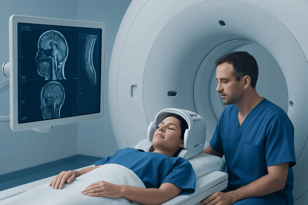
MRI is one of the most detailed imaging tools for the nervous system. It is especially useful for seeing soft tissues, including the brain, spinal cord, intervertebral discs, nerves, ligaments, and areas of inflammation. Because of this, MRI is often used to investigate persistent headaches, seizures, unexplained weakness, numbness, multiple sclerosis, suspected tumors, spinal disc disease, and many chronic neurological symptoms.
A brain MRI can show changes in tissue that may not appear well on CT, including small strokes, demyelinating disease, infection, inflammation, and some tumors. A spine MRI is helpful for evaluating slipped discs, spinal canal narrowing, nerve root compression, spinal cord injury, or suspected infection. In many cases, MRI gives the clearest overall picture of how soft tissues are affected.
Some MRI scans use contrast dye to improve the visibility of certain tissues or blood vessels. Patients usually lie still on a table that slides into the scanner. The test is painless, but it can be noisy and may feel confined for some people. Before MRI, the care team will ask about metal implants, pacemakers, aneurysm clips, shrapnel, or other devices, because not all metal is safe in the MRI environment.
When appropriate, MRI may be part of the evaluation for conditions such as brain tumors or spinal cord disorders. Depending on the findings, it may also help guide next steps, including medical therapy, surgery, or further vascular imaging.
CT scans: Fast imaging for urgent answers
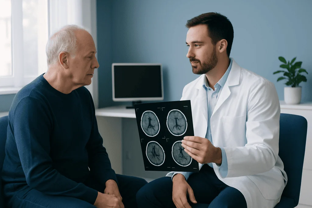
CT scans use X-rays to create detailed cross-sectional images of the body. In neuroradiology, CT is widely used because it is fast, accessible, and very useful in emergencies. A CT scan of the head is often one of the first tests ordered when a person has a sudden severe headache, head injury, suspected stroke, loss of consciousness, or signs of bleeding in the brain.
CT is particularly good at showing fresh bleeding, skull fractures, calcifications, and major swelling. CT of the spine can also help detect fractures, alignment problems, and some bone-related causes of pain or nerve compression. In emergency settings, speed matters, and CT can provide critical information within minutes.
Some CT examinations use contrast material given through a vein to better show blood vessels, tumors, or areas of inflammation. A CT angiogram is a special type of CT focused on arteries and veins. Although CT involves radiation, the amount is carefully controlled, and the benefits usually outweigh the risks when the test is medically necessary.
CT may be the first step before further tests such as MRI or catheter angiography. For example, if a stroke is suspected, doctors may begin with CT to look for bleeding and then choose the next scan based on the results and the treatment plan.
Angiography: Looking closely at blood vessels
Angiography is used when doctors need a detailed view of the blood vessels in the brain, neck, or sometimes the spine. It can help detect aneurysms, arteriovenous malformations, narrowing, blockages, dissections, and other vascular abnormalities. There are different forms of angiography, including MR angiography (MRA), CT angiography (CTA), and catheter angiography, also called digital subtraction angiography.
MRA and CTA are less invasive because they use MRI or CT technology to create vessel images. Catheter angiography is more invasive but remains the most detailed test for some vascular conditions. In this procedure, a thin tube is guided through a blood vessel, contrast dye is injected, and X-ray images are taken in real time. It can be used not only for diagnosis but sometimes for treatment during the same session.
Doctors may request angiography if a person has symptoms of stroke, a suspected aneurysm, unexplained bleeding, or signs that a blood vessel is narrowed or blocked. It is also important in planning procedures such as interventional neuroradiology treatment or surgery for complex vascular conditions.
Because blood vessel disorders can sometimes affect the brain suddenly, angiography plays an important role in timely and accurate care. In selected cases, it may be used to assess diseases such as stroke or aneurysm-related problems when precise vessel mapping is needed.
How doctors choose between MRI, CT, and angiography
The choice of scan depends on the medical question. If a doctor needs to know whether there is acute bleeding after a head injury or signs of a stroke in the first moments, CT is often preferred because it is quick. If the concern is a spinal disc problem, inflammation, or a subtle brain abnormality, MRI often provides more detail. If the main question involves arteries or veins, angiography may be the best option.
Other factors matter too. MRI may not be suitable for some patients with certain implanted devices or severe claustrophobia, although many modern systems and support strategies can help. CT may be limited when repeated radiation exposure is a concern. Contrast dye, whether used in CT, MRI, or angiography, may require special caution in people with kidney problems, prior contrast reactions, or certain other health conditions.
Doctors also consider whether the scan is intended for diagnosis, treatment planning, or follow-up. A patient may need more than one type of scan over time, because each test offers different information. For example, CT may identify an emergency problem quickly, MRI may define the tissue changes more clearly, and angiography may show whether a vessel procedure is needed.
- MRI: best for soft tissues, spinal cord, discs, brain detail, inflammation, and many tumors
- CT: best for speed, acute bleeding, fractures, and urgent assessment
- Angiography: best for arteries, veins, aneurysms, narrowing, and treatment planning for vascular disease
What patients can expect before, during, and after the scan
Preparation depends on the type of scan. Patients are usually asked about allergies, kidney function, pregnancy, implanted devices, medications, and any previous reactions to contrast dye. For MRI, they may need to remove jewelry, hearing aids, or other metal objects. For some scans, eating and drinking may need to be limited for a few hours beforehand, especially if contrast or sedation is planned.
During the test, staying still is important because movement can blur the images. MRI scanners make loud tapping noises, so ear protection is usually provided. CT scans are typically much quicker and are often completed within minutes. Catheter angiography takes longer and may involve local anesthesia, a recovery period, and temporary monitoring afterward.
After most MRI and CT scans, patients can usually return to normal activities quickly unless they received sedation or special medications. After catheter angiography, they may need to rest for several hours and follow instructions about activity and hydration. Results are interpreted by a radiologist or neuroradiologist and then discussed by the treating doctor in the context of symptoms and examination findings.
If a scan shows a condition that may need treatment, the next step could include medication, surgery, rehabilitation, or a minimally invasive procedure such as brain aneurysm treatment or spine surgery, depending on the diagnosis.
Benefits, limits, and safety considerations
Each neuro scan has important strengths, but no single test answers every question. MRI offers excellent soft tissue detail and does not use ionizing radiation, but it takes longer and may not be suitable for everyone. CT is rapid and highly valuable in emergencies, though it uses radiation. Angiography provides crucial vessel information, but catheter angiography is invasive and carries a small risk of bleeding, bruising, or vessel-related complications.
Contrast agents are generally safe when used appropriately, but they can occasionally cause allergic-type reactions or require caution in people with reduced kidney function. Patients should tell their doctor about any past reactions to contrast dye, asthma, kidney disease, diabetes, or pregnancy. This helps the team choose the safest imaging approach.
It is also helpful for patients to know that a scan is only one part of diagnosis. Symptoms, neurological examination, blood tests, and medical history all matter. Sometimes a scan is normal even when symptoms are real, and sometimes small findings need follow-up rather than immediate treatment. Careful interpretation by specialists is essential.
Near the end of the diagnostic process, some patients may be referred to centers with multidisciplinary neurological and imaging expertise. Acibadem International’s multidisciplinary specialists and JCI-accredited hospitals diagnose and treat brain and spine conditions for international patients when advanced neuroradiology evaluation is needed.
When to seek medical attention for brain or spine symptoms
Urgent medical attention is important if a person develops sudden weakness, facial drooping, speech difficulty, loss of vision, a sudden severe headache unlike previous headaches, seizure, confusion, head trauma with neurological symptoms, or loss of consciousness. These symptoms can signal bleeding, stroke, or another emergency that may require immediate imaging.
Prompt medical review is also recommended for persistent or worsening back pain with leg weakness, numbness around the groin, loss of bladder or bowel control, progressive balance problems, unexplained numbness, or ongoing severe neck pain after injury. These symptoms may suggest spinal cord or nerve compression and should not be ignored.
Not every headache or episode of back pain requires a scan, and many common symptoms improve with routine care. Still, if symptoms are new, unusually intense, keep returning, or are affecting daily life, a doctor can help decide whether MRI, CT, or angiography is appropriate. Early assessment often leads to clearer answers and more suitable treatment.
Frequently asked questions
What is the difference between MRI and CT for brain and spine problems?
MRI is usually better for showing soft tissues such as the brain, spinal cord, discs, and nerves in fine detail. CT is faster and is often used in emergencies to look for bleeding, fractures, or major swelling. The right choice depends on the symptoms and the clinical question.
Is angiography the same as a regular CT or MRI scan?
Not exactly. Angiography is focused on blood vessels and may be done using CT, MRI, or a catheter-based technique. It is especially useful when doctors suspect aneurysms, narrowing, blockages, or other vascular problems.
Are neuro scans painful?
Most MRI and CT scans are painless. Patients mainly need to lie still, and some may feel uncomfortable because of noise, the enclosed MRI space, or the contrast injection. Catheter angiography is more invasive, but local anesthesia is used to reduce discomfort.
Do all brain and spine scans require contrast dye?
No. Many MRI and CT scans can be done without contrast, depending on what the doctor is looking for. Contrast is used when it is likely to improve the visibility of blood vessels, inflammation, tumors, or certain other abnormalities.
How long do these scans usually take?
A CT scan is often completed within minutes, which is why it is common in emergencies. MRI usually takes longer, often around 20 to 60 minutes depending on the area being examined. Catheter angiography can take longer because it includes preparation and recovery time.
Are MRI, CT, and angiography safe?
These tests are generally safe when used appropriately and when the medical team knows the patient’s history. MRI does not use radiation, CT does, and angiography may involve contrast dye and, in some cases, a catheter procedure. Doctors weigh the benefits and risks carefully before recommending a scan.
References
- World Health Organization
- National Institute of Neurological Disorders and Stroke
- Radiological Society of North America
- American College of Radiology
- National Health Service
This article is for general information only and is not a substitute for professional medical advice. Please consult a qualified doctor about your individual situation.
Joint and spine care in Turkey — expert assessment & treatment
JCI-accredited · board-certified surgeons · reply within 24h
Add us as a Preferred Source to see more of our trusted health content across Google Search, AI Overviews and Discover.