Neuroendovascular Treatment for AVM and AV Fistula: When Embolization Is Used

Embolization blocks abnormal blood flow through a thin catheter placed inside blood vessels. It may be used for arteriovenous malformations (AVMs) and arteriovenous fistulas (AVFs) in the brain or spine.
Key Takeaways
- Embolization blocks abnormal blood flow through a thin catheter placed inside blood vessels.
- It may be used for arteriovenous malformations (AVMs) and arteriovenous fistulas (AVFs) in the brain or spine.
- Treatment goals include lowering bleeding risk, reducing symptoms, and making other treatments safer or more effective.
- Not every AVM or AV fistula needs embolization; the best plan depends on the lesion’s size, location, and blood flow pattern.
- Care is usually planned by a multidisciplinary team that may include interventional neuroradiology, neurology, and neurosurgery specialists.
Neuroendovascular treatment for AVM and AV fistula is a minimally invasive approach that can close or reduce abnormal blood vessel connections in the brain or spine. Embolization may be used on its own in selected cases or as part of a wider treatment plan that includes surgery or radiosurgery.
Overview
Neuroendovascular treatment for AVM and AV fistula refers to minimally invasive procedures performed from inside the blood vessels to treat abnormal connections between arteries and veins. In an arteriovenous malformation, or AVM, blood passes directly from arteries into veins through a tangled network of abnormal vessels. In an arteriovenous fistula, or AVF, there is one or more direct abnormal connections between an artery and a vein without the usual tiny capillaries in between.
These conditions can occur in the brain or spinal region. Some are discovered after symptoms such as headache, seizure, weakness, pulsating noise in the ear, or bleeding. Others are found incidentally during imaging for another reason. The health risks vary widely depending on the exact type, location, and flow pattern of the abnormal vessels.
Embolization is the main neuroendovascular technique used for these conditions. During embolization, a specialist guides a very thin tube called a catheter through the blood vessels to the target area and releases materials that block the abnormal connection. This can reduce blood flow, seal the lesion completely in selected cases, or prepare it for surgery or focused radiation treatment.
Because AVMs and AV fistulas are complex, treatment is highly individualized. The decision to use embolization depends on whether the abnormal vessels are causing symptoms, how likely they are to bleed or worsen, and whether the expected benefits outweigh the risks of intervention.
Symptoms and possible complications
Symptoms depend on where the AVM or AV fistula is located and how it affects nearby tissue or blood flow. Brain AVMs may cause headaches, seizures, weakness, numbness, vision problems, speech changes, or difficulty with balance. Some people have no symptoms at all until the lesion is found on a scan.
AV fistulas can present differently. A dural arteriovenous fistula in the head may cause a whooshing or pulsing sound in one ear, headache, eye redness, double vision, facial swelling, or neurologic symptoms if blood drainage is impaired. Spinal AV fistulas may cause leg weakness, back pain, numbness, trouble walking, or bladder and bowel symptoms because of congestion around the spinal cord.
The most important complication in some cases is bleeding, especially for certain AVMs and high-risk fistulas. Bleeding in or around the brain can cause sudden severe headache, vomiting, weakness, confusion, seizure, or loss of consciousness. Even without bleeding, abnormal blood flow may irritate brain tissue, raise pressure in nearby veins, or reduce normal circulation to important areas.
It is important to remember that not every lesion behaves the same way. Some remain stable for years, while others need prompt attention. Careful imaging and specialist review help identify which findings are low risk and which require treatment sooner.
Why embolization is used

Embolization is used when blocking the abnormal vessels is expected to improve safety or symptoms. In some AVMs, embolization can reduce the size of the nidus, which is the central tangle of abnormal vessels, and lower the amount of blood flowing through it. In certain AV fistulas, especially when the connection can be reached clearly through the blood vessels, embolization may close the fistula completely.
Doctors may recommend embolization for several reasons. It may be used as a primary treatment, as a step before surgery to reduce bleeding risk during an operation, or before radiosurgery to simplify the lesion. It can also be used urgently when a fistula is causing dangerous venous congestion or after bleeding when the anatomy suggests a treatable source.
The materials used for embolization vary by case. They may include medical glue, liquid embolic agents, tiny coils, or other devices designed to block abnormal flow. The choice depends on the structure of the lesion, the size of the feeding arteries, and the need to protect nearby normal vessels.
Not all AVMs are suitable for curative embolization, and in some cases treatment is aimed at reducing high-risk features rather than eliminating the entire lesion. This is why the procedure is best planned in the context of a broader evaluation of arteriovenous malformation and arteriovenous fistula anatomy, symptoms, and long-term goals.
How doctors decide who is a candidate
Doctors consider many factors before recommending embolization. These include the type of lesion, whether it is in the brain or spine, the presence or absence of prior bleeding, the pattern of venous drainage, the size and location of the abnormal vessels, and the person’s age and general health. Symptoms such as seizures, progressive weakness, severe pulsatile tinnitus, or signs of spinal cord dysfunction may also make treatment more urgent.
Imaging plays a central role in decision-making. Magnetic resonance imaging and computed tomography help show the surrounding brain or spinal tissue, while catheter angiography gives the most detailed map of the blood vessels. This map shows where the lesion is supplied, how it drains, and whether a catheter can be positioned safely for treatment.
Some people benefit more from observation than intervention, particularly if the lesion is found incidentally and appears lower risk. In others, the anatomy suggests a meaningful chance of complete closure or a clear reduction in risk with embolization. A balanced discussion about expected benefits, alternatives, and possible complications is essential.
Many cases are reviewed by a team that may include neurologists, neurosurgeons, radiation specialists, and interventional neuroradiology experts. This collaborative approach helps determine whether embolization alone is appropriate or whether it should be combined with other options such as Gamma Knife radiosurgery or brain surgery.
What happens during the procedure
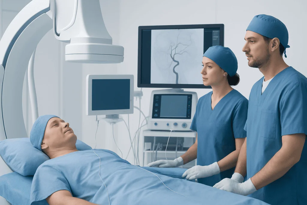
Embolization is usually performed in an angiography suite. The patient receives anesthesia or sedation depending on the situation and the treatment plan. The specialist typically inserts a catheter through a blood vessel in the groin or wrist and navigates it carefully to the arteries supplying the AVM or AV fistula using live X-ray guidance and contrast dye.
Once the catheter is in the correct position, the embolic material is delivered to block the abnormal connection or reduce flow through the lesion. The doctor may treat one part of the lesion or several feeders during the same session. Some complex AVMs require more than one procedure staged over time to improve safety.
After the procedure, the patient is monitored closely. A short hospital stay is common, especially if the lesion is in the brain or spinal region or if the procedure was extensive. Doctors watch for headache, nausea, changes in strength or speech, access-site bleeding, and blood pressure changes while the body adjusts to altered blood flow.
Recovery is often quicker than with open surgery, but rest and follow-up are still important. The care team explains activity limits, medicines, and warning signs to watch for at home. Follow-up imaging helps confirm how much of the lesion was closed and whether further treatment is needed.
Benefits, risks, and treatment results
The potential benefits of embolization include reducing abnormal blood flow, relieving certain symptoms, lowering the chance of bleeding in selected cases, and making surgery or radiosurgery more manageable. For some AV fistulas, embolization can be definitive treatment with complete closure of the abnormal connection. For AVMs, success depends heavily on size, location, and vessel anatomy.
Like all invasive procedures, embolization carries risks. Possible complications include stroke, bleeding, injury to a normal blood vessel, allergic reaction to contrast, infection, or changes in brain or spinal cord function if normal tissue is affected. The exact risk profile is different for each patient and is discussed in detail before treatment.
Results are assessed by symptoms, neurologic examination, and follow-up imaging. Sometimes the goal is complete cure, while in other cases the realistic aim is partial reduction of dangerous flow or preparation for another treatment. It is common for patients to need repeat angiography to confirm whether the lesion remains closed over time.
Because these lesions are uncommon and technically demanding, experience matters. Centers with coordinated vascular neurology, neurosurgery, and imaging expertise are often best equipped to guide treatment choices and long-term follow-up.
Aftercare, self-care, and follow-up
After embolization, patients are usually advised to rest, drink fluids if allowed, and avoid strenuous activity for a short period. The exact instructions depend on the access site used, the complexity of the procedure, and whether there were any immediate concerns after treatment. Headache or mild tiredness may occur and should be discussed with the care team if persistent or severe.
Long-term follow-up is an important part of care. Even when symptoms improve, the abnormal vessels may need repeat imaging because some lesions can reopen or residual flow may remain. Follow-up may include MRI, CT angiography, or repeat catheter angiography depending on what was treated and how complete the closure appeared.
Self-care also means managing general vascular health. This may include controlling blood pressure, avoiding smoking, taking prescribed medicines exactly as directed, and attending all scheduled reviews. People who have had seizures or neurologic symptoms should follow any activity restrictions recommended by their doctor, such as guidance about driving or high-risk activities.
For international patients seeking specialist care, Acibadem International’s multidisciplinary teams and JCI-accredited hospitals diagnose and treat complex neurovascular conditions with individualized planning and follow-up.
When to seek medical attention
Urgent medical attention is needed for sudden severe headache, new weakness or numbness, difficulty speaking, seizure, loss of consciousness, sudden vision change, or severe back pain with rapidly worsening leg weakness or bladder symptoms. These may signal bleeding or a serious change in blood flow and should not be ignored.
Patients recovering from embolization should also contact their doctor promptly if they develop worsening headache, persistent vomiting, fever, confusion, increasing drowsiness, new neurologic symptoms, or significant bleeding or swelling at the catheter entry site. These signs do not always mean a major problem, but they need timely evaluation.
Even without emergency symptoms, specialist review is important when a person has pulsatile tinnitus, unexplained seizures, progressive neurologic symptoms, or an imaging report suggesting an AVM or AV fistula. Early assessment helps clarify the diagnosis and identify whether monitoring or treatment is the safest next step.
Because treatment decisions are nuanced, patients and families should feel comfortable asking questions about the goals of embolization, alternatives, expected recovery, and the need for future procedures. A clear understanding of the plan can make care less stressful and more effective.
Frequently asked questions
What is the difference between an AVM and an AV fistula?
An AVM is usually a tangle of abnormal blood vessels connecting arteries and veins, while an AV fistula is a more direct connection between an artery and a vein. Both can disturb normal blood flow, but their structure, symptoms, and treatment approach may differ.
Is embolization always the only treatment needed?
No. In some cases, embolization can completely treat the lesion, especially certain AV fistulas. In other cases, it is one part of a treatment plan and may be combined with surgery, radiosurgery, or careful monitoring.
How long does recovery after embolization usually take?
Recovery varies depending on the lesion treated, the complexity of the procedure, and the patient's overall health. Many people recover faster than they would after open surgery, but they still need observation, rest, and follow-up imaging.
Can an AVM or AV fistula come back after treatment?
Sometimes residual or recurrent blood flow can be seen on follow-up imaging, especially if treatment was staged or only partial closure was intended. That is why ongoing follow-up is important even when symptoms improve.
Is embolization safe?
Embolization is a well-established procedure, but like any invasive treatment it has risks. Safety depends on the lesion’s anatomy, the patient’s condition, and the experience of the treating team, so decisions are made carefully and individually.
What tests are usually needed before treatment?
Doctors commonly use MRI or CT scans to look at surrounding tissue and catheter angiography to map the blood vessels in detail. These tests help define the lesion and guide the safest and most effective treatment plan.
References
- World Health Organization
- American Heart Association
- National Institute of Neurological Disorders and Stroke
- Society of NeuroInterventional Surgery
- Radiological Society of North America
This article is for general information only and is not a substitute for professional medical advice. Please consult a qualified doctor about your individual situation.
Joint and spine care in Turkey — expert assessment & treatment
JCI-accredited · board-certified surgeons · reply within 24h
Add us as a Preferred Source to see more of our trusted health content across Google Search, AI Overviews and Discover.