Neurointerventional Radiology: How Minimally Invasive Brain Vessel Procedures Work

Neurointerventional radiology uses catheters and real-time imaging to reach blood vessels without open brain surgery. It can help diagnose and treat conditions such as aneurysms, stroke due to blocked arteries, arteriovenous malformations, and carotid artery narrowing.
Key Takeaways
- Neurointerventional radiology uses catheters and real-time imaging to reach blood vessels without open brain surgery.
- It can help diagnose and treat conditions such as aneurysms, stroke due to blocked arteries, arteriovenous malformations, and carotid artery narrowing.
- Common procedures include cerebral angiography, thrombectomy, embolization, stenting, and aneurysm coiling.
- Not every patient is a candidate; treatment depends on the condition, timing, anatomy, and overall health.
- These procedures are usually performed by highly specialized teams including interventional neurologists, neuroradiologists, and neurosurgeons.
- Prompt medical attention is important for symptoms such as sudden weakness, severe headache, speech difficulty, or vision changes.
Neurointerventional radiology is a medical specialty that uses imaging guidance and thin catheters to diagnose and treat conditions affecting the blood vessels of the brain, head, neck, and spine. These procedures are designed to be minimally invasive, often offering shorter recovery times than open surgery for selected patients.
Overview
Neurointerventional radiology is a highly specialized field focused on diagnosing and treating blood vessel conditions that affect the brain, spinal cord, head, and neck. It is also called endovascular neurology, interventional neuroradiology, or neurointerventional surgery, depending on the team and healthcare system. The central idea is to treat problems from inside the blood vessels using very small instruments rather than making a large surgical opening.
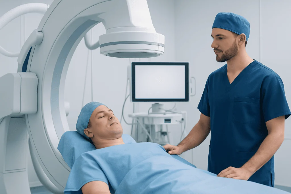
During these procedures, a doctor inserts a thin tube called a catheter into a blood vessel, usually in the wrist or groin, and guides it to the target area with live X-ray imaging. Through that catheter, the team can inject contrast dye to map the vessels, remove a clot, place a stent, or block an abnormal vessel. This approach can be used for both emergency and planned care.
Because the procedure is minimally invasive, it may reduce pain, hospital stay, and recovery time for some patients compared with open surgery. However, it is still a serious medical treatment that requires advanced imaging, careful planning, and experienced specialists. The best option depends on the exact diagnosis, the patient’s symptoms, and how urgent the situation is.
What Conditions Can It Treat?
Neurointerventional radiology is used for several important vascular and structural conditions. One of the best-known examples is acute ischemic stroke caused by a large blood clot blocking blood flow to the brain. In selected patients, a catheter-based procedure called thrombectomy may remove the clot and help restore circulation.
It is also commonly used to treat brain aneurysms, which are weakened, bulging areas in an artery wall. Depending on the aneurysm’s size, shape, and location, treatment may involve coils, flow-diverting stents, or other devices placed through a catheter. Some patients with carotid artery disease may also be treated with angioplasty or stenting to improve blood flow and lower stroke risk.
Other conditions include arteriovenous malformations and dural arteriovenous fistulas, where blood vessels connect abnormally and may bleed or cause neurological symptoms. Neurointerventional teams may also treat certain tumors by blocking their blood supply before surgery, evaluate unexplained bleeding, and investigate vessel narrowing, vasculitis, or spinal vascular disorders. In some cases, these procedures are used mainly to diagnose a problem accurately before a treatment plan is chosen.
How Minimally Invasive Brain Vessel Procedures Work

The process usually starts with detailed imaging such as CT, MRI, or ultrasound, followed by a discussion of the risks and benefits. If a neurointerventional procedure is recommended, the patient is prepared in a specialized angiography suite. Monitoring equipment tracks blood pressure, heart rate, oxygen level, and neurological status throughout the procedure.
After numbing the skin, the doctor inserts a catheter into an artery, often through the wrist or groin. Using real-time imaging, the catheter is carefully navigated through the vascular system to the arteries supplying the brain or spine. Contrast dye may be injected so the team can see the shape of the vessels and identify a blockage, weak spot, or abnormal connection.
Once the catheter reaches the target area, tiny devices can be passed through it. For a blocked artery, a stent retriever or suction device may remove the clot. For an aneurysm, very small coils or a special stent may be placed to redirect blood flow and reduce the risk of rupture. For abnormal vessels, materials such as liquid embolic agents, particles, or coils may be used to close them from the inside, a method broadly known as embolization.
At the end of the procedure, the catheter is removed and pressure or a closure device is used at the entry site. The patient is then observed in a recovery area, stroke unit, or intensive care setting, depending on the procedure and overall condition. Some people go home the same day after diagnostic testing, while others need several days of monitoring after more complex treatment.
Common Procedures in Neurointerventional Radiology
One of the most common diagnostic procedures is cerebral angiography. This test provides a detailed map of the brain’s blood vessels and can help confirm aneurysms, vessel narrowing, malformations, or sources of bleeding. It is often performed when noninvasive imaging suggests a vascular problem but more precise information is needed.
Treatment procedures vary by diagnosis. Mechanical thrombectomy is used in selected stroke cases to remove a clot from a large artery. Aneurysm coiling places tiny platinum coils inside the aneurysm sac so blood no longer flows into it as easily. Flow-diverting stents can be placed in some aneurysms to change the direction of blood flow and encourage healing of the vessel wall.
Angioplasty and stent placement may be used to open narrowed arteries in carefully selected situations. Embolization is used for arteriovenous malformations, fistulas, some tumors, or active bleeding. In certain patients, these procedures are combined with neurosurgery or radiation therapy as part of a broader treatment plan. This is why care is often coordinated by a multidisciplinary team rather than a single specialist.
Benefits, Risks, and Recovery
The main benefit of neurointerventional radiology is that it can treat delicate brain and spine vessel problems through a very small access point. For many patients, this means less tissue disruption than open surgery, smaller scars, and potentially faster recovery. In emergency stroke care, the technique can be time-sensitive and may improve the chance of preserving brain function when performed in the right patient at the right time.
Still, every procedure has risks. Possible complications include bleeding, infection, allergic reaction to contrast dye, blood vessel injury, kidney strain from contrast in susceptible patients, or stroke caused by clot movement or vessel spasm. The level of risk varies depending on the condition being treated, the patient’s medical history, and the complexity of the anatomy.
Recovery depends on the procedure performed. After a diagnostic angiogram, a patient may need only several hours of observation and a short period of reduced activity. After treatment for stroke, aneurysm, or malformation, recovery may involve hospital monitoring, repeat imaging, medications to prevent clotting or vessel spasm, and rehabilitation if there has been neurological injury. The care team explains what to expect before discharge, including when to resume work, exercise, and travel.
Who May Be a Candidate and How Doctors Decide
Not every brain or spine blood vessel condition can be treated with a neurointerventional approach. Doctors consider many factors, including the exact diagnosis, the size and shape of the affected vessel, the patient’s age, symptoms, timing, and any other health problems. In emergency stroke, for example, symptom onset time and imaging findings are especially important when deciding whether thrombectomy is appropriate.
Before recommending treatment, the team reviews brain scans, blood tests, medications, and any history of bleeding or kidney disease. They also consider whether an open neurosurgical operation, medical management alone, or observation might be safer or more effective. Shared decision-making is important, especially when treatment is elective rather than urgent.
Many hospitals discuss complex cases in multidisciplinary meetings. Interventional neurologists, neuroradiologists, neurosurgeons, stroke physicians, anesthesiologists, and intensive care specialists may all contribute to the plan. Near the end of the care journey, some international patients choose centers such as Acibadem International, where multidisciplinary specialists in JCI-accredited hospitals evaluate and treat neurovascular conditions using coordinated medical and procedural care.
Preparing for the Procedure and When to Seek Medical Help
Preparation depends on whether the procedure is urgent or scheduled. For planned treatment, patients are usually told when to stop eating and drinking, which regular medicines to take, and whether blood thinners or diabetes medications need adjustment. It is important to mention any allergies, kidney problems, pregnancy, or prior reactions to contrast dye before the procedure.
After the procedure, patients should follow instructions about hydration, wound care, activity limits, and medications. They should also watch for warning signs such as increasing pain or swelling at the catheter site, new weakness, trouble speaking, chest pain, shortness of breath, fever, or severe headache. These symptoms do not always mean there is a serious complication, but they deserve prompt medical review.
Emergency help is especially important if there are signs of stroke or bleeding in the brain. Sudden facial drooping, arm weakness, numbness, confusion, loss of balance, vision changes, or the worst headache of a person’s life should be treated as urgent. Fast treatment can make a meaningful difference in outcomes, so it is best not to wait and see if symptoms go away.
Frequently asked questions
Is neurointerventional radiology the same as brain surgery?
No. Neurointerventional radiology is a minimally invasive approach that treats certain brain and spine vessel conditions from inside the blood vessels using catheters and imaging guidance. Open brain surgery may still be needed in some situations, but many patients can be treated without a large incision.
What is the difference between cerebral angiography and treatment?
Cerebral angiography is mainly a diagnostic test that creates a detailed map of the brain’s blood vessels. Treatment procedures use similar catheter techniques, but they also place devices or remove blockages to correct the problem.
Is neurointerventional radiology used for stroke?
Yes, it can be used for selected patients with ischemic stroke caused by a large vessel blockage. In these cases, a thrombectomy may remove the clot and restore blood flow, but eligibility depends on timing, imaging, and the patient’s overall condition.
Are these procedures painful?
Most patients feel only minor discomfort at the catheter insertion site, and many procedures are done with sedation or anesthesia. The care team works to keep the patient comfortable and closely monitored throughout the procedure.
How long does recovery take?
Recovery varies widely depending on the reason for the procedure and whether it was diagnostic or therapeutic. Some people return home the same day after an angiogram, while others need a longer hospital stay and rehabilitation after stroke or treatment of a complex vascular condition.
What conditions are commonly treated this way?
Common examples include brain aneurysms, acute ischemic stroke due to a large clot, arteriovenous malformations, dural fistulas, and certain cases of carotid artery narrowing. Neurointerventional radiology may also be used to investigate bleeding or prepare some tumors for surgery.
References
- World Health Organization
- American Stroke Association
- National Institute of Neurological Disorders and Stroke
- Society of NeuroInterventional Surgery
- Radiological Society of North America
This article is for general information only and is not a substitute for professional medical advice. Please consult a qualified doctor about your individual situation.
Minimally Invasive in Turkey — costs, top hospitals & a free quote
JCI-accredited · board-certified surgeons · reply within 24h
Add us as a Preferred Source to see more of our trusted health content across Google Search, AI Overviews and Discover.
Check your numbers in seconds
BMI, calories, due date, blood pressure and 30+ more clinical calculators — free, instant, doctor-reviewed ranges.

Robotic Surgery
Robot-assisted procedures across urology, gynecology and general surgery for greater precision and faster recovery.