Neurointerventional Radiology: What Happens During Diagnosis and Treatment Planning

Neurointerventional radiology combines imaging with catheter-based techniques to assess and treat certain brain and spinal vascular conditions. Diagnosis and treatment planning often involve MRI, CT, ultrasound, and cerebral angiography.
Key Takeaways
- Neurointerventional radiology combines imaging with catheter-based techniques to assess and treat certain brain and spinal vascular conditions.
- Diagnosis and treatment planning often involve MRI, CT, ultrasound, and cerebral angiography.
- The care team reviews symptoms, medical history, medications, and imaging findings to choose the safest approach.
- Many procedures are minimally invasive and performed through a small blood vessel access point.
- Patients usually receive detailed instructions about fasting, medications, contrast dye, and recovery before the procedure.
Neurointerventional radiology is a specialized field that uses advanced imaging and minimally invasive techniques to diagnose and plan treatment for disorders affecting the brain, spine, head, and neck blood vessels. Understanding what happens before, during, and after evaluation can help patients feel more prepared and confident.
Overview of Neurointerventional Radiology
Neurointerventional radiology, also called interventional neuroradiology, is a medical subspecialty that diagnoses and treats conditions involving the blood vessels of the brain, spinal cord, head, and neck. It uses imaging guidance to navigate very small instruments through blood vessels, often avoiding the need for large surgical incisions. This approach may be used for conditions such as stroke, aneurysms, arteriovenous malformations, fistulas, and narrowing of certain arteries.
The specialty is highly collaborative. Neurointerventional radiologists often work closely with neurologists, neurosurgeons, stroke specialists, anesthesiologists, and imaging experts. Together, they review a patient’s symptoms, scan results, and overall health to decide whether a minimally invasive procedure is appropriate and how it should be performed.
For many patients, the first step is not treatment itself but a careful diagnostic evaluation and treatment plan. This planning process is important because brain and spinal blood vessel disorders can vary greatly in size, location, severity, and urgency. A personalized plan helps the team balance effectiveness, safety, and recovery needs.
When Neurointerventional Evaluation May Be Needed
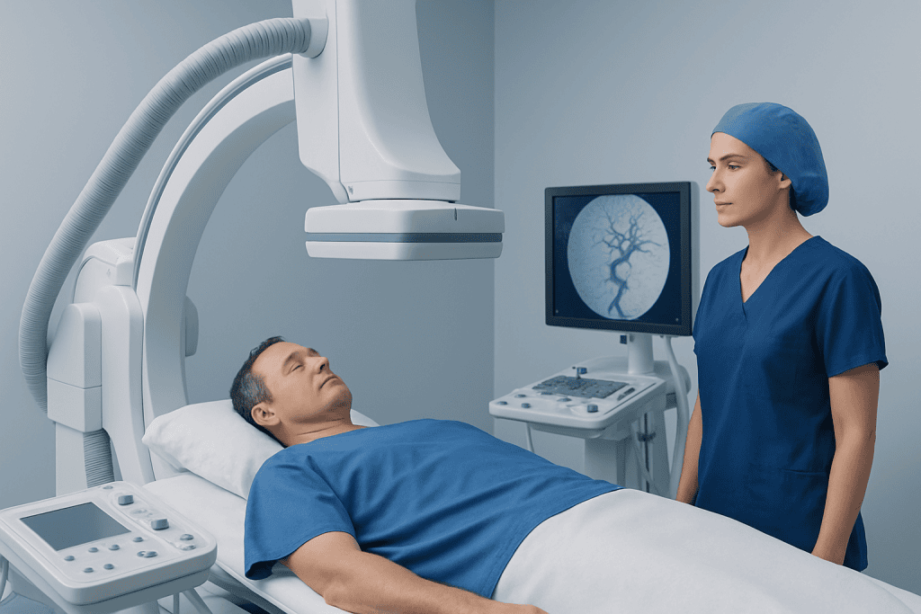
A patient may be referred for neurointerventional radiology after symptoms suggest a blood vessel problem in the brain or spine. Examples include sudden weakness, speech difficulty, severe headache, vision changes, dizziness, facial drooping, pulsating noise in the ear, seizures, or unexplained neurological symptoms. Sometimes the referral happens after an emergency event, such as a suspected stroke. In other cases, it follows an abnormal finding on a routine scan.
Some people are evaluated even when they feel well, because imaging has revealed a possible aneurysm, vessel narrowing, or vascular malformation. In these situations, the goal is to understand whether the finding needs monitoring, medical management, or an intervention. Diagnosis and planning are especially important because not every abnormality requires immediate treatment.
Neurointerventional assessment may also help guide care for people with complex vascular conditions that overlap with other specialties. For example, a patient with a suspected brain aneurysm or a vascular cause of stroke may benefit from detailed vessel imaging and a multidisciplinary review. The focus is always on matching the right test and the right treatment to the individual patient.
What Happens During Diagnosis

Diagnosis begins with a detailed medical review. The specialist asks about symptoms, when they started, how they have changed, and whether there are any triggers. Medical history matters as well, including high blood pressure, diabetes, kidney disease, prior stroke, smoking, bleeding problems, allergies, and current medications such as blood thinners.
The next step is imaging. Depending on the clinical question, the team may use CT, MRI, CT angiography, MR angiography, carotid ultrasound, or other studies to look at the brain, spinal cord, and blood vessels. These tests can show bleeding, blocked arteries, vessel malformations, narrowing, or changes in blood flow. They help clarify what problem may be present and whether a procedure is likely to help.
In some cases, the most informative test is cerebral angiography. During this procedure, a doctor guides a thin catheter through a blood vessel, usually from the wrist or groin, and injects contrast dye while taking detailed X-ray images. Cerebral angiography can provide a highly precise map of the blood vessels and is often used when treatment decisions depend on exact anatomy. Although it is invasive, it is usually far less invasive than open surgery.
Patients are often asked to complete blood tests before angiography or another planned procedure. These tests may check kidney function, blood counts, and clotting status. This preparation helps the team use contrast safely, reduce bleeding risk, and plan sedation or anesthesia appropriately.
How Treatment Planning Is Decided
After reviewing symptoms, examination findings, and imaging, the care team develops a treatment plan tailored to the patient’s condition. Planning includes deciding whether treatment is urgent, whether a minimally invasive technique is suitable, and what the goals are. In some situations, the best plan is observation with follow-up scans. In others, medical treatment or a catheter-based intervention offers the best balance of risk and benefit.
Several factors influence the plan. These include the type of vascular problem, its size and shape, where it is located, whether it has caused bleeding or neurological symptoms, and the patient’s age and general health. The team also considers practical issues such as medication use, allergy to contrast dye, kidney function, pregnancy status, and how well the patient may tolerate sedation or anesthesia.
Treatment planning also includes discussing possible procedural options in understandable terms. For example, an aneurysm may be approached with techniques such as aneurysm embolization, while a blocked artery in an acute stroke setting may call for mechanical thrombectomy. The doctor explains why a particular option is recommended, what alternatives exist, and what outcomes and risks should be considered.
Importantly, informed consent is part of planning. Patients and families have the opportunity to ask questions about the purpose of the procedure, how long it may take, what recovery is like, and what signs to watch for afterward. Clear communication can make the process feel more manageable and less overwhelming.
What to Expect on the Day of the Procedure
Before the procedure, the patient usually receives instructions about fasting, medications, and arrival time. Some medicines may need to be continued, adjusted, or temporarily stopped, especially blood thinners, diabetes medicines, or drugs that affect kidney function. The team may also ask about allergies to contrast material, previous reactions to anesthesia, and whether there is any chance of pregnancy.
At the hospital, nurses and physicians prepare the patient by checking vital signs, placing an intravenous line, and reviewing consent and test results. The procedure itself takes place in a specialized imaging suite. Depending on the situation, the patient may receive local anesthesia with sedation or, less commonly, general anesthesia. The skin is cleaned carefully, and a small access point is made, often in the wrist or groin.
Using real-time imaging, the specialist guides a catheter through the blood vessels to the area of interest. Diagnostic angiography may end after detailed images are obtained, or treatment may be performed during the same session if this has been planned. For certain vascular malformations or tumors, the team may use techniques such as embolization to reduce abnormal blood flow or prepare for further treatment.
Afterward, the catheter is removed and pressure or a closure device is used to help prevent bleeding. The patient is observed in a recovery area, where the team checks blood pressure, neurological status, and the access site. Some people go home the same day, while others stay overnight for monitoring, depending on the procedure and their overall condition.
Benefits, Risks, and Recovery
One of the main advantages of neurointerventional radiology is that it can diagnose and treat certain conditions through a minimally invasive approach. Compared with open surgery, this may mean smaller incisions, less pain, shorter hospital stays, and faster recovery for selected patients. In urgent settings such as stroke, rapid imaging and intervention can also be critical for preserving brain function.
Like all medical procedures, neurointerventional tests and treatments carry risks. These may include bleeding at the access site, bruising, contrast reactions, kidney strain from dye, blood vessel injury, infection, or, in some situations, stroke-related complications. The exact level of risk depends on the procedure, the condition being treated, and the patient’s individual health factors.
Recovery advice is personalized, but patients are commonly told to rest, drink fluids if allowed, and avoid strenuous activity for a short period. The care team explains what symptoms are expected, such as mild soreness at the access site, and which symptoms need urgent attention, such as worsening weakness, severe headache, heavy bleeding, or trouble speaking. Follow-up appointments and repeat imaging may be needed to confirm that the treatment has worked as intended.
Questions to Ask and When to Seek Medical Care
Patients often feel more comfortable when they know what questions to ask. Helpful topics include why the test or procedure is being recommended, whether there are alternatives, what the benefits and risks are, what type of anesthesia will be used, and how long recovery may take. Asking whether the procedure is diagnostic only or whether treatment might happen during the same session can also help with planning.
Medical attention should be sought promptly for sudden neurological symptoms such as new weakness, facial drooping, severe headache, confusion, loss of vision, seizures, or difficulty speaking. These can signal an emergency, especially if stroke is a concern. After a procedure, patients should also contact their doctor if they develop increasing pain, swelling, fever, heavy bleeding from the access site, or any new neurological change.
Ongoing care may involve more than one specialist, particularly for complex vascular disorders. A coordinated approach supports safe diagnosis, clear treatment planning, and appropriate follow-up. Near the end of the care journey, some international patients may choose centers such as Acibadem International, where multidisciplinary specialists in JCI-accredited hospitals evaluate and treat neurovascular conditions with individualized planning.
Frequently asked questions
Is neurointerventional radiology the same as brain surgery?
No. Neurointerventional radiology uses imaging guidance and thin catheters placed through blood vessels, usually from the wrist or groin, rather than a large surgical opening. It is considered minimally invasive, although some conditions still require neurosurgery or combined care.
What conditions can neurointerventional radiology help diagnose or treat?
It can help with conditions affecting the blood vessels of the brain, spine, head, and neck. Examples include aneurysms, some types of stroke, arteriovenous malformations, fistulas, and narrowed arteries. The exact role depends on the condition and the patient’s overall health.
Is cerebral angiography painful?
Most patients feel only minor discomfort from the numbing injection and the blood vessel access point. Sedation may be used to help with comfort, and many people do not feel the catheter moving inside the blood vessels. The team monitors the patient closely throughout the procedure.
How should a patient prepare for a neurointerventional procedure?
Preparation often includes fasting for a period of time, reviewing medications, and having blood tests. Patients should tell their doctor about allergies, kidney problems, bleeding disorders, pregnancy, and all medicines they take, especially blood thinners or diabetes medications. Following the team’s instructions carefully helps improve safety.
Will treatment always happen on the same day as diagnosis?
Not always. Sometimes a procedure is done only to obtain detailed images and confirm the diagnosis. In other situations, if it is appropriate and already planned, diagnosis and treatment may happen during the same session.
How long does recovery usually take?
Recovery depends on the type of procedure, the reason for treatment, and the patient’s health. Some people go home the same day after a short period of monitoring, while others stay overnight or longer. The doctor will explain activity limits, follow-up, and warning signs before discharge.
This article is for general information only and is not a substitute for professional medical advice. Please consult a qualified doctor about your individual situation.
Joint and spine care in Turkey — expert assessment & treatment
JCI-accredited · board-certified surgeons · reply within 24h
Add us as a Preferred Source to see more of our trusted health content across Google Search, AI Overviews and Discover.
Check your numbers in seconds
BMI, calories, due date, blood pressure and 30+ more clinical calculators — free, instant, doctor-reviewed ranges.

Neurosurgery
Surgical treatment of the brain, spine and nervous system, including minimally invasive and functional procedures.
70 specialists in this unitMore from the Health Library

Cerebral Aneurysm Embolization: Recovery Timeline and Aftercare
Cerebral Angiography for Stroke and Aneurysm Care: What Patients Should Expect

Who Is a Good Candidate for Neurointervention?

Stroke Thrombectomy Recovery: What to Expect After Clot Removal

When to See an Interventional Neuroradiologist for Brain or Spine Vessel Problems