When to See an Interventional Neuroradiologist for Brain or Spine Vessel Problems

Interventional neuroradiologists treat brain and spine vessel conditions through tiny catheters guided by advanced imaging. Urgent symptoms such as sudden severe headache, stroke signs, seizures, or sudden weakness need prompt medical evaluation.
Key Takeaways
- Interventional neuroradiologists treat brain and spine vessel conditions through tiny catheters guided by advanced imaging.
- Urgent symptoms such as sudden severe headache, stroke signs, seizures, or sudden weakness need prompt medical evaluation.
- Common reasons for referral include aneurysms, arteriovenous malformations, carotid narrowing, acute stroke, and spinal vascular malformations.
- Diagnosis often involves CT, MRI, vascular imaging, and sometimes catheter angiography.
- Treatment may include clot removal, embolization, stenting, or coiling, depending on the condition and the patient’s overall health.
An interventional neuroradiologist is a doctor who diagnoses and treats certain blood vessel problems in the brain, head, neck, and spine using minimally invasive, image-guided procedures. Seeing this specialist may be important when symptoms, emergency findings, or imaging suggest a condition such as an aneurysm, stroke, arteriovenous malformation, or spinal vascular disorder.
Overview: What an Interventional Neuroradiologist Does
An interventional neuroradiologist is a physician with specialized training in diagnosing and treating blood vessel disorders that affect the brain, spinal cord, head, and neck. This specialist uses imaging tools such as X-ray guidance, CT, MRI, and angiography to perform precise procedures through very small tubes called catheters, usually inserted through an artery in the wrist or groin.
These procedures are often described as minimally invasive because they avoid large surgical openings. In many cases, they can help restore blood flow, block abnormal blood vessels, prevent bleeding, or reduce the risk of future neurological injury. Interventional neuroradiology works closely with neurology, neurosurgery, stroke medicine, and vascular specialists.
People may be referred to this specialist in an emergency, such as during a stroke, or after a scan finds an abnormal vessel problem. Referral can also happen when symptoms are ongoing but unexplained, including certain headaches, pulsating noises in the ear, seizures, nerve symptoms, or weakness related to vascular changes in the brain or spine.
Which Brain and Spine Vessel Problems May Need This Specialist
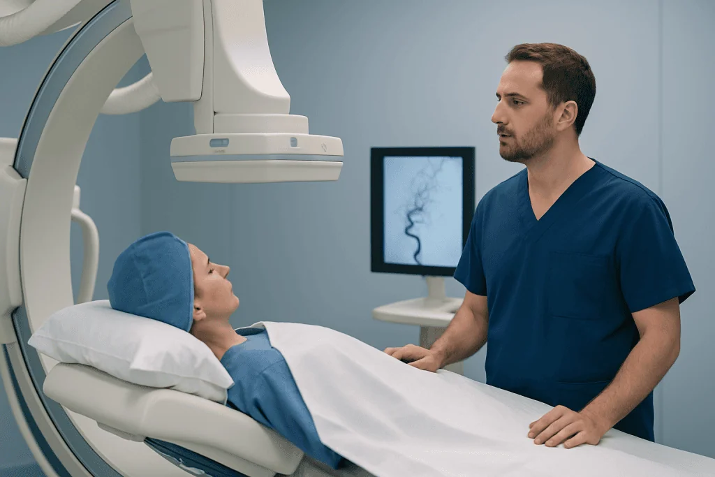
Interventional neuroradiologists commonly evaluate and treat conditions involving narrowed, blocked, weakened, or abnormal blood vessels. In the brain, these may include cerebral aneurysms, arteriovenous malformations (AVMs), dural arteriovenous fistulas, carotid or intracranial artery narrowing, and blood clots causing acute ischemic stroke. Some patients are referred after a scan suggests a brain aneurysm or another abnormality that needs expert review.
Spinal vascular disorders are less common but can be important because they may gradually affect walking, leg strength, sensation, or bladder function. These conditions include spinal dural arteriovenous fistulas and other malformations that alter normal blood flow around the spinal cord. Early diagnosis matters because symptoms can resemble more common spine problems and may worsen over time if the underlying vessel issue is missed.
Some conditions are discovered by chance during imaging done for another reason. Even when there are no symptoms, specialist assessment may still be useful to understand whether the finding is harmless, needs monitoring, or should be treated to reduce future risks. The decision depends on the location, size, blood flow pattern, and the person’s age, health, and symptoms.
Symptoms and Warning Signs That Should Prompt Evaluation

Urgent medical attention is needed for sudden neurological symptoms. These include facial drooping, arm or leg weakness, difficulty speaking, sudden confusion, vision loss, severe dizziness, loss of balance, or a sudden severe headache unlike usual headaches. These can be signs of stroke or bleeding in the brain and should be treated as emergencies.
Other symptoms may be less dramatic but still deserve prompt evaluation, especially when they are new, persistent, or unexplained. Examples include seizures, double vision, a whooshing or pulsating sound in one ear, frequent unexplained headaches with neurological symptoms, numbness, fainting, or episodes of temporary weakness. Some vascular conditions can also cause nerve pressure symptoms depending on where the abnormal vessel is located.
Spinal vessel problems may cause progressive back pain, leg weakness, walking difficulty, tingling, numbness, or changes in bladder or bowel control. These symptoms can overlap with orthopedic or neurological disorders, so imaging and specialist review are often needed to identify the true cause. If symptoms are rapidly worsening, medical evaluation should not be delayed.
- Call emergency services for stroke symptoms or a sudden thunderclap headache.
- Seek prompt review for new seizures or unexplained neurological changes.
- Discuss incidental vascular findings with a specialist rather than ignoring them.
Causes, Risk Factors, and Who Might Be Referred
Brain and spine vessel problems can develop for different reasons. Some are congenital, meaning a person is born with the abnormal vessel pattern, as may occur with some AVMs. Others develop later because of atherosclerosis, high blood pressure, connective tissue disorders, head or neck trauma, age-related changes, or clotting tendencies. Not every condition has a clear single cause.
People with hypertension, smoking exposure, diabetes, high cholesterol, heart rhythm disorders, or a family history of aneurysm or stroke may be at higher risk for some vascular problems. Certain symptoms, previous strokes, known vascular disease, or abnormal findings on carotid ultrasound, CT angiography, or MRI may also lead to referral. For example, a patient with acute ischemic stroke may need urgent catheter-based mechanical thrombectomy to restore blood flow.
Referrals also come from neurologists, emergency physicians, neurosurgeons, spine specialists, or primary care doctors when a scan suggests a vascular lesion that might benefit from a less invasive approach. In some cases, the interventional neuroradiologist helps decide not only whether a procedure is needed, but also whether monitoring, medication, surgery, or another strategy would be safer and more appropriate.
How Diagnosis Is Made
Diagnosis usually begins with a medical history, neurological examination, and review of symptoms. The doctor considers how symptoms started, whether they are sudden or gradual, and whether there are risk factors such as high blood pressure, smoking, or previous stroke. This clinical picture helps determine how urgent the evaluation is and which imaging tests are most useful.
Common imaging tests include CT and MRI of the brain or spine, along with vascular studies such as CT angiography, MR angiography, or Doppler ultrasound in selected cases. These tests can show bleeding, blocked arteries, aneurysms, narrowed vessels, or abnormal vascular connections. For stroke, rapid imaging is especially important to decide whether time-sensitive treatment is possible.
In some situations, the most detailed test is a catheter angiogram, also called cerebral or spinal angiography. During this procedure, a thin catheter is guided into the blood vessels and contrast dye is used to create highly detailed images. Angiography can help confirm a diagnosis, define anatomy, and guide treatment planning for conditions such as arteriovenous malformation or aneurysm. Although invasive, it is a standard and valuable tool when noninvasive scans do not give enough detail.
Treatment Options an Interventional Neuroradiologist May Offer
Treatment depends on the exact condition, symptoms, anatomy, and overall health of the patient. In emergencies such as large-vessel ischemic stroke, the priority may be reopening a blocked artery as quickly as possible. In other situations, the goal may be preventing bleeding, reducing pressure on nearby structures, or stopping abnormal blood flow within a malformation or fistula.
Common procedures include clot retrieval, aneurysm coiling, stent placement, angioplasty, and embolization. Embolization means blocking an abnormal or harmful blood vessel from inside using special materials delivered through a catheter. This may be used for AVMs, fistulas, tumors with high blood supply, or certain sources of bleeding. Some patients may be treated with endovascular aneurysm treatment or embolization as part of a planned, individualized approach.
Not every patient needs a procedure. Some vascular findings are best monitored with periodic imaging, while others are managed with medicines that reduce stroke risk, such as blood pressure treatment or antithrombotic therapy prescribed by the treating team. Care decisions are often made in a multidisciplinary meeting involving neuroradiology, neurology, neurosurgery, and other specialists to choose the safest option.
Recovery and follow-up vary by procedure and condition. Patients may need repeat imaging to confirm that a vessel remains closed, repaired, or open as intended. Ongoing management of risk factors remains important even after successful treatment, especially in conditions related to atherosclerosis or stroke.
What to Expect at the Appointment and Questions to Ask
At an interventional neuroradiology appointment, the specialist typically reviews symptoms, prior scans, medications, allergies, and overall health. The consultation often focuses on what the abnormality is, whether it explains the symptoms, the short- and long-term risks, and whether urgent treatment is necessary. If more imaging is needed, the doctor explains why and how it may affect decisions.
Patients may find it helpful to bring prior imaging reports, a list of medications, and information about past medical problems such as stroke, heart disease, kidney disease, or bleeding disorders. Family members can also be useful at the visit, particularly when complex treatment decisions are being discussed. Clear communication is important because several reasonable options may exist.
Helpful questions include: What exactly was found? Is it dangerous now, or mainly something to monitor? What are the benefits and risks of treatment compared with observation? How experienced is the team with this procedure, and what follow-up will be needed? For international patients, centers such as Acibadem International have multidisciplinary specialists and JCI-accredited hospitals that diagnose and treat these conditions with coordinated care.
When to See a Doctor Right Away and How to Protect Vascular Health
Immediate medical care is important for signs of stroke, sudden loss of consciousness, a thunderclap headache, new seizures, or rapidly worsening weakness or numbness. These symptoms do not always mean a vessel problem, but they do require urgent evaluation because timing can affect treatment choices and outcomes. It is safer to seek emergency help promptly than to wait for symptoms to pass.
For non-emergency concerns, an appointment should be arranged if there is a newly discovered aneurysm or AVM, unexplained pulsatile tinnitus, recurrent transient neurological symptoms, or progressive leg weakness and walking difficulty that could suggest a spinal vascular disorder. Early specialist review can clarify whether watchful monitoring or intervention is the better path.
General prevention focuses on vascular health. Managing blood pressure, avoiding tobacco, controlling diabetes and cholesterol, staying physically active, following prescribed medications, and attending follow-up imaging can all help reduce risk. People with a family history of aneurysm, stroke, or vascular malformations should discuss individualized screening or referral decisions with a qualified doctor.
Frequently asked questions
What is the difference between an interventional neuroradiologist and a neurosurgeon?
An interventional neuroradiologist treats many brain and spine vessel problems from inside the blood vessels using imaging-guided catheters. A neurosurgeon typically performs open or microscopic surgery when an operation is the best option. These specialists often work together, and some patients may need evaluation by both.
Does seeing an interventional neuroradiologist mean surgery is definitely needed?
No. Many referrals are made to better understand a scan finding or to compare treatment choices with careful monitoring. In some cases, no procedure is needed, and the safest plan is follow-up imaging and management of risk factors.
Are all aneurysms emergencies?
No. Some aneurysms are found before they cause symptoms and can be monitored or treated electively depending on their size, shape, and location. An aneurysm becomes an emergency if it ruptures or causes sudden severe symptoms, especially a sudden intense headache.
When is stroke treated by an interventional neuroradiologist?
This specialist may be involved when imaging shows a blocked large artery that could respond to catheter-based clot removal. The decision depends on the time symptoms started, the type of stroke, the patient’s condition, and the imaging findings. Stroke care is time-sensitive, so emergency evaluation is essential.
Can spinal blood vessel problems be mistaken for regular back problems?
Yes. Spinal vascular disorders can sometimes look like more common spine conditions because they may cause back discomfort, leg weakness, numbness, or difficulty walking. When symptoms do not fit the usual pattern or keep worsening, further imaging may be needed.
Is angiography safe?
Catheter angiography is a commonly used diagnostic test and is generally safe when performed by experienced teams, but it does carry some risks. These may include bleeding at the access site, contrast reactions, kidney strain, or rarely stroke. The doctor weighs these risks against the value of getting detailed information that may change treatment.
This article is for general information only and is not a substitute for professional medical advice. Please consult a qualified doctor about your individual situation.
Joint and spine care in Turkey — expert assessment & treatment
JCI-accredited · board-certified surgeons · reply within 24h
Add us as a Preferred Source to see more of our trusted health content across Google Search, AI Overviews and Discover.

Neurosurgery
Surgical treatment of the brain, spine and nervous system, including minimally invasive and functional procedures.
70 specialists in this unitMore from the Health Library

Neurointervention in Turkey: How to Choose the Right Hospital for Complex Brain Vessel Care

Cerebral Aneurysm Symptoms: When a Sudden Headache Needs Emergency Imaging

Neurointerventional Radiology Recovery: Hospital Stay, Aftercare, and Warning Signs

Interventional Neurology vs Neurointerventional Radiology: What Is the Difference?

Stroke Thrombectomy Recovery: What to Expect After Clot Removal