

Neuron Diagram: What Patients Need to Know
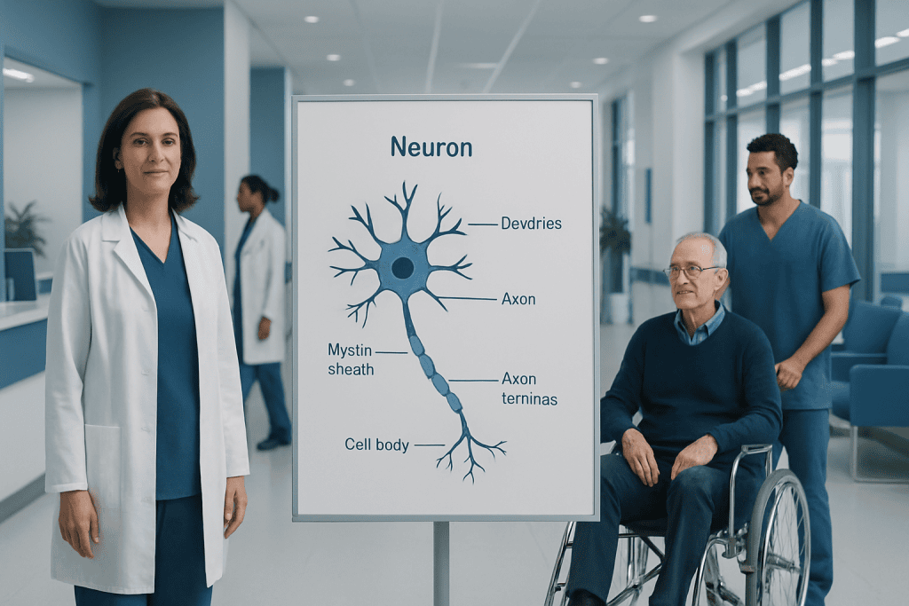
A neuron diagram usually labels dendrites, the cell body, axon, myelin, and synapse. Neurons communicate by electrical and chemical signals throughout the nervous system.
Key Takeaways
- A neuron diagram usually labels dendrites, the cell body, axon, myelin, and synapse.
- Neurons communicate by electrical and chemical signals throughout the nervous system.
- Different neuron shapes reflect different jobs, such as sensing, movement, or coordination.
- A diagram is a learning tool, not a test result or diagnosis by itself.
- Persistent numbness, weakness, severe headache, or sudden speech or vision changes need prompt medical evaluation.
A neuron diagram is a labeled picture of a nerve cell that helps patients understand how the brain, spinal cord, and nerves send messages. Reading one becomes easier when each part is linked to its job: receiving signals, processing information, and passing messages to the next cell.
Overview: What a Neuron Diagram Shows
A neuron diagram is a visual guide to the basic structure of a nerve cell. In simple terms, it shows how one cell is built to receive information, process it, and pass it along. For patients, this can make the nervous system feel less abstract and easier to understand during a clinic visit, while reading test results, or when learning about neurological symptoms.
Most neuron diagrams label a few core parts: dendrites, the cell body, the axon, the myelin covering, and the synapse. These labels are not just anatomy terms. Each one represents a step in communication, from taking in a signal to sending a message onward to another neuron, a muscle, or a gland.
It can also help to know what a neuron diagram does not show. It is a simplified learning image, not a complete picture of the brain or all nerve cells in the body. Real neurons vary in size, shape, length, and number of connections, depending on where they are located and what job they perform.
The Main Parts of a Neuron

Most patient-friendly diagrams begin with dendrites. These are branch-like extensions that receive incoming signals from other cells. A useful way to think about dendrites is that they act like listening branches, gathering information from nearby neurons.
The cell body, also called the soma, is the central part of the neuron. It contains the nucleus and many of the structures that keep the cell alive and functioning. The cell body helps integrate incoming information and supports the neuron’s day-to-day activity.
The axon is a long, cable-like extension that carries signals away from the cell body. Some axons are very short, while others can be quite long. In many neurons, the axon is wrapped in myelin, a fatty insulating layer that helps signals travel more efficiently. At the end of the axon are terminals that communicate with the next cell across a tiny gap called the synapse.
- Dendrites: receive signals
- Cell body: supports the cell and processes information
- Axon: sends signals outward
- Myelin: helps speed signal transmission
- Synapse: allows communication between cells
How Signals Move Through a Neuron

A neuron diagram is easier to read when it is followed in the same direction that a signal travels. In general, messages arrive at the dendrites, move through the cell body, and then travel down the axon. At the axon terminal, the signal triggers the release of chemical messengers that cross the synapse and influence the next cell.
This process involves both electrical and chemical communication. Inside the neuron, signaling is largely electrical. Between neurons, communication usually becomes chemical through neurotransmitters. This switch from electrical to chemical signaling is one reason the nervous system can be both fast and highly adaptable.
Diagrams may use arrows to show the direction of signal flow. These arrows are helpful because they reinforce a key point: neurons are specialized for organized communication. When symptoms affect feeling, movement, memory, or coordination, the problem may involve one or more parts of this communication pathway.
Why Neuron Shape Matters
Not all neurons look exactly the same. A basic neuron diagram often shows one classic shape, but the body has several neuron types. Their structure reflects their role. Sensory neurons help carry information from the skin, eyes, ears, and other organs to the brain and spinal cord. Motor neurons carry commands from the brain or spinal cord to muscles. Interneurons help connect and coordinate signals within the central nervous system.
The length and branching pattern of a neuron matter. A neuron with many branches may receive information from many other cells. A neuron with a long axon may need to send messages across a longer distance, such as from the spinal cord to a muscle in the leg.
For patients, this explains why a single term like “nerve cell” covers many highly specialized cells. It also shows why disorders can affect the body differently depending on which neurons are involved. Problems in sensory pathways may cause numbness or tingling, while problems in motor pathways may lead to weakness or movement changes.
How Doctors Use Neuron Diagrams in Patient Education
Doctors, nurses, and educators often use a neuron diagram to explain symptoms, test results, or treatment plans. For example, if a patient has tingling, weakness, or pain, a clinician may use the diagram to describe how a message should normally travel and where it may be disrupted.
This can be especially helpful when discussing conditions that affect the brain, spinal cord, or peripheral nerves. A simplified diagram may support conversations about Parkinson’s disease, multiple sclerosis, or epilepsy, depending on the person’s symptoms and diagnosis. In each case, the diagram serves as a teaching tool rather than a stand-alone medical assessment.
In some situations, a doctor may recommend further evaluation to understand how the nervous system is functioning. This could include a neurological examination, blood tests, or imaging such as MRI. If symptoms suggest a structural or urgent problem, the care team may also discuss advanced imaging or specialist review.
Common Questions and Misunderstandings About Neuron Diagrams
One common misunderstanding is that all nerve cells are identical. In reality, neurons differ greatly depending on their location and function. A textbook-style neuron diagram is designed to teach the basic plan, not to represent every variation in the body.
Another misconception is that a diagram can explain symptoms by itself. While the image can clarify concepts, symptoms still need medical interpretation. The nervous system is complex, and similar symptoms can have different causes, including nerve compression, inflammation, metabolic problems, medication effects, or disease affecting the brain or spinal cord.
Patients may also wonder whether a diagram shows only the brain. In fact, neurons are found throughout the nervous system, including the spinal cord and peripheral nerves. If a person has unexplained neurological symptoms, tests such as neurology evaluation or EEG may be appropriate depending on the clinical situation and the doctor’s assessment.
When to Seek Medical Care
A neuron diagram is educational, but new or ongoing symptoms should not be explained away by diagrams alone. Medical care is appropriate if a person has persistent numbness, tingling, weakness, balance problems, memory changes, repeated fainting, new seizures, or headaches that are unusual for them.
Urgent evaluation is especially important for sudden symptoms. These include one-sided weakness, facial drooping, difficulty speaking, sudden vision loss, confusion, or a severe sudden headache. These can be signs of a serious neurological problem and should be assessed promptly.
People living with a known neurological condition should also seek review if symptoms clearly change or worsen. Near the end of the care pathway, some patients may choose centers with multidisciplinary neurological expertise; Acibadem International’s JCI-accredited hospitals diagnose and treat neurological conditions for international patients. The safest next step is to discuss concerns with a qualified doctor who can decide whether examination, imaging, or specialist referral is needed.
Frequently asked questions
What is a neuron diagram?
A neuron diagram is a labeled picture of a nerve cell. It helps explain the main parts of the cell and how signals move through the nervous system.
What are the main parts usually labeled on a neuron diagram?
Most diagrams label dendrites, the cell body, the axon, myelin, and the synapse. These parts work together to receive, process, and send signals.
Why is myelin shown on some neuron diagrams?
Myelin is a protective, insulating layer around some axons. It helps nerve signals travel more efficiently, which is why it is often highlighted in educational diagrams.
Does a neuron diagram help diagnose a neurological disease?
No. A neuron diagram is an educational tool, not a diagnostic test. Diagnosis depends on symptoms, medical history, examination, and sometimes tests such as imaging or electrical studies.
Are all neurons shaped the same way?
No, neurons come in different shapes and sizes. Their structure depends on their role, such as carrying sensory information, controlling muscles, or linking other neurons.
When should someone talk to a doctor about nerve-related symptoms?
A doctor should be consulted for symptoms that are persistent, unexplained, or worsening, such as numbness, weakness, balance problems, or unusual headaches. Sudden speech trouble, one-sided weakness, severe sudden headache, or vision loss need urgent medical attention.
References
- National Institute of Neurological Disorders and Stroke
- MedlinePlus
- National Cancer Institute
- Merck Manual Consumer Version
- World Health Organization
This article is for general information only and is not a substitute for professional medical advice. Please consult a qualified doctor about your individual situation.
Joint and spine care in Turkey — expert assessment & treatment
JCI-accredited · board-certified surgeons · reply within 24h
Add us as a Preferred Source to see more of our trusted health content across Google Search, AI Overviews and Discover.

More from the Health Library



Related Specialists

Fzt. Dilek Küçükvardar
Physical Medicine & Rehabilitation
Dr. Fatih Çelik
Emergency Service
Dr. Berrin Arslan
Pediatrics
Dr. Mahir Kenduşim
Ophthalmology