Neurophysiology Testing for Numbness and Weakness: What EEG and EMG Can Show
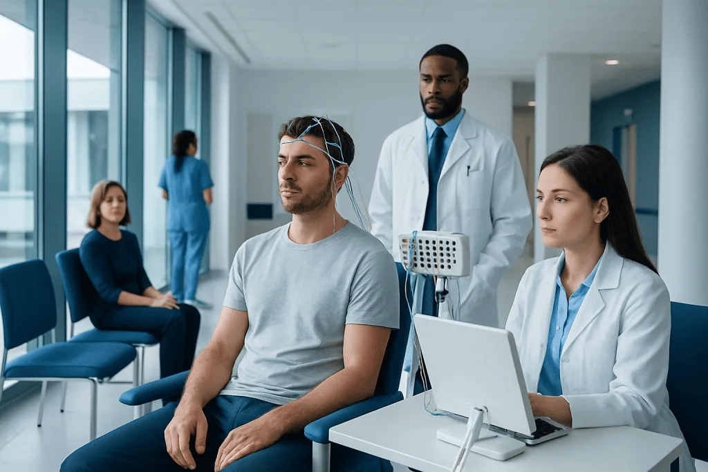
Neurophysiology testing evaluates how the brain, nerves, and muscles are working. EMG and nerve conduction studies are commonly used to investigate numbness, tingling, and weakness.
Key Takeaways
- Neurophysiology testing evaluates how the brain, nerves, and muscles are working.
- EMG and nerve conduction studies are commonly used to investigate numbness, tingling, and weakness.
- EEG is mainly used to assess brain electrical activity, especially when symptoms may relate to seizures or altered brain function.
- These tests do not always give a diagnosis on their own, but they provide important clues when combined with the medical history and examination.
- Most neurophysiology tests are done without sedation and allow patients to return home the same day.
Neurophysiology testing can help doctors understand why a person has numbness, tingling, muscle weakness, or unusual movements. Tests such as EEG, EMG, and nerve conduction studies measure electrical activity in the brain, nerves, and muscles to support an accurate diagnosis and treatment plan.
Overview: What neurophysiology testing is
Neurophysiology testing is a group of diagnostic tests that measure electrical signals in the nervous system. These signals help the brain communicate with the spinal cord, peripheral nerves, and muscles. When a person develops numbness, tingling, weakness, muscle twitching, or episodes of altered awareness, these tests can help show where the problem may be occurring.
Two of the best-known tests are electroencephalography, or EEG, and electromyography, or EMG. EEG records electrical activity in the brain through small sensors placed on the scalp. EMG measures how muscles respond to nerve signals, usually with a very thin needle electrode and often alongside nerve conduction studies, which test how fast and how strongly signals travel through peripheral nerves.
Although people often hear “EEG and EMG” mentioned together, they answer different questions. EEG is generally used when doctors need information about brain activity. EMG and nerve conduction studies are more often used for symptoms such as hand numbness, foot tingling, leg weakness, muscle cramps, or suspected nerve compression.
These tests are tools, not final answers by themselves. Doctors interpret the results together with the patient’s symptoms, medical history, physical examination, and sometimes imaging or blood tests. This combined approach helps guide the next steps in care.
When these tests may be recommended
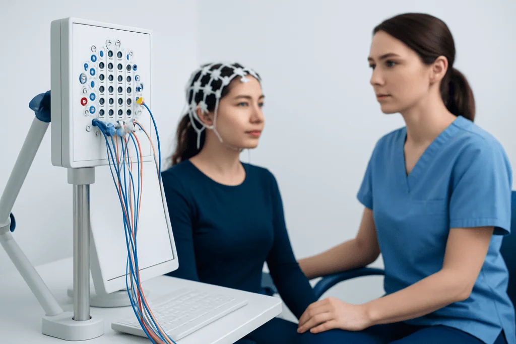
A doctor may recommend neurophysiology testing when symptoms suggest a problem in the brain, spinal cord, peripheral nerves, nerve roots, or muscles. Common reasons include persistent numbness, pins-and-needles sensations, muscle weakness, unexplained muscle wasting, tremor, muscle twitching, balance problems, or episodes that may represent seizures.
EMG and nerve conduction studies are often requested when symptoms point to a peripheral nerve disorder. Examples include suspected carpal tunnel syndrome, nerve injury after trauma, sciatica, or conditions that affect many nerves at once, such as peripheral neuropathy. They may also help evaluate whether weakness comes from a nerve problem, a muscle disorder, or a disorder involving the connection between nerve and muscle.
EEG is used in a different setting. It may be recommended if a person has episodes of loss of awareness, unexplained staring, convulsions, unusual movements, or concern for seizure activity. It can also support the evaluation of some sleep-related events or altered brain function in certain medical situations.
In many cases, the aim is not simply to confirm that symptoms are real, but to localize the source. For example, hand numbness could come from a compressed wrist nerve, a pinched nerve in the neck, or a more widespread nerve condition. Neurophysiology testing can help narrow these possibilities.
What EEG can show

EEG records the brain’s electrical activity using electrodes placed on the scalp. It does not measure thoughts or intelligence. Instead, it shows patterns of brain waves that may appear normal, slowed, or abnormally synchronized. These patterns can provide clues about seizures, epilepsy, or other conditions that affect brain function.
For numbness and weakness, EEG is not usually the first test unless the symptoms may be linked to the brain. For example, brief episodes of one-sided numbness, shaking, or weakness may sometimes be investigated with EEG if there is concern that the events could be seizures rather than a problem in a peripheral nerve or muscle. EEG may also help when symptoms come with blackouts, confusion, or sudden changes in awareness.
During the test, the patient usually sits or lies comfortably while the scalp electrodes record brain activity. The technician may ask the person to open and close the eyes, breathe deeply for a short period, or look at flashing lights. These standard steps can help reveal certain abnormal patterns. The test itself is painless, though the scalp paste or cleaning process may feel slightly inconvenient.
A normal EEG does not completely rule out a seizure disorder, and an abnormal EEG does not always mean a person has epilepsy. The result must be interpreted in context. When needed, doctors may recommend longer recordings, sleep-deprived EEG, or video EEG monitoring for more detailed information.
What EMG and nerve conduction studies can show
EMG and nerve conduction studies are often the most useful neurophysiology tests for evaluating numbness and weakness in the arms or legs. Nerve conduction studies use small electrical pulses on the skin to measure how quickly and effectively nerves carry signals. EMG uses a thin needle electrode inserted into selected muscles to assess their electrical activity at rest and during contraction.
These tests can help identify whether symptoms are caused by damage to a peripheral nerve, irritation of a nerve root leaving the spine, a disorder affecting many nerves, or a primary muscle problem. They may support the diagnosis of conditions such as carpal tunnel syndrome, ulnar nerve compression, radiculopathy, peripheral neuropathy, or some muscle diseases.
The pattern of findings matters. Slowed conduction in a specific nerve can suggest compression or entrapment. Reduced signal strength may point to axonal nerve injury. EMG changes in muscles supplied by one spinal nerve root can suggest a pinched nerve in the neck or lower back. In other cases, the findings may raise concern for disorders of the neuromuscular junction or broader diseases that affect the motor nerves.
These tests can also help doctors understand severity and timing. For instance, they may show whether a nerve injury appears recent or longstanding, mild or more advanced. That information may help decide whether conservative care, follow-up testing, rehabilitation, or procedures such as physical therapy and rehabilitation are likely to be helpful.
How to prepare and what to expect during testing
Preparation is usually simple. Patients are often advised to arrive with clean skin and hair and to avoid lotions, oils, or heavy hair products, because these can interfere with electrode placement. Comfortable clothing is helpful, especially if the arms or legs will be tested. The care team may also ask about pacemakers, implanted devices, blood thinners, bleeding disorders, or skin infections.
During a nerve conduction study, small electrodes are placed on the skin and brief electrical impulses are delivered to the nerve. These feel like quick taps or shocks and can be uncomfortable, but they are generally well tolerated. During EMG, a fine needle is inserted into selected muscles. Some people describe this as similar to an injection or brief muscle ache. The needle is used for recording only and does not give an electric shock.
During EEG, electrodes are attached to the scalp with a special paste or adhesive. The recording itself is painless. The patient is usually asked to stay still and relaxed. Depending on the reason for the test, EEG may take a short routine recording or a longer session. Afterward, the scalp may simply need gentle washing to remove the paste.
Most patients can return to normal daily activities after testing. There may be mild soreness in muscles after EMG for a day or two, but significant side effects are uncommon. If there are concerns about pain, anxiety, or special medical circumstances, it is helpful to discuss them with the doctor in advance.
How doctors use the results
Neurophysiology results help doctors answer practical questions: Is the problem in the brain, nerve root, peripheral nerve, neuromuscular junction, or muscle? Is one area involved or are symptoms more widespread? Are the findings consistent with compression, inflammation, injury, or a chronic degenerative process? These details can shape the next stage of evaluation.
Often, neurophysiology testing is combined with other diagnostic tools. If symptoms suggest a structural issue in the spine or brain, the doctor may also recommend MRI to look at the anatomy in more detail. Blood tests may be used to check for diabetes, vitamin deficiencies, thyroid disease, autoimmune conditions, or infection. In some cases, ultrasound or additional imaging of nerves and muscles may also be useful.
Results can support treatment planning as well. If the pattern suggests a compressed nerve, treatment may focus on splinting, posture changes, medication, injections, or surgery depending on severity. If testing suggests a more generalized nerve disorder such as peripheral neuropathy, management may include identifying the underlying cause, symptom relief, and rehabilitation.
Sometimes the findings are normal or only mildly abnormal even though symptoms are real and bothersome. This can happen if a condition is very early, intermittent, involves small nerve fibers that standard studies do not measure well, or comes from a cause outside the area tested. In that situation, follow-up and further evaluation may still be important.
Treatment and follow-up after diagnosis
Neurophysiology testing does not treat symptoms directly, but it can point toward the right treatment. The best option depends on the cause. Nerve compression may improve with activity changes, ergonomic adjustments, splints, anti-inflammatory strategies, or surgery in selected cases. Muscle disorders, autoimmune nerve conditions, and seizure-related problems need different approaches tailored by a specialist.
Rehabilitation is an important part of care for many patients with weakness or nerve-related symptoms. A structured program may improve strength, balance, coordination, and daily function. Depending on the diagnosis, doctors may suggest neurology rehabilitation or broader physical therapy and rehabilitation to support recovery and reduce the risk of falls or further strain.
Follow-up matters because nerve and muscle conditions can change over time. A doctor may repeat testing if symptoms progress, spread, or fail to improve as expected. Repeat studies can show whether a nerve is healing, whether damage appears stable, or whether the pattern now suggests a different diagnosis.
Near the end of the diagnostic journey, some patients benefit from multidisciplinary care involving neurology, physical medicine and rehabilitation, orthopedics, neurosurgery, or internal medicine. Acibadem International’s multidisciplinary specialists and JCI-accredited hospitals diagnose and treat neurological and neuromuscular conditions for international patients when further evaluation is needed.
When to seek medical attention
Numbness and weakness are not always emergencies, but they should not be ignored if they are persistent, worsening, or unexplained. A medical review is especially important when symptoms affect walking, hand function, bladder or bowel control, or daily activities. Progressive weakness, visible muscle wasting, or repeated falls also deserve prompt assessment.
Urgent care is needed if weakness comes on suddenly, affects one side of the body, or occurs with facial drooping, trouble speaking, severe headache, chest pain, or new confusion. These symptoms may signal a stroke or another emergency. Immediate medical help is also important after major trauma or if symptoms are accompanied by loss of consciousness.
People with diabetes, autoimmune disease, recent infection, cancer history, or exposure to medications known to affect nerves should mention this to their doctor, as it can help guide testing. Even when symptoms seem mild, early evaluation may make diagnosis clearer and treatment more effective.
If a doctor recommends neurophysiology testing, patients can usually feel reassured that the goal is to gain more precise information. Understanding whether symptoms arise from the brain, nerve, or muscle is often the key step toward a safe and effective treatment plan.
Frequently asked questions
What is the difference between EEG and EMG?
EEG measures electrical activity in the brain using electrodes placed on the scalp. EMG evaluates muscle activity and is often performed together with nerve conduction studies to assess how well nerves and muscles are working. They are used for different symptoms and answer different clinical questions.
Can EMG diagnose the cause of numbness and weakness?
EMG can provide important clues, especially when symptoms are caused by nerve injury, nerve compression, radiculopathy, or muscle disease. However, it is usually interpreted together with the physical examination, medical history, and sometimes imaging or blood tests. It helps narrow the diagnosis rather than replacing the full clinical evaluation.
Is neurophysiology testing painful?
EEG is painless, though the scalp electrodes and paste can feel slightly inconvenient. Nerve conduction studies cause brief electrical pulses that may be uncomfortable, and EMG needle testing can cause short-lasting soreness or a cramp-like feeling. Most people tolerate the tests well and can go home the same day.
How long do EEG and EMG tests take?
The exact timing depends on the reason for the test and how many areas need to be assessed. Routine EEG often takes less than an hour, while EMG and nerve conduction studies may take around 30 to 90 minutes. Some EEG studies are longer if extended monitoring is needed.
Can test results be normal even if symptoms are real?
Yes. Some nerve and muscle conditions are intermittent, very early, or affect structures that standard studies do not detect well. A normal result does not mean symptoms should be dismissed, and the doctor may recommend follow-up, additional testing, or monitoring over time.
Do I need to prepare for an EMG or EEG?
Preparation is usually simple. Patients are often asked to avoid lotions, oils, or heavy hair products and to wear comfortable clothing. It is also important to tell the doctor about blood thinners, implanted devices, skin problems, or any medical concerns before the test.
References
- National Institute of Neurological Disorders and Stroke
- American Association of Neuromuscular & Electrodiagnostic Medicine
- Epilepsy Foundation
- National Institute for Health and Care Excellence
- Mayo Clinic
This article is for general information only and is not a substitute for professional medical advice. Please consult a qualified doctor about your individual situation.
Neurophysiology in Turkey — costs, top hospitals & a free quote
JCI-accredited · board-certified surgeons · reply within 24h
Add us as a Preferred Source to see more of our trusted health content across Google Search, AI Overviews and Discover.