Neuroradiology: When MRI, CT, and Advanced Brain Imaging Are Used
Neuroradiology focuses on imaging of the brain, spine, head, neck, and nerves. MRI and CT are the most commonly used neuroradiology tests, but advanced techniques may also be needed.
Key Takeaways
- Neuroradiology focuses on imaging of the brain, spine, head, neck, and nerves.
- MRI and CT are the most commonly used neuroradiology tests, but advanced techniques may also be needed.
- The best imaging test depends on the symptoms, the urgency, and what condition is suspected.
- Some scans are used for diagnosis, while others help plan treatment or monitor recovery over time.
- Most neuroradiology tests are noninvasive, though contrast dye may sometimes be recommended.
Neuroradiology is the medical specialty that uses imaging tests to examine the brain, spine, head, neck, and nervous system. MRI, CT, and other advanced scans help doctors diagnose symptoms, guide treatment decisions, and monitor many neurological conditions.
Overview: What neuroradiology is
Neuroradiology is a branch of diagnostic imaging focused on the brain, spinal cord, head, neck, and nervous system. It helps doctors look inside the body without surgery and can reveal changes in anatomy, blood flow, inflammation, bleeding, tumors, infection, or injury. These images are interpreted by radiologists with advanced training in neurological conditions.
Common neuroradiology tests include magnetic resonance imaging (MRI), computed tomography (CT), and blood vessel imaging such as CT angiography or MR angiography. In some situations, specialized studies such as functional MRI, perfusion imaging, or diffusion imaging are also used. Each test gives different information, so the choice depends on the person’s symptoms and medical history.
Neuroradiology plays an important role in both urgent and planned care. In emergencies such as stroke or head trauma, imaging may help doctors act quickly. In non-emergency settings, it can help evaluate headaches, seizures, dizziness, memory changes, back pain, weakness, numbness, and many other neurological symptoms.
When MRI, CT, and advanced brain imaging are used
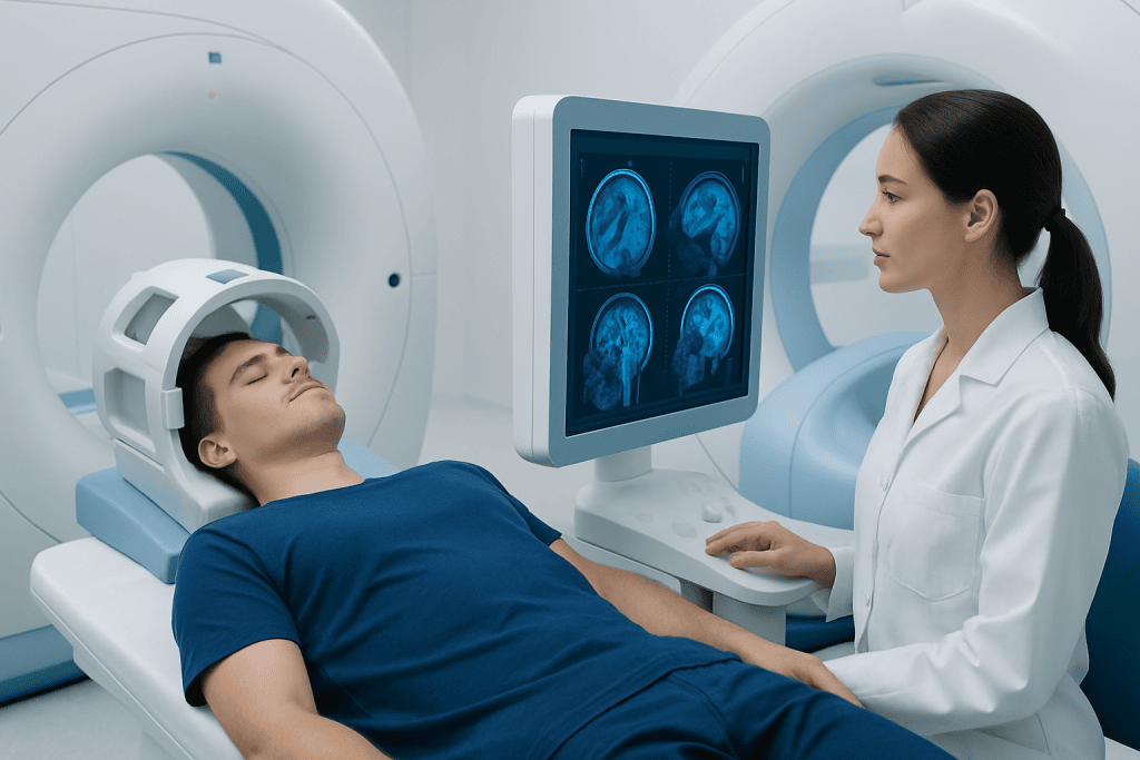
Doctors request neuroradiology tests when they need a clearer view of possible problems affecting the nervous system. Imaging may be used to investigate new symptoms, confirm a suspected diagnosis, check whether a condition is stable or progressing, or plan surgery or other treatment. It can also be used after treatment to assess recovery or look for recurrence.
MRI is often chosen when detailed images of soft tissues are needed. It is especially helpful for examining the brain, spinal cord, discs, nerves, and subtle tissue changes. CT is often preferred when speed matters, such as after trauma or when bleeding is suspected. CT also shows bone very well and can be useful for skull, facial, and spine injuries.
Advanced brain imaging may be used when standard MRI or CT does not answer the full clinical question. For example, perfusion imaging can assess blood flow, diffusion imaging can detect early stroke-related changes, and angiographic techniques can show narrowing, blockage, or abnormal blood vessels. Functional MRI may help map language or movement areas before brain surgery.
- Sudden severe headache or suspected stroke
- Head injury or loss of consciousness
- Seizures or unexplained fainting
- Persistent dizziness, balance problems, or hearing-related neurological symptoms
- Weakness, numbness, facial droop, or vision changes
- Memory problems, confusion, or behavior changes
- Neck pain, back pain, or suspected nerve compression
Understanding the main tests
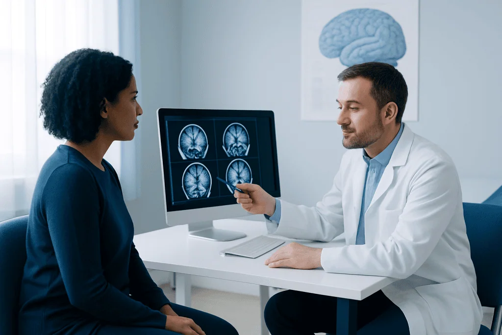
MRI uses a magnetic field and radio waves to create detailed images. It does not use ionizing radiation. MRI is often the preferred test for many brain and spine conditions because it can show soft tissues with great clarity. It may be done with or without contrast dye, depending on whether doctors need more information about inflammation, blood vessels, or possible tumors.
CT uses X-rays to produce cross-sectional images and is widely available and fast. This makes it especially useful in emergency care. A head CT may quickly identify bleeding, skull fractures, swelling, or large structural changes. CT can also be combined with contrast to evaluate blood vessels and blood flow.
Other neuroradiology studies include MR angiography, CT angiography, venography, perfusion imaging, and sometimes PET-CT in selected cases. These tests can provide more detail about circulation, tissue activity, or treatment response. For spinal symptoms, imaging may focus on discs, nerves, vertebrae, or the spinal cord itself using MRI scanning or CT scanning when appropriate.
Doctors choose among these tests based on the question they need answered. A person with sudden neurological symptoms may need rapid CT-based imaging first, while someone with long-standing nerve-related symptoms may benefit more from MRI. In many cases, the tests complement each other rather than compete.
Conditions neuroradiology can help diagnose
Neuroradiology can support the diagnosis of a wide range of conditions affecting the central and peripheral nervous systems. In the brain, imaging may help identify stroke, bleeding, aneurysms, tumors, infection, inflammation, hydrocephalus, and changes linked to degenerative disorders. In the spine, it can reveal disc herniation, spinal stenosis, fractures, cord compression, infection, or tumors.
It is also useful when symptoms are non-specific. For example, persistent headaches, unexplained weakness, numbness, or changes in speech may prompt imaging to look for structural causes. In people with seizures, scans can help search for lesions, scarring, vascular abnormalities, or other changes that may be contributing to episodes.
Advanced imaging is often valuable for cerebrovascular disease. It may help detect problems such as brain aneurysm, narrowing of blood vessels, or patterns of injury related to reduced blood flow. In other cases, MRI may support the evaluation of suspected multiple sclerosis by showing characteristic changes in the brain or spinal cord.
Imaging is only one part of diagnosis. Doctors usually combine scan results with a medical history, physical examination, neurological assessment, and sometimes blood tests or other investigations. A normal scan can also be important, because it helps rule out serious structural causes and guide the next step in care.
What to expect before, during, and after a scan
Preparation depends on the type of test. Many CT and MRI scans require little preparation, but a person may be asked not to eat for a few hours if contrast dye is planned. It is important to tell the medical team about kidney problems, allergies, pregnancy, implanted devices, pacemakers, metal fragments, or claustrophobia. These details help determine the safest and most suitable scan.
During an MRI, the person lies on a table that moves into the scanner. The machine makes loud tapping sounds, so ear protection is usually provided. The test is painless, but keeping still is important for clear images. CT scans are usually quicker and quieter. If contrast is used for either test, a temporary warm sensation or metallic taste may occur.
After the scan, most people can return to normal activities straight away. If sedative medication was used, rest and transportation assistance may be needed. A radiologist studies the images and sends a report to the referring doctor, who then explains what the findings mean in the context of symptoms and next steps.
When imaging shows a condition that may need further care, doctors may recommend follow-up tests, medication, rehabilitation, surgery, or minimally invasive procedures. For example, vascular findings may lead to assessment by a specialist in interventional neuroradiology if treatment through the blood vessels is being considered.
Benefits, limits, and safety considerations
The main benefit of neuroradiology is that it helps doctors make more accurate decisions without invasive procedures. It can detect problems early, identify emergencies, guide biopsies or surgery, and monitor whether treatment is working. In many cases, it helps avoid unnecessary interventions by showing when symptoms are not caused by a structural neurological problem.
At the same time, every imaging method has limits. CT is excellent for speed and bone detail but uses ionizing radiation. MRI offers high soft-tissue detail without radiation, but it takes longer and may not be suitable for everyone with certain implanted devices or severe claustrophobia. Some abnormalities are subtle, and not every symptom can be explained by imaging alone.
Contrast dyes can be very helpful, but they are not always needed. Doctors weigh the benefits against possible risks, especially in people with kidney disease or a history of allergic reactions. Safety checks before the scan are an important part of care and are designed to reduce risk as much as possible.
Results must always be interpreted in context. A scan may show an incidental finding that is unrelated to symptoms, or it may miss very early disease that becomes clearer later. For that reason, doctors sometimes repeat imaging or use a different technique when symptoms continue or change.
How neuroradiology guides treatment and follow-up
Neuroradiology does more than identify disease. It also helps doctors decide which treatment is most appropriate and when it should begin. In stroke care, imaging can help determine whether there is a blocked artery or bleeding and whether urgent treatment may be suitable. In spine problems, it can show whether pain is more likely coming from a disc, the joints, the bones, or nerve compression.
For brain and spine tumors, imaging helps define size, location, and relation to nearby structures. This information is important for planning surgery, radiotherapy, or medical treatment. MRI can also be used over time to monitor whether a tumor is stable, shrinking, or returning after therapy. Similar follow-up imaging may be used after infection, injury, or inflammatory disease.
In some cases, advanced imaging supports treatment planning in highly targeted ways. Functional MRI may help surgeons avoid critical areas controlling speech or movement. Perfusion or angiographic studies may guide decisions in vascular conditions. When clinicians suspect acute stroke, rapid multimodal imaging can be central to time-sensitive care.
Near the end of the care pathway, some patients need coordinated expertise from neurology, neurosurgery, oncology, rehabilitation, and imaging specialists. Acibadem International’s multidisciplinary teams and JCI-accredited hospitals diagnose and treat neurological conditions for international patients, using advanced imaging as part of individualized care.
When to seek medical attention
Many neurological symptoms are not emergencies, but some require urgent evaluation. Immediate medical attention is important for sudden weakness, numbness on one side, difficulty speaking, new confusion, sudden vision loss, severe unexplained headache, seizures, or major head injury. These symptoms can be caused by serious problems that benefit from prompt imaging and treatment.
A non-urgent medical review is still important for symptoms that persist or gradually worsen. These may include ongoing headaches, dizziness, balance problems, memory changes, hearing-related neurological symptoms, unexplained tingling, back pain with nerve symptoms, or weakness that affects daily life. A doctor can decide whether neuroradiology is appropriate and which test is most useful.
People should not request imaging on their own without guidance if symptoms are complex or unclear. The most helpful scan depends on the medical question, and unnecessary imaging can lead to confusion or incidental findings. A qualified clinician can match the right test to the right situation and explain the results clearly.
Frequently asked questions
What is the difference between neuroradiology and radiology?
Radiology is the broader medical field that uses imaging to diagnose and sometimes treat disease throughout the body. Neuroradiology is a subspecialty focused specifically on the brain, spine, head, neck, and nervous system.
Is MRI better than CT for brain imaging?
Neither test is always better in every situation. MRI usually gives more detail about soft tissues, while CT is faster and often preferred in emergencies such as trauma or suspected bleeding. The best choice depends on the symptoms and the clinical question.
Why might a doctor order contrast dye for a brain or spine scan?
Contrast dye can make certain tissues, blood vessels, inflammation, or abnormal growths easier to see. It is not needed for every scan, and doctors only recommend it when the added information is likely to be useful.
Are neuroradiology scans safe?
Most neuroradiology scans are safe and routine. MRI does not use radiation, while CT does use X-rays, so doctors consider the expected benefit before ordering it. Safety screening is especially important if contrast dye or implanted medical devices are involved.
Can neuroradiology detect stroke?
Yes, neuroradiology is central to stroke diagnosis. CT can quickly identify bleeding, while MRI and vessel imaging can show areas of reduced blood flow or blocked arteries. Fast imaging helps doctors decide on the most appropriate treatment.
Do all headaches need brain imaging?
No, many headaches do not require imaging. Doctors usually recommend scans when headaches are new, severe, unusual, associated with neurological symptoms, or accompanied by warning signs such as weakness, seizures, or confusion.
This article is for general information only and is not a substitute for professional medical advice. Please consult a qualified doctor about your individual situation.
Neuroradiology in Turkey — costs, top hospitals & a free quote
JCI-accredited · board-certified surgeons · reply within 24h
Add us as a Preferred Source to see more of our trusted health content across Google Search, AI Overviews and Discover.

More from the Health Library
What Is a CT Scan and When Is It Needed?
Echocardiogram: Heart Ultrasound for Valves, Pump Function, and Symptoms

Diagnostic Testing Abroad: What International Patients Should Ask Before Traveling
Quest Diagnostics Appointment: How to Book, Prepare, and Check In
Diagnostic Mammogram: Who Needs It and What Results Mean