Nuclear Medicine Scan: How It Works, Safety, and What Results Can Show
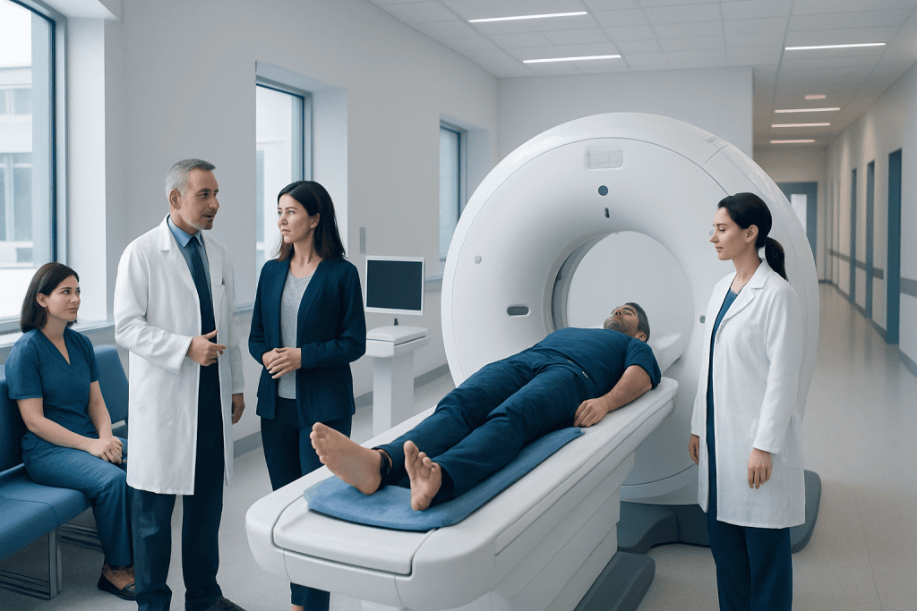
A nuclear medicine scan shows organ function as well as structure. It uses a small amount of radioactive tracer that is usually injected, swallowed, or inhaled.
Key Takeaways
- A nuclear medicine scan shows organ function as well as structure.
- It uses a small amount of radioactive tracer that is usually injected, swallowed, or inhaled.
- Common types include PET and SPECT scans, often combined with CT for more detail.
- These scans can help diagnose cancer, heart disease, bone disorders, thyroid problems, and more.
- The amount of radiation is carefully controlled, and the test is generally well tolerated.
- Preparation depends on the scan type and may include fasting or temporarily avoiding certain medicines.
A nuclear medicine scan is an imaging test that uses a small amount of radioactive material to show how organs and tissues function. It can help doctors detect disease, assess treatment response, and guide care in a safe, targeted way.
Overview: What Is a Nuclear Medicine Scan?
A nuclear medicine scan is a medical imaging test that helps doctors see how the body is working from the inside. Unlike standard X-rays, ultrasound, or some CT scans that mainly show anatomy, nuclear imaging focuses on function. This means it can reveal how blood flows, how active a tissue is, or how well an organ is performing.
To do this, a very small amount of radioactive material, called a tracer or radiotracer, is introduced into the body. The tracer collects in certain organs, bones, or tissues, depending on the type of scan. A special camera then detects the radiation coming from the tracer and creates images that doctors can interpret.
Nuclear medicine scans are used in many areas of healthcare. They can help evaluate the heart, thyroid, bones, kidneys, lungs, brain, and digestive system. They are also widely used in cancer care, including cancer diagnosis, staging, and monitoring, and may be part of a wider imaging plan that includes PET-CT imaging.
How Nuclear Medicine Imaging Works
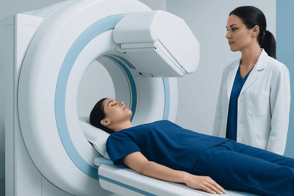
The key principle behind nuclear medicine is that different tissues take up tracers in different ways. For example, a tracer designed for bone imaging collects in areas of active bone turnover, while another tracer may highlight heart muscle blood flow or areas of high metabolic activity. This gives doctors information that cannot always be seen on routine structural imaging alone.
After the tracer is given, there is often a waiting period so it can travel through the body and reach the target area. Imaging is then performed using a gamma camera, SPECT scanner, or PET scanner. Some examinations are combined with CT to match functional findings with detailed anatomy, such as SPECT-CT or PET-CT.
There are several common forms of nuclear imaging:
- Bone scan: looks for changes in bone activity.
- Thyroid scan: assesses thyroid structure and function.
- Myocardial perfusion scan: evaluates blood flow to the heart.
- Renal scan: checks kidney function and drainage.
- PET scan: often used in oncology, neurology, and cardiology.
- SPECT scan: provides 3D functional imaging for selected organs.
Because these scans show function, they may detect abnormalities before structural changes become obvious. This can support earlier diagnosis or more precise follow-up when symptoms, lab tests, or other scans do not give a complete picture.
What Conditions and Results a Scan Can Show

A nuclear medicine scan can help identify whether an organ is working normally, too slowly, too actively, or unevenly. Depending on the tracer used, the images may show inflammation, infection, areas of reduced blood supply, abnormal cell activity, or changes in bone metabolism. The exact meaning of results always depends on the clinical question and the type of scan performed.
In cancer care, nuclear imaging can help detect suspicious areas, determine whether disease has spread, and assess how treatment is working. PET scans are especially useful in many cancers because they can highlight tissues with increased metabolic activity. In some situations, doctors may use these scans alongside biopsy or other tests to confirm a diagnosis.
In cardiology, nuclear scans can show whether the heart muscle is receiving enough blood during rest or exercise. In bone health, they may help find fractures, infection, arthritis-related changes, or areas affected by tumors. In endocrinology, a thyroid scan may help explain overactivity, nodules, or unusual uptake patterns related to thyroid cancer.
Results are usually described as normal, reduced uptake, increased uptake, or uneven distribution of tracer. A “hot spot” may indicate increased activity in a tissue, while a “cold spot” may suggest reduced function or blood flow. These terms are descriptive rather than diagnostic on their own, so the radiologist or nuclear medicine physician interprets them together with symptoms, examination findings, and other test results.
What to Expect Before, During, and After the Test
Preparation varies by scan type. Some tests require fasting for several hours, while others may ask the patient to drink extra water, avoid caffeine, or temporarily stop certain medications if a doctor advises it. It is important to tell the care team about all medicines, allergies, recent imaging tests, and whether there is any possibility of pregnancy or if the patient is breastfeeding.
The tracer may be injected into a vein, swallowed, or inhaled. Most people feel little more than a brief needle prick if an injection is used. There may then be a short or longer waiting period before imaging begins, depending on how quickly the tracer needs to circulate and collect in the target tissue.
During imaging, the patient usually lies still on a table while the camera moves around or over the body. The scan itself is painless, though staying still can be uncomfortable for some people. The test may take anywhere from less than an hour to several hours in total, especially if there are waiting periods between tracer administration and image acquisition.
After the scan, most people can return to usual daily activities unless told otherwise. Drinking fluids may help the body clear the tracer more quickly. If special precautions are needed, such as for close contact with infants or during breastfeeding, the healthcare team will explain them clearly in advance.
Safety, Radiation Exposure, and Possible Risks
Nuclear medicine scans are generally considered safe when used appropriately. The amount of radioactive tracer is small and carefully selected for the specific test. In most cases, the radiation exposure is within accepted medical limits and is balanced against the benefit of obtaining important diagnostic information.
Allergic reactions to radiotracers are uncommon, and serious reactions are rare. Some people may have mild discomfort at the injection site. The tracer naturally loses radioactivity over time and is usually cleared from the body through urine, stool, or, less commonly, perspiration.
Radiation exposure is an understandable concern for many patients. The healthcare team chooses the lowest amount needed to produce useful images. Doctors consider age, medical history, and the reason for testing before recommending a scan, especially for children and for people who are pregnant or may be pregnant.
Breastfeeding may need to be paused for a short time after certain tracers, depending on the test. Patients should always ask for individualized guidance. In experienced centers, nuclear medicine professionals follow strict safety procedures for preparation, handling, and imaging to protect both patients and staff.
How Doctors Interpret Nuclear Medicine Results
The images from a nuclear medicine scan are reviewed by a radiologist or nuclear medicine physician with specialized training. They examine where the tracer has collected, how much uptake there is, and whether the pattern is symmetrical or abnormal. This is then matched with the person’s symptoms, blood tests, physical examination, and other imaging findings.
Not every abnormal result means a serious disease. Increased uptake may reflect inflammation, healing, infection, or a benign growth depending on the organ involved. Reduced uptake may point to poor blood supply, scarring, or low activity in a tissue. Because there can be overlap between conditions, results often guide the next step rather than provide the whole answer alone.
Sometimes a nuclear scan is used to monitor known disease over time. For example, it may help show whether a cancer has responded to treatment, whether heart blood flow improves after therapy, or whether bone changes are progressing. If needed, doctors may recommend additional imaging such as MRI or a follow-up scan to clarify uncertain findings.
Patients often receive a report after the images are analyzed, but the most useful discussion happens with the referring doctor. That conversation places the findings in context and explains what, if anything, should happen next.
When to See a Doctor and Questions to Ask
A doctor may recommend a nuclear medicine scan when symptoms, blood tests, or other imaging suggest that more information about organ function is needed. This can include unexplained bone pain, chest pain, thyroid concerns, possible infection, neurological symptoms, or follow-up for a known diagnosis. It may also be ordered to check whether a treatment is working as expected.
Before the appointment, patients may find it helpful to ask what the scan is looking for, how they should prepare, how long it will take, and whether they need someone to accompany them. They can also ask about medications, eating and drinking instructions, and any special advice related to pregnancy or breastfeeding.
After the scan, patients should contact their doctor if they do not understand the results or if symptoms persist despite a normal report. Prompt follow-up is also sensible if there is fever, worsening pain, shortness of breath, or another new concerning symptom, because imaging is only one part of a full medical evaluation.
For people seeking care across borders, Acibadem International’s multidisciplinary specialists and JCI-accredited hospitals diagnose and treat conditions evaluated with nuclear medicine imaging for international patients. The best approach is always individualized and based on the patient’s history, examination, and test findings.
Frequently asked questions
Is a nuclear medicine scan the same as a CT or MRI scan?
No. A nuclear medicine scan mainly shows how organs and tissues function, while CT and MRI are primarily used to show body structure in detail. In some cases, nuclear imaging is combined with CT to provide both types of information together.
Are nuclear medicine scans safe?
Yes, they are generally safe when performed for an appropriate medical reason. They use a small, carefully measured amount of radioactive tracer, and the benefits of accurate diagnosis usually outweigh the risks. The care team will review special considerations such as pregnancy or breastfeeding before the test.
How long does a nuclear medicine scan take?
The timing depends on the type of scan. Some tests are completed within an hour, while others involve a waiting period after the tracer is given and may take several hours in total. The imaging team usually explains the schedule beforehand.
Will the scan hurt?
The scan itself is usually painless. If the tracer is injected, there may be a brief needle prick, and some people feel mild discomfort from lying still. Most patients tolerate the procedure well.
Do patients need to prepare for a nuclear medicine scan?
Often, yes, but the instructions vary by scan type. Preparation may include fasting, avoiding caffeine, drinking more water, or temporarily changing certain medicines if a doctor advises it. Following the provided instructions helps improve image quality and accuracy.
What can a nuclear medicine scan detect?
It can help detect changes in organ function, blood flow, bone activity, thyroid activity, infection, inflammation, and some cancers. It is also useful for assessing treatment response in certain conditions. The exact information depends on the tracer used and the body area being examined.
References
- International Atomic Energy Agency
- Radiological Society of North America
- Society of Nuclear Medicine and Molecular Imaging
- American College of Radiology
- National Cancer Institute
This article is for general information only and is not a substitute for professional medical advice. Please consult a qualified doctor about your individual situation.
Explore treatments in Turkey — costs, top hospitals & a free quote
JCI-accredited · board-certified surgeons · reply within 24h
Add us as a Preferred Source to see more of our trusted health content across Google Search, AI Overviews and Discover.

More from the Health Library



Related Specialists

Dr. Veli Mehmet Kindan
Emergency Service
Dr. Serap Gülşeni
Check Up & Wellness Center
Prof. Dr. Gül Esen İçten
Breast Clinic Clinical Service
Prof. Dr. Bora Peynircioğlu
Interventional Radiology