Obturator Trach: A Complete Medical Overview

An obturator trach is not a long-term part of the tracheostomy tube; it is used only during insertion. The obturator creates a smooth tip that helps guide the trach tube through the stoma.
Key Takeaways
- An obturator trach is not a long-term part of the tracheostomy tube; it is used only during insertion.
- The obturator creates a smooth tip that helps guide the trach tube through the stoma.
- After placement, the obturator must be removed so air can pass through the tracheostomy tube.
- Safe tracheostomy care includes cleaning, humidification, suction when needed, and monitoring for tube blockage or infection.
- Urgent medical attention is needed for breathing difficulty, heavy bleeding, tube displacement, or signs of severe infection.
Obturator trach usually refers to the obturator used during tracheostomy tube placement. It is a smooth, rounded guide that helps the trach tube enter the airway more safely and comfortably, and it is removed immediately after the tube is in place.
Overview: What an Obturator Trach Means
The term obturator trach most often refers to the obturator used with a tracheostomy tube. A tracheostomy is an opening created in the front of the neck into the windpipe, and a tracheostomy tube is placed into that opening to help maintain breathing. The obturator is a firm inner guide with a rounded tip that fits inside the tube during insertion.
Its main purpose is to make the leading edge of the trach tube smoother. This helps reduce friction as the tube is guided through the tracheostomy opening, also called the stoma. Once the tube is correctly positioned, the obturator is removed right away, because air cannot move normally through the tube while the obturator is still in place.
This distinction is important because patients and caregivers may hear several tracheostomy terms used together. The trach tube remains in the airway, while the obturator is a temporary placement tool. Understanding that difference can make conversations with clinicians clearer and can support safer daily tracheostomy care at home or in the hospital.
How the Obturator Is Used During Tracheostomy Tube Placement
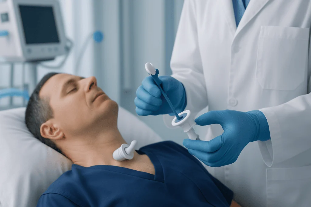
Before a tracheostomy tube is inserted, the obturator is placed inside the outer cannula of the tube. Its rounded tip extends just beyond the edge of the tube, creating a smoother surface for insertion. This can help the clinician guide the tube into the stoma with less resistance than the tube alone.
During the procedure, the healthcare professional advances the trach tube into the airway while keeping the obturator in place. As soon as the tube is correctly seated, the obturator is removed immediately. The inner opening of the trach tube must be clear so the person can breathe through the tube and so attachments such as oxygen or ventilation support can be used if needed.
Because the obturator blocks airflow, it should never be left inside the tracheostomy tube after placement. For this reason, trained staff check tube position and remove the obturator without delay. Families and caregivers are often shown what the obturator looks like, where it is stored, and why it may be needed again if a tube has to be changed in a clinical or home-care setting.
People who are learning about tracheostomy may also benefit from understanding the broader procedure of tracheostomy care and tube placement, especially if they or a loved one will need ongoing airway support.
Why a Tracheostomy May Be Needed

A tracheostomy may be recommended when a person needs a more stable airway for breathing over a longer period or when the upper airway is blocked. Common reasons include prolonged need for mechanical ventilation, severe swelling in the throat, injury to the face or neck, certain neurologic conditions, or tumors affecting the airway.
Some people need a tracheostomy after major surgery or critical illness. Others may need it because of chronic conditions that weaken the muscles used for breathing or swallowing. In children and adults, the decision is individualized and based on how well the person can protect the airway and breathe effectively.
Airway narrowing from conditions such as laryngeal cancer or advanced obstructive processes may make a tracheostomy necessary to bypass the blockage. In other cases, tracheostomy is part of supportive care while another problem is treated, such as serious lung disease, trauma, or neurological illness.
The need for a tracheostomy does not always mean it will be permanent. Some tubes are temporary and removed once breathing improves, while others are needed for ongoing airway protection or long-term ventilation support.
Symptoms, Problems, and Possible Complications to Watch For
The obturator itself is not usually associated with symptoms because it is only used briefly during tube insertion or tube changes. Most day-to-day concerns relate instead to the tracheostomy tube and the stoma. Patients and caregivers are typically advised to watch for changes in breathing, unusual secretions, skin irritation, and signs that the tube may be obstructed or displaced.
Possible warning signs include noisy breathing, difficulty passing a suction catheter, thick mucus, increasing cough, redness around the stoma, foul-smelling drainage, fever, or pain that is getting worse. Small amounts of mucus are common, but heavy bleeding, sudden shortness of breath, or a tube that appears to have moved out of place needs prompt evaluation.
Complications can include blockage from dried secretions, local infection, skin breakdown, accidental decannulation, and, less commonly, airway injury. Some people may also experience granulation tissue, which is extra healing tissue that forms around the stoma or tube and may lead to irritation or bleeding.
- Breathing difficulty or bluish lips requires emergency care.
- Persistent bleeding should not be treated as routine trach irritation.
- A clogged tube can become serious quickly, especially in children or people dependent on ventilation.
- Regular follow-up can help identify preventable problems early.
Diagnosis, Tube Checks, and Follow-Up Care
When clinicians assess a person with a tracheostomy, they look at both the airway and the device. They check whether the tube is the right size, whether it is positioned correctly, and whether the stoma appears healthy. If there is concern about blockage, infection, or structural problems, additional testing may be needed.
Evaluation may include physical examination, pulse oximetry to measure oxygen levels, chest imaging, and direct airway examination. In some cases, specialists use a small camera to inspect the trachea and surrounding structures. This type of assessment can help identify narrowing, injury, mucus plugs, granulation tissue, or tube-related pressure effects.
Patients who have complex airway conditions may be reviewed by ear, nose, and throat specialists, pulmonologists, intensivists, speech and swallowing specialists, or rehabilitation teams. When the airway needs closer visual assessment, procedures related to bronchoscopy may be used to examine the breathing passages and guide treatment decisions.
Regular follow-up is especially important after a new tracheostomy, after a tube change, or if the person has repeated infections or secretion problems. A structured plan for cleaning, humidification, suction, and emergency backup equipment can make home care safer and more manageable.
Treatment Options and Daily Tracheostomy Care
Treatment depends on the reason for the tracheostomy and any complications that develop. Routine care usually includes humidification, cleaning around the stoma, changing dressings as instructed, suctioning secretions when necessary, and monitoring tube ties or securement devices. Good hand hygiene and correct technique are central to reducing infection risk.
If the tube becomes blocked, the care team may suction the airway, replace the inner cannula if the tube has one, or perform a tube change. If there is infection, treatment may involve local wound care and sometimes medication based on the suspected cause. Skin breakdown may improve with better moisture control, dressing changes, and adjustment of tube positioning or straps.
Some people also need evaluation of swallowing and voice. A tracheostomy can affect airflow through the upper airway, speech, and secretion management. Depending on the person’s condition, speech-language therapy, cuff adjustments, or specialized speaking valves may be considered under professional guidance.
If the tracheostomy is no longer needed, decannulation may be possible after careful assessment. In more complex airway disorders, treatment may also involve support from respiratory specialists or surgeons. For international patients, Acibadem International’s multidisciplinary specialists in JCI-accredited hospitals diagnose and treat tracheostomy-related conditions as part of coordinated airway care.
Prevention, Self-Care, and Safe Home Management
Not every tracheostomy problem can be prevented, but careful daily care lowers the risk of complications. Secretions tend to become thicker when air is dry, so adequate humidification and hydration can help. Following the care plan exactly as taught by the healthcare team is especially important in the first weeks after surgery and after any tube change.
Caregivers are often advised to keep essential supplies nearby, such as suction equipment, spare trach tubes in the recommended sizes, and the correct obturator for emergency tube replacement. Storage should be clean, organized, and easy to access. Families should also know who to call and where to go if the tube becomes blocked or comes out unexpectedly.
General self-care measures may include avoiding smoke exposure, washing hands before trach care, and checking the skin around the stoma every day. If another airway or lung condition is present, managing that condition can reduce stress on breathing. Some patients may also have related respiratory concerns such as sleep apnea or chronic airway disease that need separate follow-up.
Education is a key part of prevention. People usually do best when they receive repeated demonstrations, written instructions, and supervised practice. Asking questions about what is normal and what is not can improve confidence and safety at home.
When to Seek Medical Care
Medical advice should be sought promptly if there is increasing redness, swelling, discharge, fever, worsening pain, bad odor from the stoma, or difficulty clearing secretions. These may suggest infection, irritation, or an early tube problem that should be checked before it becomes more serious.
Urgent or emergency care is needed for severe shortness of breath, inability to pass air through the trach tube, sudden heavy bleeding, accidental tube removal with breathing trouble, or a marked drop in oxygen levels if home monitoring is being used. Anyone who depends on a tracheostomy for breathing should have an emergency plan made with their healthcare team.
If there are concerns about recurrent chest infections, aspiration, or underlying lung disease, clinicians may also recommend assessment by respiratory specialists and treatments related to chronic respiratory care when appropriate. Seeking care early is usually the safest approach when a change in breathing or stoma appearance is noticed.
Frequently asked questions
What is an obturator trach?
An obturator trach usually means the obturator used with a tracheostomy tube during insertion. It is a smooth, rounded guide placed inside the tube to help it enter the stoma more easily. It is removed immediately after the tube is in place.
Does the obturator stay inside the tracheostomy tube?
No. The obturator is only used briefly during tube placement or tube changes. It must be removed right away because it blocks the inner opening of the tracheostomy tube and prevents normal airflow.
Why is an obturator important during a trach tube change?
The obturator helps create a smooth leading edge, which can make reinsertion of the trach tube safer and easier. This is especially helpful when the stoma is still relatively new or when tube changes are technically difficult. Tube changes should be performed only by trained professionals or caregivers who have been properly instructed.
What problems can happen with a tracheostomy tube?
Possible problems include mucus blockage, infection around the stoma, bleeding, skin irritation, accidental tube displacement, and breathing difficulty. Many issues can be reduced with proper cleaning, humidification, suctioning when needed, and regular follow-up. Any sudden change in breathing should be treated urgently.
How should an obturator be stored at home?
It should be kept clean, dry, and easy to find with the person's emergency tracheostomy supplies. The exact storage method depends on the type of trach tube and the care team's instructions. Caregivers should always know which obturator matches the current spare tube.
When should someone with a tracheostomy go to the emergency room?
Emergency care is needed for severe breathing trouble, a blocked tube, heavy bleeding, bluish lips, or accidental tube removal with distress. It is also important to seek urgent help if secretions suddenly become difficult to clear or if oxygen levels drop. When in doubt, contacting emergency services or the treating team is the safest choice.
References
- American Thoracic Society
- American Academy of Otolaryngology–Head and Neck Surgery
- National Health Service
- MedlinePlus
- National Institute for Health and Care Excellence
This article is for general information only and is not a substitute for professional medical advice. Please consult a qualified doctor about your individual situation.
Explore treatments in Turkey — costs, top hospitals & a free quote
JCI-accredited · board-certified surgeons · reply within 24h
Add us as a Preferred Source to see more of our trusted health content across Google Search, AI Overviews and Discover.