Optic Neuritis and Multiple Sclerosis: When Vision Changes Need Urgent Care

Optic neuritis often causes pain with eye movement, blurred vision, and reduced color vision. It can be associated with multiple sclerosis, but not everyone with optic neuritis develops MS.
Key Takeaways
- Optic neuritis often causes pain with eye movement, blurred vision, and reduced color vision.
- It can be associated with multiple sclerosis, but not everyone with optic neuritis develops MS.
- Urgent evaluation is important when vision loss, eye pain, or changes in color vision happen suddenly.
- Diagnosis may involve an eye exam, MRI, blood tests, and sometimes other neurological tests.
- Treatment may include observation, corticosteroids, and follow-up care based on the cause and MS risk.
- Early specialist care can help protect vision and guide long-term monitoring.
Optic neuritis is inflammation of the optic nerve that can cause eye pain and sudden vision changes, often in one eye. It may occur on its own, but it can also be an early sign of multiple sclerosis, so timely medical assessment is important.
Overview
Optic neuritis is inflammation of the optic nerve, the structure that carries visual information from the eye to the brain. When this nerve becomes inflamed, vision may become blurred, dim, or distorted, and many people feel pain, especially when moving the affected eye. Symptoms often develop over hours to days rather than all at once.
One reason optic neuritis receives special attention is its connection to multiple sclerosis (MS). MS is a condition in which the immune system mistakenly attacks the protective covering of nerves in the brain, spinal cord, and optic nerves. In some people, optic neuritis is the first noticeable event that leads doctors to investigate multiple sclerosis.
At the same time, optic neuritis does not always mean MS. It can also occur after infections, with other inflammatory conditions, or less commonly with disorders such as neuromyelitis optica spectrum disorder and MOG antibody-associated disease. Because the causes differ, a careful assessment helps guide the right treatment and follow-up.
Prompt medical care matters. Sudden vision changes should not be ignored, especially when they are accompanied by eye pain, reduced color vision, or a dark spot in the center of sight. Early evaluation helps confirm whether optic neuritis is present and whether another urgent eye or neurological problem needs treatment.
Symptoms of Optic Neuritis
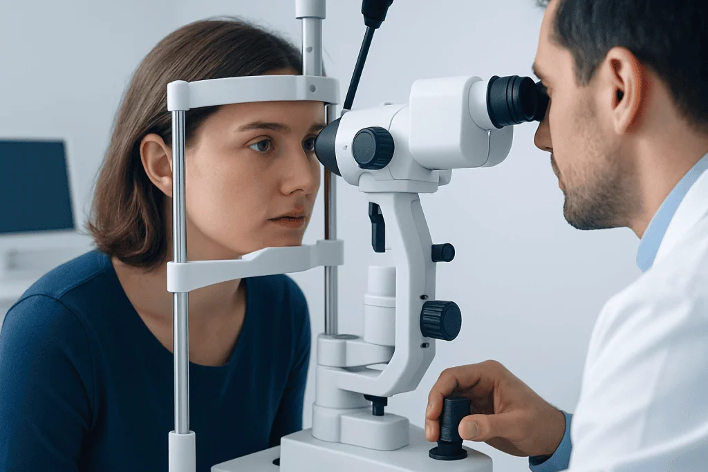
Symptoms usually affect one eye, although both eyes can be involved in some conditions. Vision often becomes blurry or dim, and some people notice that colors, especially red, look faded or less vivid. A central blind spot or patchy areas of missing vision may also appear.
Pain is another common feature. Many people describe discomfort behind the eye or pain that becomes worse with eye movement. This symptom can help distinguish optic neuritis from some other causes of vision change, but only an examination can confirm the reason.
Common symptoms can include:
- Blurred or reduced vision in one eye
- Pain with eye movement
- Loss of color brightness or washed-out colors
- A central dark spot or areas of missing vision
- Reduced contrast sensitivity
- Flashing lights or visual changes with heat or exercise in some cases
Symptoms can vary from mild to severe. Some people recover much of their vision over weeks to months, but others may have lingering problems such as reduced contrast sensitivity, mild color vision changes, or visual fatigue. Any sudden drop in vision deserves prompt assessment, even if symptoms seem to improve.
Causes and Risk Factors

Optic neuritis most often results from inflammation caused by the immune system. In many adults, especially younger adults, it may be related to a demyelinating process, meaning the insulating layer around the optic nerve is damaged. This is why doctors think carefully about MS when optic neuritis is diagnosed.
However, MS is only one possible cause. Other autoimmune and inflammatory disorders can affect the optic nerve, including neuromyelitis optica spectrum disorder, MOG antibody-associated disease, sarcoidosis, lupus, and other systemic conditions. Infections, certain medications, and, more rarely, nutritional or toxic causes may also lead to optic nerve inflammation or similar symptoms.
Risk factors for MS-associated optic neuritis may include age, sex, and the presence of white matter lesions on brain MRI. A person with optic neuritis who has MRI changes typical of demyelination may have a higher chance of later being diagnosed with MS. This is one reason MRI is a key part of the work-up.
Not every painful red eye or vision problem is optic neuritis. Doctors also consider glaucoma, retinal disease, migraine, stroke-related visual symptoms, and other eye or brain conditions. Because the symptom overlap can be significant, it is safest not to self-diagnose vision loss.
How Doctors Diagnose It
Diagnosis starts with a detailed history and examination. The doctor asks how quickly symptoms began, whether pain is present, whether one or both eyes are involved, and whether there have been previous neurological symptoms such as numbness, weakness, balance problems, or bladder changes. An eye examination may include visual acuity, color vision, visual fields, pupil responses, and inspection of the optic nerve.
MRI of the brain and orbits is often one of the most important tests. It can show inflammation in the optic nerve and can also reveal lesions in the brain that suggest MS or another inflammatory disorder. In some settings, specialists may recommend MRI scanning to help assess both the optic nerve and the broader nervous system.
Additional testing depends on the clinical picture. Blood tests may look for antibodies linked to neuromyelitis optica spectrum disorder or MOG antibody-associated disease, as well as signs of infection or systemic autoimmune disease. Optical coherence tomography, visual evoked potentials, or lumbar puncture may also be used in selected cases.
The main goals of diagnosis are to confirm that the optic nerve is involved, identify the underlying cause, and estimate the risk of future neurological disease. If MS is suspected, referral to a neurologist or neuroimmunology specialist is often recommended. In some patients, an integrated evaluation may include neurology consultation alongside ophthalmology or neuro-ophthalmology care.
Treatment Options
Treatment depends on severity, timing, and the underlying cause. Some people with typical optic neuritis begin to improve on their own, but corticosteroids are often used when symptoms are more significant or when faster recovery is important. These medicines can reduce inflammation, though they may not fully determine the final amount of vision recovery.
If optic neuritis is linked to MS or another neuroimmune disorder, longer-term treatment may also be considered. For people who are found to have MS, disease-modifying therapy may help reduce future relapses and new inflammatory activity. Treatment decisions are individualized based on MRI findings, neurological history, and test results.
When the cause is not typical optic neuritis, management may differ. Conditions such as neuromyelitis optica spectrum disorder or MOG antibody-associated disease may need other immune therapies and closer monitoring because they can behave differently from MS-related optic neuritis. This is why accurate diagnosis is so important before making long-term treatment plans.
In selected cases, patients may be evaluated for broader MS care, including multiple sclerosis treatment. Near the end of the care pathway, international patients may also seek coordinated assessment at centers such as Acibadem International, where multidisciplinary specialists in JCI-accredited hospitals evaluate and treat optic neuritis, MS, and related neuroimmunological conditions.
Recovery, Monitoring, and Self-care
Many people begin to notice improvement within several weeks, but recovery is gradual. Vision may continue to improve over months, and color or contrast changes can linger longer than standard eye-chart measurements suggest. Follow-up visits help doctors monitor healing and check for signs of recurrence or a broader neurological condition.
If MRI or other findings raise concern for MS, regular monitoring becomes especially important. Follow-up imaging and neurological review may be advised even if the first episode improves well. This can help detect new inflammatory activity early and support decisions about preventive treatment.
Self-care does not replace medical treatment, but a few practical steps may help during recovery:
- Rest the eyes as needed, especially during periods of visual fatigue
- Avoid delaying follow-up appointments and test reviews
- Keep a record of new symptoms such as numbness, weakness, or balance changes
- Manage general health factors, including sleep, smoking cessation, and stress reduction
- Use caution with driving or detailed work if vision is reduced
Some people notice temporary worsening of vision with heat, fever, or strenuous exercise. This does not always mean new damage has occurred, but it should be mentioned to the doctor. Any fresh drop in vision, new eye pain, or symptoms in the other eye should be assessed promptly.
When Vision Changes Need Urgent Care
Urgent medical attention is appropriate when vision loss develops suddenly, pain is severe, or symptoms progress over a short period. Prompt assessment is also important if both eyes are affected, if there is marked swelling, or if the person has other neurological symptoms such as weakness, numbness, trouble speaking, or difficulty walking. These features can suggest a broader neurological issue or another eye emergency.
It is also wise to seek care quickly if vision changes are accompanied by fever, severe headache, a very red eye, halos around lights, or trauma. These features may point to conditions other than optic neuritis that need immediate treatment. The safest approach is to have sudden unexplained vision symptoms evaluated rather than waiting for them to settle.
Children, older adults, and people with a known immune disorder may need especially careful assessment because the range of possible causes can differ. Pregnant individuals should also contact their clinician promptly if new neurological or visual symptoms occur, so testing and treatment can be planned safely.
Early diagnosis can protect vision and clarify whether MS or another inflammatory disease is involved. If the symptoms fit optic neuritis, seeing an ophthalmologist, neurologist, or neuro-ophthalmologist without delay can make the path to recovery clearer and safer.
Frequently asked questions
Is optic neuritis always a sign of multiple sclerosis?
No. Optic neuritis can occur with multiple sclerosis, but it can also happen with other inflammatory disorders, infections, or sometimes without a clear long-term condition being identified. A careful evaluation helps determine the cause and the likelihood of MS.
How quickly should someone seek help for sudden vision changes?
Sudden vision loss, eye pain with movement, or rapid changes in color vision should be assessed promptly, ideally the same day or as soon as possible. Early evaluation helps rule out other urgent eye or neurological conditions and allows timely treatment when needed.
Can vision return to normal after optic neuritis?
Many people recover a large part of their vision over weeks to months. However, some may continue to notice subtle problems such as reduced color brightness, lower contrast sensitivity, or visual fatigue even after the main symptoms improve.
What tests are usually done if doctors suspect optic neuritis?
The evaluation often includes an eye examination and MRI of the brain and orbits. Depending on the situation, doctors may also order blood tests, visual field testing, optical coherence tomography, visual evoked potentials, or sometimes a lumbar puncture.
If MRI shows lesions, does that mean the person definitely has MS?
Not necessarily. MRI lesions can raise the suspicion for MS, especially when they have a typical appearance and distribution, but the diagnosis depends on the full clinical picture and formal diagnostic criteria. A neurologist or neuroimmunology specialist usually interprets these findings in context.
Can optic neuritis happen more than once?
Yes. Some people have a single episode, while others may experience recurrences, especially if there is an underlying neuroimmune condition such as MS, neuromyelitis optica spectrum disorder, or MOG antibody-associated disease. Recurrent episodes should always be reviewed by a specialist.
References
- National Eye Institute
- National Institute of Neurological Disorders and Stroke
- American Academy of Ophthalmology
- Multiple Sclerosis International Federation
- Mayo Clinic
This article is for general information only and is not a substitute for professional medical advice. Please consult a qualified doctor about your individual situation.
Multiple Sclerosis in Turkey — costs, top hospitals & a free quote
JCI-accredited · board-certified surgeons · reply within 24h
Add us as a Preferred Source to see more of our trusted health content across Google Search, AI Overviews and Discover.

Physical Medicine & Rehabilitation
Restoring movement and function after injury, surgery or neurological conditions.
150 specialists in this unit