Optic Neuropathy: Vision Symptoms, Causes, and Neuro-Ophthalmology Workup

Optic neuropathy can cause blurred vision, dim vision, color fading, blind spots, or vision loss in one or both eyes. Some forms develop suddenly, while others progress slowly and may be noticed only during an eye examination.
Key Takeaways
- Optic neuropathy can cause blurred vision, dim vision, color fading, blind spots, or vision loss in one or both eyes.
- Some forms develop suddenly, while others progress slowly and may be noticed only during an eye examination.
- Diagnosis often includes a detailed eye exam, visual field testing, optic nerve imaging, blood tests, and sometimes MRI.
- Treatment depends on the cause and may involve managing inflammation, blood vessel risk factors, pressure on the optic nerve, toxins, or underlying neurologic disease.
- Sudden vision loss, eye pain with vision changes, or new neurologic symptoms should be assessed urgently.
Optic neuropathy describes damage or dysfunction of the optic nerve, the cable that carries visual information from the eye to the brain. Because causes range from inflammation and reduced blood flow to compression, toxins, inherited conditions, and glaucoma-related damage, a careful neuro-ophthalmology workup is important.
Overview
Optic neuropathy is a broad term for damage to the optic nerve. The optic nerve is made of more than a million nerve fibers that transmit signals from the retina at the back of the eye to the brain. When these fibers are injured, vision may become blurred, dim, distorted, or partly missing, even when the front structures of the eye appear healthy.
The condition is not a single disease. Instead, it is a pattern of optic nerve dysfunction that can result from inflammation, reduced blood supply, pressure on the nerve, inherited nerve vulnerability, nutritional deficiency, toxic exposure, trauma, tumors, or chronic eye pressure damage. The cause matters because treatment and expected recovery differ widely.
Many patients with suspected optic neuropathy are evaluated by a neuro-ophthalmologist, a specialist who focuses on conditions at the intersection of the eyes, optic nerves, brain, and nervous system. The aim of evaluation is to confirm that the optic nerve is involved, identify the likely cause, and decide whether urgent treatment or further neurologic investigation is needed.
Common Vision Symptoms
Optic neuropathy often affects central vision, side vision, color perception, or the brightness of what a person sees. Some people describe vision as looking washed out, gray, or less sharp. Others notice a dark or blurred spot in the center of vision, difficulty reading, or reduced contrast, especially in low light.
Symptoms may appear in one eye or both eyes. In optic neuritis, an inflammatory form of optic neuropathy, vision loss can develop over hours to days and may be associated with pain when moving the eye. In ischemic optic neuropathy, which relates to reduced blood flow, vision loss is often sudden and may be noticed on waking. Compressive or hereditary optic neuropathies may progress more gradually.
Possible symptoms include:
- Blurred, dim, or missing vision in one or both eyes
- Reduced color brightness, especially red appearing faded
- Blind spots or areas of missing side vision
- Difficulty reading, recognizing faces, or seeing in dim lighting
- Pain with eye movement in some inflammatory cases
- A difference in pupil reaction between the two eyes
Because symptoms can overlap with retinal disease, cataract, migraine, stroke, and other eye or brain conditions, a medical examination is needed to determine the source. A person should not assume that painless vision loss is harmless, as some serious optic nerve conditions cause little or no pain.
Causes and Risk Factors

Several categories of disease can injure the optic nerve. Inflammatory optic neuropathy, often called optic neuritis, may occur after a viral illness or in association with conditions such as multiple sclerosis, neuromyelitis optica spectrum disorder, or myelin oligodendrocyte glycoprotein antibody-associated disease. These conditions require careful assessment because treatment and follow-up can differ.
Ischemic optic neuropathy occurs when the blood supply to the optic nerve is reduced. It is more common in older adults and may be associated with high blood pressure, diabetes, sleep apnea, high cholesterol, smoking, or crowded optic nerve anatomy. A specific urgent form, arteritic ischemic optic neuropathy, is linked to giant cell arteritis and needs prompt treatment to protect vision and general health.
Other causes include compression from an orbital or brain mass, trauma, glaucoma-related optic nerve damage, certain medications or toxins, severe nutritional deficiencies, and inherited conditions such as Leber hereditary optic neuropathy. The pattern of vision loss, age of onset, speed of progression, pain, optic nerve appearance, and medical history all help narrow the possibilities.
Risk factors vary by cause, but they may include autoimmune disease, vascular risk factors, smoking, heavy alcohol use, exposure to specific toxins or drugs, family history of optic nerve disease, prior cancer or radiation exposure, and symptoms suggesting systemic inflammation. A complete medication and supplement history is important, including over-the-counter products.
Neuro-Ophthalmology Workup and Diagnosis
A neuro-ophthalmology workup begins with a detailed history. The clinician asks when symptoms started, whether vision loss was sudden or gradual, whether pain is present, and whether there are headaches, jaw pain, scalp tenderness, weakness, numbness, double vision, or other neurologic symptoms. Past medical conditions, family history, and medication or toxin exposures are reviewed carefully.
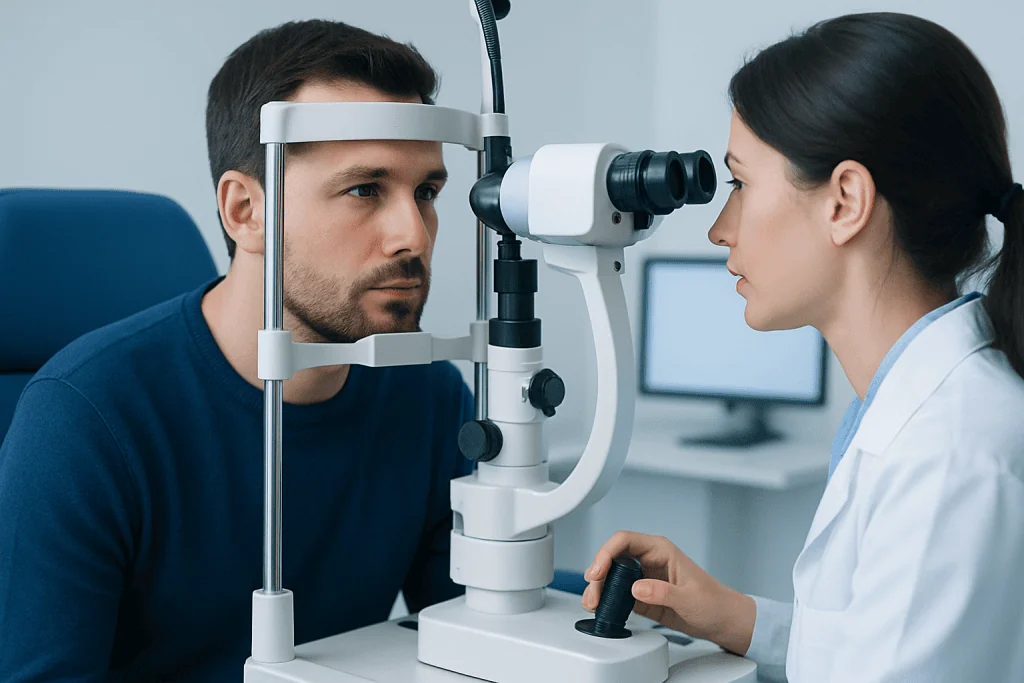
The eye examination usually includes visual acuity testing, color vision testing, pupil examination, eye pressure measurement, and a dilated examination of the retina and optic nerve. A relative afferent pupillary defect, sometimes called an abnormal swinging flashlight test, may suggest that one optic nerve is functioning less well than the other. The optic disc may look swollen, pale, cupped, or sometimes normal in early disease.
Additional tests commonly include automated visual field testing to map blind spots or side vision loss, and optical coherence tomography to measure the thickness of retinal nerve fiber and ganglion cell layers. These tests are painless and help document both the location and severity of optic nerve injury. Repeating them over time can show whether the condition is improving, stable, or progressing.
Depending on the suspected cause, the doctor may request blood tests, MRI of the brain and orbits with contrast, vascular imaging, or other neurologic studies. Blood tests may look for inflammation, infection, autoimmune markers, vitamin deficiencies, or antibodies linked to specific demyelinating disorders. The workup is individualized; not every patient needs every test.
Treatment Options
Treatment for optic neuropathy depends on the underlying cause. There is no single treatment that fits all forms. Some inflammatory conditions may require corticosteroids or other immune-directed medicines, while certain antibody-associated disorders may need urgent specialized therapy. These decisions are made by ophthalmology, neurology, or neuro-ophthalmology teams after the diagnosis is clarified.
In ischemic optic neuropathy, management focuses on identifying the type and reducing future risk. If giant cell arteritis is suspected, treatment is urgent and should not wait for all test results, because the other eye and other blood vessels may be at risk. For non-arteritic ischemic optic neuropathy, doctors often address blood pressure, diabetes, cholesterol, smoking, and sleep apnea, although vision recovery can be variable.
Compressive optic neuropathy may require treatment of the underlying mass or pressure source, such as medication, surgery, radiation, or a combination depending on the diagnosis. Toxic or nutritional optic neuropathy is managed by stopping the harmful exposure when possible and correcting nutritional deficiencies under medical supervision. Glaucomatous optic neuropathy is treated by lowering eye pressure and monitoring the optic nerve over time.
Rehabilitation is also part of care. When vision does not fully recover, low-vision services can help with magnification, contrast enhancement, lighting strategies, reading aids, and practical adaptations for work and daily life. Emotional support is important too, as changes in sight can be stressful even when the condition is stable.
Prevention, Self-Care, and Monitoring
Not all types of optic neuropathy can be prevented, especially inherited and immune-related forms. However, protecting general vascular and neurologic health may reduce risk in some cases. Patients can support optic nerve health by managing diabetes, blood pressure, cholesterol, and sleep apnea with their healthcare team, avoiding smoking, and limiting exposure to known toxins.
Safe self-care includes taking prescribed medicines as directed, keeping follow-up appointments, and reporting new symptoms promptly. Patients should avoid stopping or starting medications, high-dose vitamins, or supplements without professional guidance, particularly if optic neuropathy may be related to a drug, toxin, or deficiency. A balanced diet helps prevent nutritional problems, but medical testing is needed before treating suspected deficiencies.
People with known optic nerve disease may be asked to monitor vision at home by checking each eye separately. This can be as simple as covering one eye at a time and noticing changes in reading clarity, color brightness, or missing areas of vision. Any clear change should lead to medical contact rather than waiting for the next routine visit.
Regular eye examinations are especially important for people with glaucoma risk, diabetes, vascular risk factors, autoimmune disease, or a family history of optic nerve disease. At Acibadem International, multidisciplinary specialists in ophthalmology, neurology, radiology, and related fields evaluate and treat optic nerve disorders for international patients in JCI-accredited hospitals.
When to See a Doctor
A person should seek urgent medical care for sudden vision loss, rapidly worsening blurred vision, a new dark area in the field of vision, or vision changes with eye pain. Prompt evaluation is also important if vision symptoms occur with severe headache, scalp tenderness, jaw pain while chewing, fever, weakness, numbness, double vision, or difficulty speaking.
Even gradual symptoms deserve assessment. Slow loss of color brightness, persistent dim vision, or unexplained difficulty reading may signal optic nerve disease, retinal disease, or another eye condition that benefits from early diagnosis. If one eye is affected, patients may not notice symptoms until they cover the better eye, so subtle changes should not be ignored.
People already diagnosed with optic neuropathy should contact their doctor if symptoms worsen, if the second eye becomes affected, or if new neurologic symptoms develop. A clear plan for follow-up, test results, and warning signs can help patients feel more confident and avoid unnecessary delays in care.
Frequently asked questions
Is optic neuropathy the same as optic neuritis?
No. Optic neuropathy is the general term for optic nerve damage or dysfunction from many possible causes. Optic neuritis is one type of optic neuropathy, usually involving inflammation of the optic nerve.
Can optic neuropathy be cured?
The outcome depends on the cause, severity, and how quickly treatment begins when treatment is available. Some inflammatory forms improve substantially, while ischemic, hereditary, toxic, compressive, or glaucoma-related damage may have different recovery patterns. A specialist can explain the most realistic expectations after the workup.
Why is MRI sometimes needed for optic neuropathy?
MRI can show inflammation, compression, tumors, demyelinating disease, or other changes affecting the optic nerves and brain. It is especially helpful when symptoms, examination findings, or age suggest a neurologic or orbital cause. Not every patient needs MRI, so the decision is individualized.
What does color vision have to do with optic nerve disease?
The optic nerve is important for transmitting color information, and many optic neuropathies make colors look faded or less bright. Doctors often compare color perception between the two eyes because a difference can be an early clue. Red desaturation is a common symptom patients may notice.
Is optic neuropathy always painful?
No. Some forms, such as optic neuritis, may cause pain with eye movement, but many other types are painless. Sudden painless vision loss can still be medically important and should be evaluated promptly.
Can both eyes be affected?
Yes. Some optic neuropathies affect one eye, while others affect both eyes at the same time or at different times. Bilateral or sequential symptoms can help doctors consider causes such as inflammatory, hereditary, toxic, nutritional, compressive, or systemic vascular conditions.
What should a patient bring to a neuro-ophthalmology appointment?
It is helpful to bring prior eye records, visual field or OCT results, MRI or CT images and reports, a medication and supplement list, and details about symptom timing. Patients should also mention family history, autoimmune disease, vascular risk factors, and any toxin or occupational exposures. This information can make the diagnostic process more efficient.
References
- American Academy of Ophthalmology
- North American Neuro-Ophthalmology Society
- National Eye Institute
- Mayo Clinic
- European Academy of Neurology
This article is for general information only and is not a substitute for professional medical advice. Please consult a qualified doctor about your individual situation.
Explore treatments in Turkey — costs, top hospitals & a free quote
JCI-accredited · board-certified surgeons · reply within 24h
Add us as a Preferred Source to see more of our trusted health content across Google Search, AI Overviews and Discover.
Check your numbers in seconds
BMI, calories, due date, blood pressure and 30+ more clinical calculators — free, instant, doctor-reviewed ranges.