

Pediatric Retina Specialist: An Evidence-Based Patient Guide
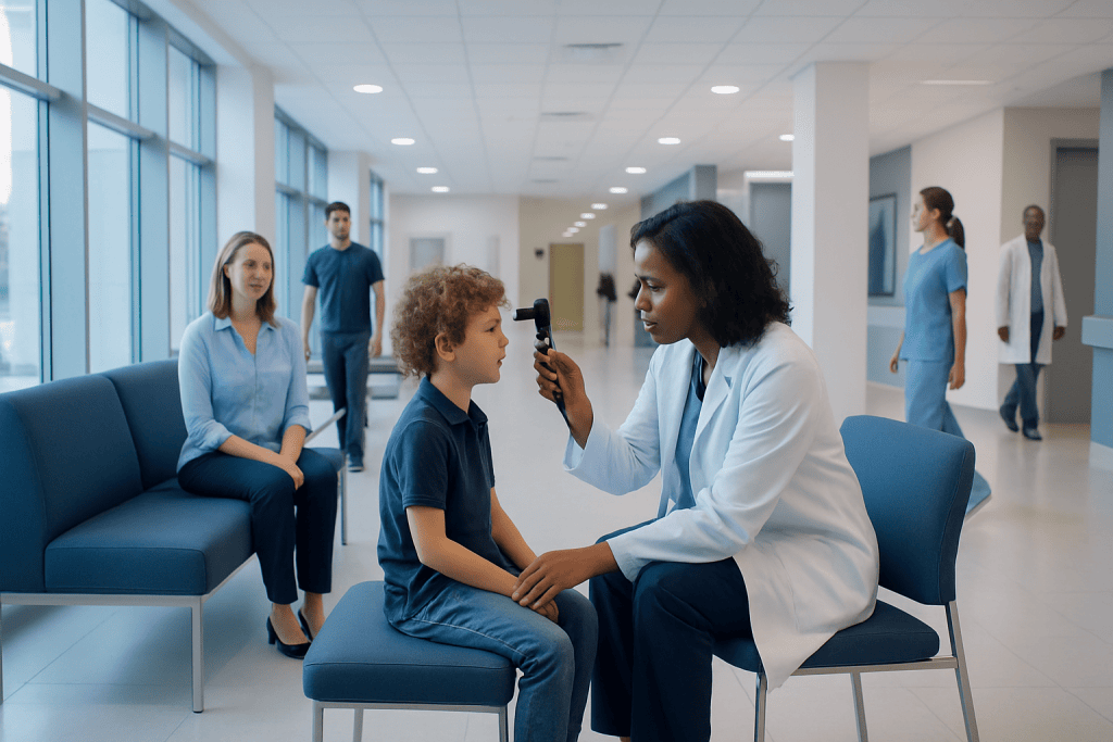
A pediatric retina specialist evaluates retinal conditions in infants, children, and teenagers using child-friendly examination techniques. Prompt assessment is important for warning signs such as a white pupil, new eye misalignment, reduced vision, flashes, floaters, or a curtain-like loss of vision.
Key Takeaways
- A pediatric retina specialist evaluates retinal conditions in infants, children, and teenagers using child-friendly examination techniques.
- Prompt assessment is important for warning signs such as a white pupil, new eye misalignment, reduced vision, flashes, floaters, or a curtain-like loss of vision.
- Some retinal disorders need observation, while others may require laser treatment, injections, cryotherapy, surgery, glasses, low-vision support, or genetic counselling.
- Retinopathy of prematurity screening is an important part of care for eligible premature babies.
- Parents should not wait for a routine check-up if they notice sudden vision changes, eye injury, or concerning changes in the appearance of a child’s eye.
A pediatric retina specialist is an ophthalmologist with advanced expertise in disorders affecting the retina, the light-sensitive tissue at the back of a child’s eye. Early specialist assessment can help protect vision when a child has an inherited retinal condition, retinal detachment, retinopathy of prematurity, eye injury, or unexplained changes in sight.
Overview: What Does a Pediatric Retina Specialist Do?
A pediatric retina specialist is an eye doctor with additional expertise in diagnosing and treating conditions of the retina in babies, children, and adolescents. The retina is a thin layer of light-sensitive nerve tissue lining the back of the eye. It converts light into signals that travel through the optic nerve to the brain, making clear vision possible.
Children may be referred to this specialist because of a finding during a routine eye examination, a family history of an inherited eye condition, complications of premature birth, an eye injury, or a change noticed at home. A specialist works to identify the cause, assess how the retina is functioning, and recommend monitoring or treatment that is appropriate for the child’s age and needs.
Care is often coordinated with pediatric ophthalmologists, neonatologists, genetic specialists, neurologists, pediatricians, low-vision professionals, and other members of the child’s care team. This multidisciplinary approach is especially helpful when a retinal condition is part of a broader health or genetic condition.
Conditions a Pediatric Retina Specialist May Assess
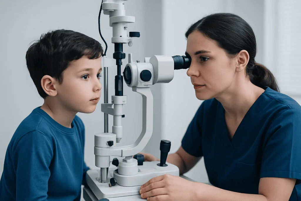
Pediatric retinal conditions may be present at birth, develop during childhood, or occur after trauma or inflammation. Some affect one eye, while others affect both. The impact on vision varies widely: some conditions remain stable and require regular observation, whereas others need timely treatment to reduce the chance of lasting visual impairment.
A common reason for specialist involvement in newborn care is retinopathy of prematurity, or ROP. This condition can affect babies born very early or with very low birth weight, because retinal blood vessels are still developing. Screening and follow-up are arranged by the neonatal team according to established clinical criteria.
Other conditions that may require pediatric retinal expertise include retinal tears or detachment, Coats disease, familial exudative vitreoretinopathy, persistent fetal vasculature, retinal changes related to high myopia, retinal infection or inflammation, and inherited retinal diseases. A child may also need assessment for a tumour-like lesion or retinoblastoma concern; a white reflection in the pupil should always receive urgent medical assessment.
- Inherited retinal disorders may cause night-vision difficulty, reduced central vision, colour-vision changes, or progressive visual symptoms.
- Retinal detachment may occur after significant eye trauma, severe short-sightedness, prior eye surgery, or certain inherited conditions.
- Some retinal findings are first detected through school screening, an optometrist’s examination, or photographs in which one pupil appears unusually white.
Symptoms and Signs Parents May Notice

Young children cannot always describe a visual symptom, so parents and caregivers often play an important role in identifying potential concerns. A child may move closer to objects, have trouble recognizing faces at a distance, bump into furniture, avoid dimly lit spaces, tilt their head, cover one eye, or lose interest in visual activities they previously enjoyed.
Other signs may include a new eye turn, jerky eye movements, unusual sensitivity to light, frequent blinking, complaints of flashes or floating spots, or a sudden reduction in vision. Babies may show poor visual tracking, reduced attention to faces or lights, or an abnormal red reflex in photographs. These signs do not always indicate a retinal condition, but they warrant professional evaluation.
A white, yellowish, or absent red reflex in the pupil is particularly important to assess without delay. It can occur for several reasons, including conditions involving the lens or retina. Early evaluation helps clinicians determine the cause and arrange the appropriate care.
How a Pediatric Retinal Assessment Works
A visit begins with a detailed history. The specialist may ask about pregnancy and birth history, prematurity, developmental milestones, eye injuries, medications, family eye history, and the timing of any visual changes. Older children may complete age-appropriate vision testing, while infants and younger children are assessed through their visual behavior, fixation, tracking, and responses to light and objects.
The doctor will examine the front and back of the eye. Dilating drops are commonly used to widen the pupils so the retina can be viewed more clearly. These drops can cause temporary light sensitivity and blurred near vision. The examination is adapted to the child’s age, level of comfort, and medical needs; very young children may need a carefully planned examination under anaesthesia when a complete office examination is not possible.
Imaging and tests may include retinal photography, optical coherence tomography (OCT), ultrasound, fluorescein angiography in selected cases, visual field testing, and electrophysiology tests that measure retinal function. Genetic testing may be discussed when an inherited retinal disorder is suspected. Results are interpreted alongside the child’s symptoms and full clinical examination rather than in isolation.
Treatment Options: From Monitoring to Retinal Procedures
Treatment depends on the underlying diagnosis, the location and extent of retinal involvement, the child’s age, and whether vision is currently at risk. Some children benefit most from regular monitoring, glasses, amblyopia treatment, visual rehabilitation, or support at school. For inherited conditions, genetic counselling may help families understand testing results, inheritance patterns, and available support.
When active retinal disease needs treatment, options can include laser therapy, cryotherapy, medication injected into the eye in carefully selected situations, or retinal surgery. For example, treatment for ROP may involve laser treatment or anti-vascular endothelial growth factor medicine, with close follow-up because retinal development continues after initial treatment. The choice is individualized and guided by established pediatric retinal practice.
Retinal surgery may be considered for problems such as a retinal detachment, certain complications of trauma, or advanced retinal disease. A procedure may involve repairing retinal breaks, removing vitreous gel, using laser treatment, or placing a supportive band around the eye in selected cases. Families can learn more about retinal detachment surgery when this type of repair is being considered.
The potential benefit of treatment is to preserve or improve the best possible visual function and prevent progression where feasible. However, every procedure has limitations and risks. The specialist explains the expected goals, alternatives, anaesthesia considerations, likely follow-up, and the fact that some conditions can require more than one intervention.
Procedure Journey, Recovery, and Follow-Up
Before a retinal procedure, the care team reviews the diagnosis, imaging results, child’s general health, medications, allergies, and anaesthesia requirements. Parents are given instructions about eating and drinking before anaesthesia, arrival time, and what to bring to the appointment. Consent discussions should cover why the procedure is recommended, what it involves, possible alternatives, and expected aftercare.
On the day of treatment, children may receive local treatment, sedation, or general anaesthesia depending on the procedure and their age. The specialist examines the retina and then performs the planned treatment, such as laser, cryotherapy, an injection, or surgery. Some procedures are completed on the same day, while more complex surgery may require a hospital stay or additional observation.
Recovery varies significantly. Mild irritation, redness, tearing, temporary blurred vision, or light sensitivity can occur after eye treatment. After surgery, the child may need eye drops, a protective shield, activity restrictions, and repeated retinal examinations. If a gas bubble is used during retinal surgery, the surgeon will provide specific instructions about positioning and travel, including avoiding air travel until the gas has resolved.
Possible complications depend on the condition and treatment but can include infection, bleeding, raised eye pressure, cataract, inflammation, recurrent detachment, scarring, or incomplete visual recovery. Prompt follow-up helps the team detect and manage complications early. Parents should contact the treating team urgently if the child develops worsening pain, increasing redness, discharge, nausea or vomiting with eye pain, sudden vision loss, or a new injury to the treated eye.
Supporting Eye Health and Everyday Development
Not all pediatric retinal conditions can be prevented, particularly inherited disorders and complications related to very premature birth. Nevertheless, timely newborn and childhood eye checks, attendance at recommended ROP screening appointments, and follow-up after an eye injury can support early identification of problems.
Protective eyewear is sensible for sports and activities with a risk of eye trauma. Children should use eye protection designed for the activity, with appropriate polycarbonate lenses where recommended. Parents can also support healthy visual development by ensuring that prescribed glasses, patching plans, eye drops, or school accommodations are followed as advised.
For children with reduced vision, practical support may include larger print, high-contrast materials, classroom seating adjustments, orientation and mobility support, and low-vision aids. A child’s visual needs can change as they grow, so regular reviews are valuable even when the retinal condition is stable.
Acibadem International’s multidisciplinary specialists and JCI-accredited hospitals support diagnosis and treatment planning for international patients with pediatric retinal concerns, coordinating care with relevant pediatric and eye-health services when needed.
When to Seek Medical Care
Parents should arrange a prompt eye assessment if a child has persistent difficulty seeing, a new eye turn, unexplained clumsiness, a noticeable difference between the eyes, night-vision difficulty, repeated reports of flashes or floaters, or a family history of a serious inherited retinal condition. A pediatrician, optometrist, or general ophthalmologist can help determine whether pediatric retinal referral is appropriate.
Urgent medical care is appropriate for sudden vision loss, a curtain or shadow across vision, severe eye pain, a significant eye injury, or rapidly increasing redness and swelling. A white or yellow pupil reflex, especially if newly noticed in photographs or in normal lighting, also needs urgent assessment. These signs have several possible causes, and early evaluation is the safest way to clarify them.
For premature babies, families should attend all retinal screening appointments arranged by the neonatal team, even if the baby appears comfortable and the eyes look normal. Retinal changes in ROP may not be visible to parents, and specialist monitoring helps identify when observation or treatment is needed.
Frequently asked questions
What is the difference between a pediatric ophthalmologist and a pediatric retina specialist?
A pediatric ophthalmologist diagnoses and manages a broad range of eye conditions in children. A pediatric retina specialist has additional expertise in diseases affecting the retina and vitreous, including complex retinal conditions in premature babies and children. Children may see both specialists as part of coordinated care.
Does every child with a retinal condition need surgery?
No. Many retinal findings are monitored with regular examinations and imaging, while others can be managed with glasses, visual support, or medical treatment. Surgery is considered when the diagnosis, severity, and risk to vision suggest that repair or stabilization is needed.
Is a dilated retinal examination painful for children?
Dilating eye drops can sting briefly when applied and may cause light sensitivity and blurry near vision for several hours. The retinal examination itself is generally not painful, although it can be uncomfortable for some children. The team uses age-appropriate approaches to help children feel as comfortable as possible.
Why are premature babies screened for retinopathy of prematurity?
Babies born very early may have incompletely developed retinal blood vessels. Retinopathy of prematurity can develop without visible symptoms, so scheduled specialist examinations are used to detect changes early. Many cases are mild and improve, but some require close follow-up or treatment.
Can a retinal disorder be inherited?
Some retinal disorders are inherited and may occur in more than one family member, while others are not inherited. When a genetic condition is suspected, the specialist may recommend genetic counselling and testing. This can help clarify the diagnosis and guide family planning discussions.
What should parents bring to a pediatric retina appointment?
Useful information includes prior eye examination reports, imaging results, a list of medications, birth and neonatal records when relevant, and details of family eye conditions. Parents can also bring photos or notes showing when symptoms began, such as an unusual pupil reflection or eye alignment change. Sunglasses may help after dilating drops because children can be temporarily sensitive to light.
References
- American Academy of Ophthalmology
- American Association for Pediatric Ophthalmology and Strabismus
- National Eye Institute
- Royal College of Ophthalmologists
- World Health Organization
This article is for general information only and is not a substitute for professional medical advice. Please consult a qualified doctor about your individual situation.
Pediatric Retina in Turkey — costs, top hospitals & a free quote
JCI-accredited · board-certified surgeons · reply within 24h
Add us as a Preferred Source to see more of our trusted health content across Google Search, AI Overviews and Discover.

More from the Health Library


Related Specialists

Dr. Gizem Kaplan
Gynecology & Obstetrics
Prof. Dr. Ahmet Tayyar
Gynecology & Obstetrics
Fzt. Ece Burçak Özuyguntaş
Physical Medicine & Rehabilitation
Dr. Dilek Tomatır
Ophthalmology