

PET-CT Scan: Cancer Imaging, Preparation, and What Uptake Means
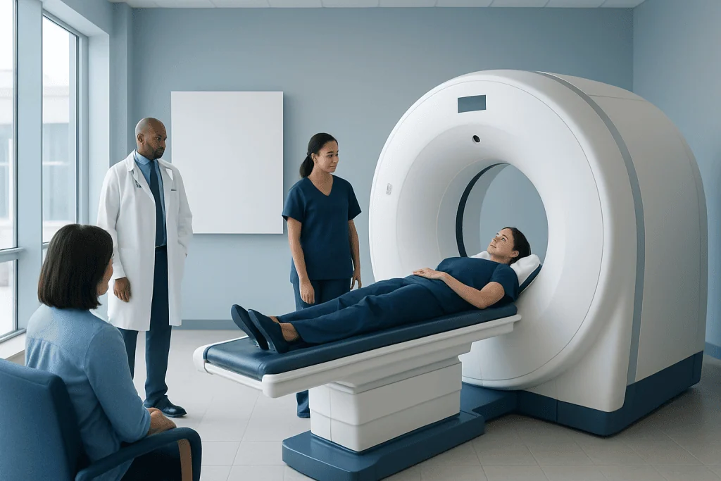
A PET-CT scan combines positron emission tomography with computed tomography to show both how tissues function and where they are located. In cancer care, PET-CT is often used for staging, treatment planning, checking treatment response, and looking for recurrence.
Key Takeaways
- A PET-CT scan combines positron emission tomography with computed tomography to show both how tissues function and where they are located.
- In cancer care, PET-CT is often used for staging, treatment planning, checking treatment response, and looking for recurrence.
- Most PET-CT scans use a small amount of radioactive glucose tracer, commonly FDG, because many cancer cells use glucose actively.
- Uptake on a PET-CT scan does not always mean cancer; infection, inflammation, healing tissue, and normal organs can also show activity.
- Preparation usually includes fasting, avoiding strenuous exercise, and careful blood sugar management when relevant.
- Results should always be interpreted by qualified specialists together with symptoms, examination findings, laboratory tests, and other imaging.
A PET-CT scan combines metabolic and anatomical imaging to help doctors detect, stage, and monitor many cancers. Understanding preparation steps and what “uptake” means can help patients feel more confident before and after the scan.
Overview: What Is a PET-CT Scan?
A PET-CT scan is an imaging test that combines two technologies in one examination. PET, or positron emission tomography, shows how active tissues are by detecting a small amount of injected radioactive tracer. CT, or computed tomography, provides detailed cross-sectional images of the body’s anatomy. When combined, the test can show both where an abnormal area is located and how metabolically active it is.
The most commonly used tracer in cancer imaging is fluorodeoxyglucose, often called FDG. FDG is a glucose-like substance that travels through the bloodstream. Cells that use more glucose may take up more FDG, and this activity can be seen on PET images. Because many cancer cells have increased glucose metabolism, FDG PET-CT can be useful in oncology.
A PET-CT scan does not diagnose every condition on its own. It is one part of a broader evaluation that may include a physical examination, blood tests, biopsy, MRI, ultrasound, or other imaging. The scan is interpreted by trained nuclear medicine and radiology specialists, who consider the pattern of tracer uptake, CT findings, and the patient’s clinical history.
Why PET-CT Is Used in Cancer Imaging
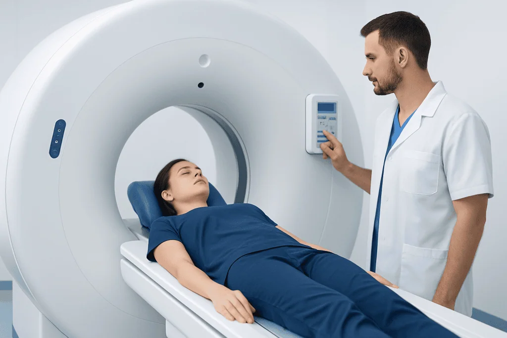
In oncology, PET-CT can help answer several important clinical questions. It may be used to evaluate whether a suspicious mass is likely to be metabolically active, to stage certain cancers, to look for spread to lymph nodes or distant organs, and to help guide treatment planning. It can also help doctors assess whether treatment is working or whether cancer has returned after therapy.
PET-CT is commonly used in selected cases of lymphoma, lung cancer, colorectal cancer, melanoma, head and neck cancers, esophageal cancer, and several other malignancies. However, its usefulness depends on the cancer type, tumor biology, size of lesions, prior treatments, and the reason for scanning. Some cancers do not take up FDG strongly, and very small lesions may be below the scan’s resolution.
The information from PET-CT can sometimes change a treatment plan. For example, it may help doctors decide whether surgery is appropriate, identify the best area for biopsy, or determine the radiation therapy target more accurately. In follow-up care, PET-CT may help distinguish scar tissue from metabolically active disease, although additional tests may still be needed.
How the PET-CT Procedure Works
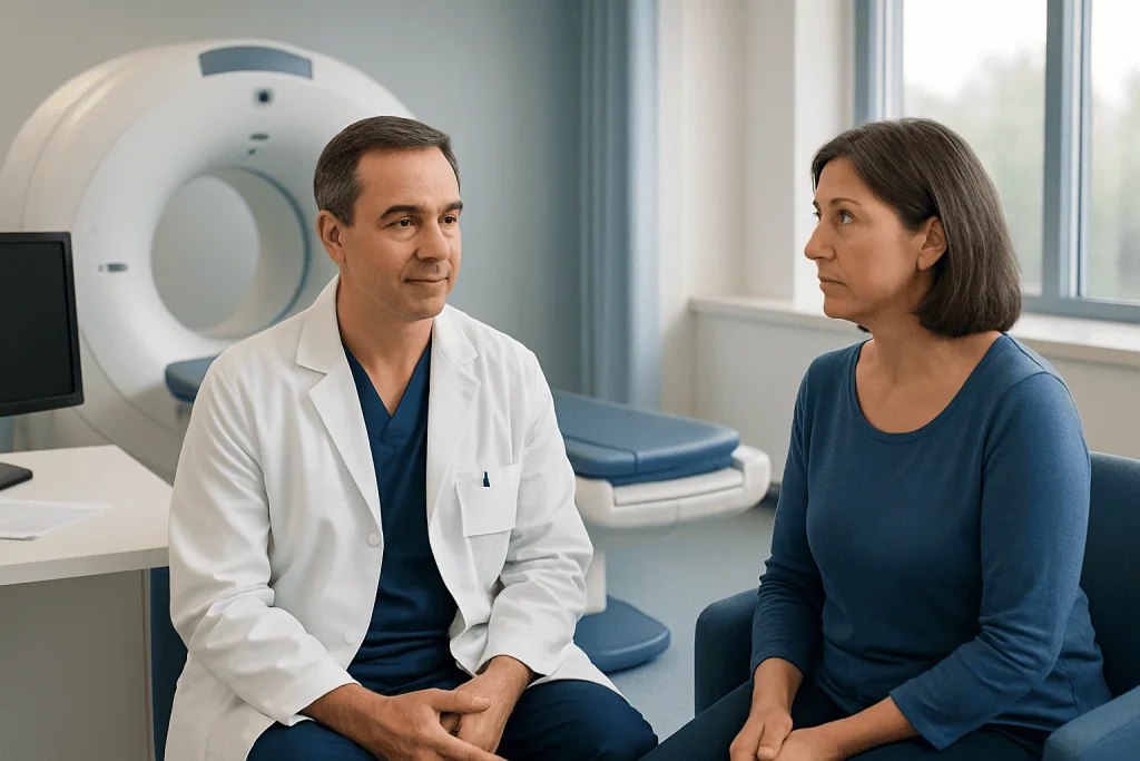
On the day of the examination, the patient is usually checked in, asked about medical history, medications, allergies, pregnancy status, breastfeeding, diabetes, and recent treatments. Blood glucose may be measured before FDG injection because high blood sugar can affect tracer distribution and image quality. The tracer is then injected into a vein, usually in the arm or hand.
After the injection, there is typically a quiet waiting period while the tracer circulates and is absorbed by tissues. During this time, patients are usually asked to rest, remain warm, avoid talking excessively, and avoid unnecessary movement. This helps reduce muscle uptake, which can make images harder to interpret.
The scan itself is painless. The patient lies on a scanning table that moves slowly through the PET-CT scanner. Some scans are performed from the base of the skull to the thighs, while others include a larger or more specific area depending on the clinical question. The CT portion may be done with or without contrast material, depending on the protocol and patient factors.
After the scan, most people can return to normal activities unless their care team gives different instructions. Drinking fluids may help the body eliminate the tracer through urine. Because the tracer contains a small amount of radioactivity, patients may receive simple instructions about limiting close contact with pregnant people or young children for a short period, depending on local practice.
PET-CT Preparation: What Patients Should Know
Preparation is important because it can affect the accuracy of PET-CT images. Instructions may vary by hospital, tracer type, and scan purpose, so patients should follow the guidance provided by their imaging team. For many FDG PET-CT scans, fasting for several hours before the appointment is required, although plain water is usually encouraged unless restricted for another medical reason.
Patients are often advised to avoid strenuous exercise before the scan. Exercise can increase glucose use in muscles and may cause muscle uptake that is unrelated to disease. It is also helpful to stay warm before and during the uptake period, because cold exposure can activate brown fat, a normal tissue that may take up FDG and complicate interpretation.
People with diabetes should receive individualized instructions. Blood sugar control matters because elevated glucose levels can compete with FDG and reduce the quality of the scan. Patients should not change diabetes medications, insulin, or fasting plans without medical advice; the imaging center and treating doctor can coordinate safe timing.
- Bring a list of medications and relevant medical records.
- Tell the team about pregnancy, possible pregnancy, or breastfeeding.
- Report recent surgery, infection, radiation therapy, chemotherapy, immunotherapy, or vaccinations, as these may influence uptake patterns.
- Wear comfortable clothing without metal when possible, or be prepared to change into a gown.
- Ask in advance whether CT contrast will be used, especially if there is a history of contrast reaction or kidney disease.
What Does FDG Uptake Mean?
“Uptake” means that a tissue has absorbed the PET tracer. On FDG PET-CT, areas with higher glucose use may appear brighter than surrounding tissues. Doctors may describe uptake as mild, moderate, intense, focal, diffuse, physiological, inflammatory, or suspicious, depending on the pattern and location.
Uptake does not automatically mean cancer. Normal organs such as the brain, heart, kidneys, bladder, liver, bowel, and muscles can show FDG activity. Inflammation, infection, healing after surgery, recent radiation, some benign tumors, and immune responses can also produce increased uptake. This is why PET findings must be matched with CT anatomy and clinical context.
Reports may include a measurement called SUV, or standardized uptake value. SUV is a semi-quantitative estimate of tracer activity in a region. It can be useful for comparison, especially when monitoring response to treatment, but it is not a standalone cancer test. SUV can be influenced by timing, body composition, blood glucose, scanner technique, lesion size, and other factors.
Sometimes PET-CT results are described as positive, negative, indeterminate, or requiring correlation. “Indeterminate” means the finding is not clearly benign or malignant based on the scan alone. In such cases, the doctor may recommend follow-up imaging, biopsy, laboratory tests, or observation depending on the overall situation.
Benefits, Limitations, and Safety
A key benefit of PET-CT is that it combines functional and anatomical information in a single examination. This can help detect disease that may not be obvious on anatomical imaging alone and may clarify whether a structural abnormality is metabolically active. For many patients, this information supports more personalized treatment decisions.
However, PET-CT has limitations. Small tumors may be missed, and some cancers have low FDG uptake. Conversely, non-cancer conditions such as infection or inflammation can look active. Recent treatments can also affect results; for example, healing tissue after surgery or radiation may show uptake, while treatment response can change the appearance of lesions over time.
PET-CT involves exposure to ionizing radiation from both the tracer and the CT scan. The amount is considered acceptable when the test is medically justified, but unnecessary repeat imaging should be avoided. Pregnant patients, people who may be pregnant, and breastfeeding patients should discuss timing and alternatives with their doctor and imaging team.
Allergic reactions to the FDG tracer are very uncommon. If CT contrast is used, there may be additional considerations, including kidney function and prior contrast reactions. The care team reviews these factors before the scan to choose the safest and most appropriate protocol.
Understanding the Report and Next Steps
A PET-CT report usually describes the clinical reason for the scan, the tracer used, the imaging technique, and the findings by body region. It may compare current images with prior scans. The impression or conclusion section summarizes the most important findings and may suggest whether the appearance is consistent with active disease, treatment response, inflammation, or another possibility.
Patients should review the results with the doctor who ordered the test, such as an oncologist, surgeon, pulmonologist, hematologist, or other specialist. The ordering doctor can explain what the findings mean for the patient’s diagnosis and treatment plan. A single phrase in a report, such as “increased uptake,” should not be interpreted in isolation.
Further steps depend on the reason for the scan and the results. Options may include biopsy, additional imaging, blood tests, treatment planning, continued therapy, surveillance, or reassurance when findings are benign or stable. When uncertainty remains, multidisciplinary review can be helpful because radiologists, nuclear medicine physicians, oncologists, surgeons, and pathologists may each add important perspective.
When to Contact a Doctor
Patients should contact their doctor or imaging center before the PET-CT scan if they are pregnant, may be pregnant, are breastfeeding, have diabetes, have kidney disease, or have had a previous reaction to contrast material. It is also important to report recent infections, surgery, biopsies, radiation therapy, chemotherapy, immunotherapy, or steroid treatment, as these may affect scheduling or interpretation.
After the scan, patients should seek medical advice if they develop unexpected symptoms, especially if CT contrast was used and they experience a rash, breathing difficulty, swelling, dizziness, or other concerning reaction. Most people do not have significant side effects from the PET tracer, but any unusual symptoms should be reported to a healthcare professional.
For international patients, coordinated evaluation can be especially important when PET-CT is part of cancer diagnosis or treatment planning. Acibadem International’s multidisciplinary specialists and JCI-accredited hospitals diagnose and treat many conditions requiring advanced imaging, including cancer, while helping patients connect imaging findings with appropriate clinical care.
Frequently asked questions
Is a PET-CT scan used only for cancer?
No. PET-CT is widely used in cancer care, but it can also be used in selected heart, brain, infection, and inflammatory conditions. The tracer and protocol depend on the medical question being investigated.
Does high FDG uptake always mean cancer?
No. High FDG uptake means that an area is metabolically active, not necessarily malignant. Infection, inflammation, healing tissue, normal organs, and muscle activity can also show increased uptake, so the result must be interpreted in context.
How should a patient prepare for a PET-CT scan?
Many FDG PET-CT scans require fasting for several hours, drinking water, and avoiding strenuous exercise beforehand. Patients with diabetes, kidney disease, pregnancy, possible pregnancy, or breastfeeding should receive individualized instructions from the imaging center.
Is PET-CT painful?
The scan itself is not painful. The main discomfort is usually the small needle used for the tracer injection and the need to lie still during imaging. Patients who have difficulty lying flat should inform the team in advance.
What is SUV on a PET-CT report?
SUV, or standardized uptake value, is a measurement that estimates how much tracer activity is present in a selected area. It can help compare lesions or monitor treatment response, but it is affected by many technical and patient-related factors and cannot diagnose cancer by itself.
Can PET-CT detect very small tumors?
PET-CT can detect many metabolically active lesions, but very small tumors may be below the scanner’s resolution. Some tumors also have low FDG uptake. Doctors may recommend other imaging tests or follow-up depending on the situation.
How soon are PET-CT results available?
Timing varies by hospital and clinical urgency. The images are reviewed by qualified specialists, often with comparison to previous studies and medical history. Patients should discuss the report with the doctor who ordered the scan to understand the next steps.
References
- Society of Nuclear Medicine and Molecular Imaging
- American College of Radiology
- National Cancer Institute
- European Association of Nuclear Medicine
- Radiological Society of North America
This article is for general information only and is not a substitute for professional medical advice. Please consult a qualified doctor about your individual situation.
Oncology care in Turkey — second opinion and treatment plan
JCI-accredited · board-certified surgeons · reply within 24h
Add us as a Preferred Source to see more of our trusted health content across Google Search, AI Overviews and Discover.

More from the Health Library



Related Specialists

Prof. Dr. Ercan Karaarslan
Radiology
Prof. Dr. Bora Peynircioğlu
Interventional Radiology
Prof. Dr. Koray Güven
Interventional Radiology
Assoc. Prof. Dr. Deniz Can Aliş
Radiology