PET-CT Scan: How It Helps Detect and Stage Cancer
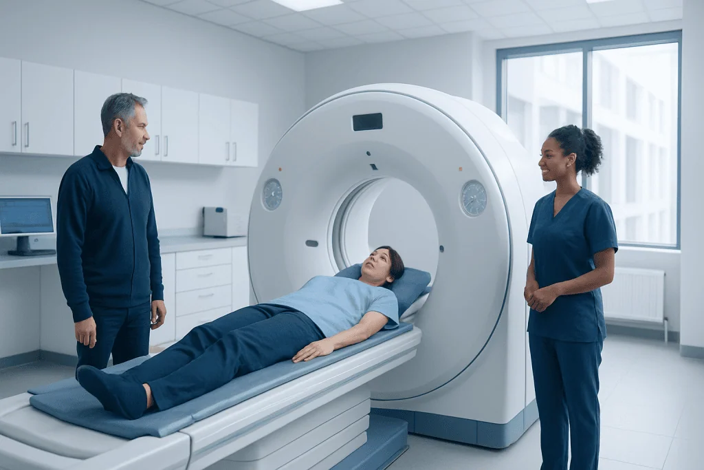
A PET-CT scan combines positron emission tomography and computed tomography in one examination. The test can show both where abnormal tissue is located and how metabolically active it is.
Key Takeaways
- A PET-CT scan combines positron emission tomography and computed tomography in one examination.
- The test can show both where abnormal tissue is located and how metabolically active it is.
- PET-CT is often used to stage cancer, guide treatment planning, monitor response, and check for recurrence.
- Preparation usually includes fasting for several hours and careful blood sugar management when needed.
- PET-CT uses a small amount of radioactive tracer and is generally safe when performed for an appropriate medical reason.
A PET-CT scan combines metabolic and anatomical imaging to help doctors find cancer, determine how far it has spread, and assess whether treatment is working. It is a commonly used, evidence-based imaging test in many areas of oncology.
Overview
A PET-CT scan is an advanced imaging test that combines two technologies: positron emission tomography, known as PET, and computed tomography, known as CT. The CT part creates detailed pictures of the body’s structures, while the PET part shows how tissues use energy. When these images are combined, doctors can see both the location and the activity of abnormal areas.
In cancer care, PET-CT is especially useful because many cancer cells use more glucose than normal cells. Before the scan, a small amount of a radioactive tracer, most commonly fluorodeoxyglucose or FDG, is injected into a vein. Areas that take up more tracer may appear as brighter or more active spots on the PET images.
A PET-CT scan is not used for every person or every type of cancer. Its value depends on the cancer type, stage, symptoms, previous test results, and the clinical question the doctor needs to answer. When used appropriately, it can provide important information for diagnosis, staging, treatment planning, and follow-up.
How PET-CT Helps Detect Cancer
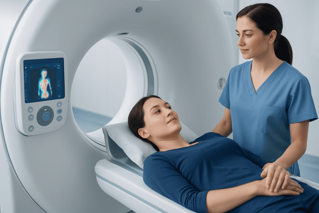
PET-CT can help identify areas of the body where cells are unusually active. Because many tumors have increased glucose metabolism, they may absorb more FDG tracer than surrounding normal tissues. This can make certain cancers more visible than they would be on anatomical imaging alone.
The scan may be recommended when a suspicious mass has been found on another test, when symptoms suggest cancer but the location is uncertain, or when doctors need to assess whether a known cancer has spread. PET-CT may also help identify the best area for a biopsy, particularly when there are several abnormal findings and one needs to be sampled.
However, PET-CT is not a perfect cancer screening test for the general population. Some cancers may not take up much FDG, especially very small or slow-growing tumors. Inflammation, infection, healing tissue, and some benign conditions can also show increased tracer uptake. For this reason, PET-CT results are interpreted together with medical history, physical examination, laboratory tests, and other imaging or biopsy results.
How PET-CT Helps Stage Cancer
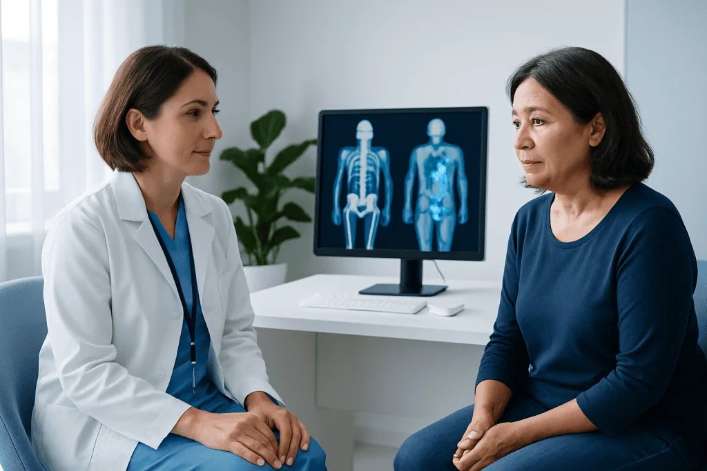
Staging describes how much cancer is present in the body and whether it has spread from its original site. Accurate staging is essential because it helps doctors choose the most appropriate treatment plan. A PET-CT scan can provide whole-body information in a single examination, which is particularly helpful for detecting disease in lymph nodes, bones, liver, lungs, and other organs.
In many cancers, PET-CT can help distinguish between localized disease, regional spread, and distant metastases. For example, it may show whether lymph nodes that look normal in size on CT are metabolically active, or whether a suspicious bone lesion is likely to represent metastatic disease. This information can influence decisions about surgery, radiotherapy, chemotherapy, immunotherapy, targeted therapy, or a combination of treatments.
PET-CT is also used in restaging. Restaging may be needed after treatment has started, after treatment is completed, or if there is concern that cancer has returned. In these situations, PET-CT can help doctors compare current findings with previous scans and evaluate changes in metabolic activity over time.
What to Expect During the Scan
Before a PET-CT scan, the patient usually receives instructions about fasting, drinking water, medications, and blood sugar control. Many people are asked not to eat for several hours before the test, because food and insulin levels can affect how FDG is distributed in the body. Patients with diabetes should receive individualized preparation instructions from their healthcare team.
At the imaging center, a technologist will check important details, such as recent meals, medications, allergies, pregnancy status, and blood glucose level when appropriate. The FDG tracer is injected through a small intravenous line. After the injection, the patient rests quietly for a period of time so the tracer can circulate and be absorbed by body tissues.
During the scan, the patient lies on a table that moves slowly through the PET-CT scanner. The examination is painless, although lying still may be uncomfortable for some people. The scan itself often takes less than an hour, but the full visit is longer because of preparation and tracer uptake time.
After the examination, most patients can return to normal activities unless their doctor gives different instructions. Drinking fluids may help the body eliminate the tracer more quickly. The images are reviewed by nuclear medicine physicians and radiologists, and the report is sent to the referring doctor.
Benefits and Limitations
The main benefit of PET-CT is that it combines functional and anatomical information. CT shows the size, shape, and location of structures, while PET shows metabolic activity. Together, these data can help doctors identify disease that may not be clear on CT alone and better understand the significance of abnormalities seen on other tests.
PET-CT can reduce uncertainty in several clinical situations. It may help confirm the extent of cancer before treatment, assess whether therapy is reducing tumor activity, and differentiate scar tissue from active disease in selected cases. It can also help avoid unnecessary procedures when findings are clearly benign or guide more targeted procedures when tissue confirmation is needed.
There are also limitations. Small tumors may be below the resolution of the scan. Some cancers, such as certain low-grade tumors, may have low FDG uptake. False-positive findings can occur with infection, inflammation, recent surgery, radiotherapy changes, or normal activity in organs such as the brain, heart, kidneys, bladder, and bowel.
Because of these limitations, PET-CT results rarely stand alone. Doctors interpret the scan in context and may recommend follow-up imaging, blood tests, endoscopy, biopsy, or multidisciplinary review before making major treatment decisions.
Safety and Preparation
A PET-CT scan involves exposure to ionizing radiation from both the CT scan and the radioactive tracer. The amount is kept as low as reasonably possible while still obtaining useful diagnostic information. For most patients, the expected medical benefit outweighs the radiation risk when the scan is clinically appropriate.
Patients should tell their healthcare team if they are pregnant, may be pregnant, or are breastfeeding. Special precautions or alternative imaging may be considered in these situations. Patients should also mention kidney disease, diabetes, allergies to contrast material, recent infections, recent surgery, or recent cancer treatment, as these can affect planning and interpretation.
Common preparation steps may include:
- Fasting for the recommended period before the scan.
- Drinking water unless fluid restriction has been advised.
- Avoiding strenuous exercise before the appointment, as muscle activity can affect tracer uptake.
- Following specific medication and insulin instructions if living with diabetes.
- Bringing previous imaging reports or discs when requested.
Some PET-CT scans include intravenous CT contrast, while others do not. If contrast is planned, the team may ask about kidney function and previous reactions to contrast agents. Patients should follow the exact instructions given by their imaging center because protocols can vary.
When to See a Doctor
A PET-CT scan is ordered by a qualified doctor when there is a clear clinical reason. Patients should speak with their doctor if they have symptoms that persist, unexplained weight loss, a new lump, abnormal blood tests, or imaging results that require further evaluation. People already diagnosed with cancer should ask whether PET-CT is appropriate for their specific cancer type and treatment stage.
Patients should also contact their healthcare team before the scan if they have difficulty fasting, blood sugar concerns, severe claustrophobia, pregnancy or breastfeeding questions, or a history of contrast reaction. These issues can often be managed safely with planning and communication.
After the scan, the referring doctor will explain what the findings mean and how they affect the next steps. If the results are unclear, additional testing may be needed. At Acibadem International, multidisciplinary specialists in JCI-accredited hospitals diagnose and treat cancer for international patients, including the use of advanced imaging such as PET-CT when clinically indicated.
Frequently asked questions
Is a PET-CT scan painful?
The scan itself is not painful. Patients may feel a brief pinch when the intravenous line is placed for the tracer injection. The main discomfort is usually lying still on the scanning table for the required time.
Does a PET-CT scan always mean cancer is present?
No. Increased tracer uptake can occur with cancer, but it can also occur with infection, inflammation, healing tissue, and some benign conditions. PET-CT results must be interpreted by specialists and correlated with other clinical information.
How long does it take to get PET-CT results?
Timing varies by hospital or imaging center. The images need to be reviewed by trained physicians, and the report is then sent to the referring doctor. Patients should ask their care team when and how they will receive the results.
Can PET-CT detect all types of cancer?
PET-CT is helpful for many cancers, but it does not detect every cancer. Some tumors are too small or do not absorb enough FDG tracer to be clearly visible. Other imaging tests or biopsy may still be needed.
Is PET-CT safe for people with diabetes?
Many people with diabetes can have PET-CT safely, but preparation may need to be adjusted. Blood glucose levels can affect image quality, so patients should follow specific instructions about meals, insulin, and diabetes medications from their healthcare team.
Why might a doctor order PET-CT after treatment?
After treatment, PET-CT can help assess whether cancer activity has decreased and whether any suspicious areas remain. It may also help distinguish active disease from scar tissue in selected cases. The timing of the scan is important and is chosen by the treating team.
This article is for general information only and is not a substitute for professional medical advice. Please consult a qualified doctor about your individual situation.
Oncology care in Turkey — second opinion and treatment plan
JCI-accredited · board-certified surgeons · reply within 24h
Add us as a Preferred Source to see more of our trusted health content across Google Search, AI Overviews and Discover.

More from the Health Library

How Doctors Choose the Right Imaging Test for Symptoms

Quest Diagnostics Appointment: How to Book, Prepare, and Check In
What Is a CT Scan and When Is It Needed?
Cardiac CT Before Coronary Bypass Surgery: Why Imaging Matters

Common Reasons Doctors Order Lab Tests: Screening, Diagnosis, and Monitoring