

PET Scan for Melanoma Cancer: Preparation, Procedure and Results
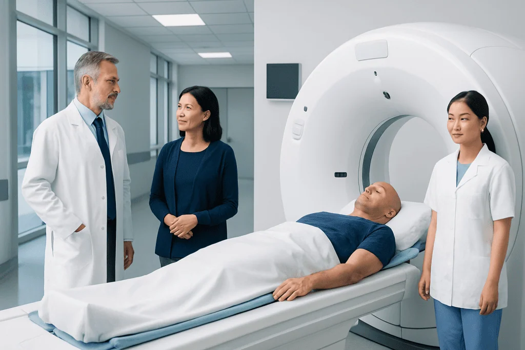
PET-CT uses a small amount of radioactive tracer to highlight tissues with increased metabolic activity. It may be recommended for higher-risk, advanced or recurrent melanoma, but is not needed for every person with early melanoma.
Key Takeaways
- PET-CT uses a small amount of radioactive tracer to highlight tissues with increased metabolic activity.
- It may be recommended for higher-risk, advanced or recurrent melanoma, but is not needed for every person with early melanoma.
- A PET scan can support cancer staging but cannot determine stage on its own.
- Preparation often includes fasting, avoiding strenuous exercise and following instructions about diabetes medicines.
- Most people return to usual activities after the scan, with short-term radiation-safety guidance from the imaging team.
- An abnormal scan result may need further imaging, follow-up or biopsy because inflammation and infection can also appear active on PET imaging.
A PET scan for melanoma cancer is an imaging test that can help doctors look for melanoma that may have spread beyond the original skin site. It is commonly combined with CT imaging and is used alongside examination, biopsy results and other tests to guide staging and treatment decisions.
PET Scan for Melanoma Cancer: An Overview
A PET scan for melanoma cancer is a nuclear medicine imaging test that helps doctors assess whether melanoma may be present in lymph nodes, organs or other areas of the body. It is usually performed as a PET-CT scan, which combines positron emission tomography (PET) with computed tomography (CT). PET shows areas using more glucose than expected, while CT provides detailed anatomical pictures to help locate those areas.
Melanoma cells can sometimes use glucose more actively than surrounding tissues. Before the scan, a small amount of a radiotracer, most often fluorodeoxyglucose (FDG), is injected into a vein. The scanner detects the tracer after it has circulated through the body. This information can help the clinical team evaluate known melanoma, investigate suspected recurrence and plan care.
A PET scan is only one part of melanoma assessment. The results are considered together with the original skin biopsy, physical examination, lymph node evaluation, blood tests when appropriate and other imaging. For information about the disease itself, readers may also find melanoma helpful.
Who May Need a PET Scan for Melanoma?
Not everyone diagnosed with melanoma needs a PET scan. For many people with a thin, early-stage melanoma that has been completely removed, routine whole-body PET imaging is unlikely to add useful information. Doctors recommend imaging when the expected benefit is greater, based on the individual features of the melanoma and the person’s symptoms.
A PET-CT may be considered when melanoma is thicker or has other higher-risk features, when cancer is found in a lymph node, when there are symptoms that could suggest spread, or when doctors need to assess possible recurrence after treatment. It may also be used to measure how melanoma responds to systemic treatment or to help plan surgery, radiotherapy or other care.
The choice of scan depends on the clinical question. Ultrasound may be particularly useful for certain lymph nodes, while CT or MRI may be preferred for specific body areas. A melanoma specialist can explain why PET-CT is, or is not, appropriate in a particular situation.
How Does a PET-CT Scan Work?
FDG is a tracer similar to glucose. After injection, it travels through the bloodstream and is taken up by tissues. Areas with higher metabolic activity may retain more tracer and appear as brighter areas on the PET images. The CT portion of the examination maps the body’s structures and helps the radiologist interpret the PET findings accurately.
Increased FDG uptake does not automatically mean cancer. Normal organs, healing tissue, infection, inflammation, recent surgery and some benign conditions can also show increased activity. Conversely, very small deposits of melanoma or lesions with low metabolic activity may not be visible. This is why scan findings are not interpreted in isolation.
The potential benefit of PET-CT is that it can survey much of the body in one examination and may identify disease that would alter management. It can also help avoid unnecessary procedures in some circumstances. However, its ability to detect microscopic disease is limited, and a normal scan does not replace recommended clinical follow-up.
What Should I Do to Prepare for a PET Scan for Cancer?
The imaging center will provide specific instructions, which should always take priority. In many cases, patients are asked not to eat for several hours before an FDG PET scan, although plain water is usually allowed. Fasting helps keep blood glucose and insulin levels more stable, which can improve image quality.
Strenuous exercise is often avoided for about 24 hours before the appointment because active muscles can take up more tracer and make interpretation harder. Patients should wear comfortable clothing, avoid metal items when possible and bring a list of medicines, allergies and recent test results. It is important to tell the team about pregnancy, possible pregnancy or breastfeeding before receiving the tracer.
People with diabetes need individualized instructions because blood glucose levels and diabetes medicines can affect the scan. They should not stop or alter prescribed medication without guidance from the imaging or diabetes team. Recent infections, vaccinations, injuries, surgery or cancer treatment should also be reported, as these can affect PET findings.
PET Scan Procedure: What Happens Step by Step?
On arrival, staff usually confirm medical information, check blood glucose when needed and place a small intravenous line. The radiotracer is injected through the vein. The injection itself is similar to a routine blood test or intravenous medication and is generally well tolerated.
After the injection, there is typically a quiet uptake period of around an hour. During this time, patients rest comfortably and are asked to limit movement, talking and reading where possible so that muscles do not take up unnecessary tracer. Drinking water may be encouraged to support hydration and later clearance of the tracer.
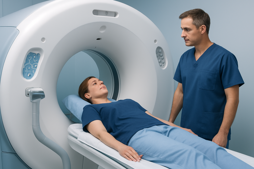
For the scan, the patient lies on a narrow table that moves slowly through the PET-CT scanner. It is important to remain as still as possible. The imaging portion often takes about 20 to 40 minutes, although the total appointment is commonly longer because of preparation and the uptake period. The scanner is open at both ends, and staff can communicate throughout the examination.
The scan does not usually cause pain. Some people find lying still uncomfortable or feel anxious in the imaging room; letting the team know in advance can help them provide practical support. If contrast is planned for the CT portion, the team will discuss possible sensations and check for relevant kidney problems or prior contrast reactions.
Recovery, Risks and Benefits After a PET Scan
Most people can return home and resume normal activities shortly after a PET scan. Drinking extra fluids, if medically appropriate, can help the body clear the tracer through urine. The amount of radiation used is carefully planned and generally leaves the body over a short period, but the department may advise temporary precautions around close, prolonged contact with pregnant people or young children.
Side effects from the FDG tracer are uncommon. The injection can cause brief discomfort, bruising or irritation at the vein. If CT contrast is used, some people experience a warm sensation or metallic taste for a short time; uncommon allergic or kidney-related risks are assessed beforehand. The radiation exposure is an important consideration, so scans are ordered when the likely clinical value outweighs the risk.
For melanoma, the main benefit is improved information for decision-making. If the scan suggests spread or recurrence, doctors may recommend targeted imaging, a repeat scan, blood tests or a biopsy to confirm the cause. Depending on the findings, care may include melanoma treatment such as surgery, immunotherapy, targeted therapy or radiotherapy.
Can You Tell What Stage Cancer Is From a PET Scan?
A PET scan can contribute important information to melanoma staging, especially by identifying suspected spread to distant lymph nodes, organs, bone or other tissues. However, it cannot establish the cancer stage by itself. Melanoma staging is based on several details, including the thickness and ulceration status of the original tumor, lymph node findings, biopsy results and whether there is confirmed distant spread.
When a PET scan identifies an unexpected area of activity, doctors assess its location, appearance and the person’s medical history. Additional imaging or a biopsy may be needed before that finding is considered melanoma. Staging should therefore be discussed with the multidisciplinary cancer team rather than inferred from a scan report alone.
Accurate staging helps guide follow-up and treatment choices. It also helps doctors discuss the likely goals of care and whether additional therapies may be appropriate after surgery or for melanoma that has spread.
How Long Does It Take to Get PET Scan Results for Cancer?
The exact timing varies by imaging center, scan complexity and whether prior images need to be compared. A radiologist or nuclear medicine physician reviews the PET and CT images carefully and sends a written report to the referring clinician. Results are often available within a few working days, but the treating team can explain the expected timeframe locally.
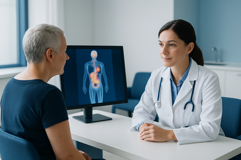
It is usually best to discuss results with the doctor who ordered the scan. They can put the findings in context, explain any uncertain areas and describe whether more tests are necessary. A report may use terms such as “uptake,” “metabolic activity” or “indeterminate,” which do not necessarily mean melanoma has spread.
Patients waiting for results may find it helpful to write down questions for their appointment. Useful questions include what the scan showed, whether any finding requires confirmation, whether the cancer stage has changed and how the result affects the recommended treatment plan.
Is It Serious if You Need a PET Scan?
Needing a PET scan does not automatically mean that melanoma has spread or that the situation is severe. Doctors may order the scan because a melanoma has certain features that warrant a more complete assessment, because symptoms need evaluation, or because it is time to monitor response after treatment.
Imaging is a tool for reducing uncertainty and making informed decisions. Many scans do not show evidence of spread, while some reveal findings unrelated to melanoma that need routine clarification. The reason for the scan is best explained by the clinician who knows the pathology results, examination findings and overall medical history.
Melanoma care may involve dermatology, surgical oncology, medical oncology, radiology, pathology and other specialties. Acibadem International’s multidisciplinary specialists and JCI-accredited hospitals diagnose and treat melanoma for international patients, with care plans based on individual clinical findings.
When to Seek Medical Care
Anyone with a new or changing mole, a dark spot that changes in size, shape or color, or a skin lesion that bleeds, crusts or does not heal should arrange a medical assessment. A clinician can examine the skin and decide whether referral to a dermatologist or biopsy is needed. Early assessment is especially important for people with a personal or family history of melanoma.
People previously treated for melanoma should contact their medical team if they notice a new lump, persistent swollen lymph node, unexplained ongoing pain, worsening breathlessness, persistent headaches, unexplained weight loss or a concerning new skin lesion. These symptoms often have causes other than melanoma, but they should be evaluated rather than self-diagnosed.
After a PET scan, urgent medical care is appropriate for signs of a severe allergic reaction, such as trouble breathing, swelling of the face or throat, or widespread hives, particularly if CT contrast was used. Such reactions are rare, and imaging teams are prepared to manage them.
Frequently asked questions
Does a PET scan detect all melanoma spread?
No. PET-CT can detect many areas of melanoma that are large enough and metabolically active enough to be visible, but it may miss microscopic disease or very small lesions. Doctors use it alongside examination, pathology and other imaging tests when appropriate.
Can inflammation cause an abnormal PET scan result?
Yes. Infection, inflammation, healing after surgery, injury and some recent vaccinations can increase FDG uptake and create an abnormal-looking area. Further assessment may be needed to determine whether a finding is related to melanoma or has another cause.
Do patients need to fast before a PET scan for melanoma cancer?
Many FDG PET scans require fasting for several hours beforehand, but the exact instructions vary by center and medical situation. Water is often permitted, while people with diabetes require personalized instructions about food and medication.
Is a PET scan painful?
The scan itself is not painful, though there is a brief needle insertion for the tracer injection. Patients need to lie still on the scanning table, which may be uncomfortable for some people but is usually manageable with support from the imaging team.
Can a PET scan replace a biopsy for melanoma?
No. A biopsy is needed to diagnose melanoma and may also be needed to confirm suspicious areas seen on imaging. PET scans provide whole-body information but cannot reliably distinguish every cancerous finding from benign inflammation or other conditions.
How soon can normal activities resume after a PET scan?
Most people return to normal daily activities immediately after the scan. The imaging team may advise drinking fluids and following short-term guidance about close contact with pregnant people and young children while the tracer leaves the body.
References
- National Cancer Institute
- American Cancer Society
- National Comprehensive Cancer Network
- Society of Nuclear Medicine and Molecular Imaging
- European Society for Medical Oncology
This article is for general information only and is not a substitute for professional medical advice. Please consult a qualified doctor about your individual situation.
Melanoma in Turkey — costs, top hospitals & a free quote
JCI-accredited · board-certified surgeons · reply within 24h
Add us as a Preferred Source to see more of our trusted health content across Google Search, AI Overviews and Discover.

Medical Oncology Department
Medical treatment of cancer with chemotherapy, immunotherapy and targeted therapies under a multidisciplinary tumor board.
60 specialists in this unitMore from the Health Library



Related Specialists

Prof. Dr. Levent Eralp
Orthopedic Surgery & Traumatology
Prof. Dr. Orhan Çınar
Emergency Service
Prof. Dr. Mehmet Cihat Ünlü
Gynecology & Obstetrics
Ody. Merve Mercan
Audiology