Red Moles — Explained by Medical Evidence, Not Myths

Most red moles are benign cherry angiomas made of small blood vessels. They often appear as bright red, pink, or purple round spots on the trunk, arms, or shoulders.
Key Takeaways
- Most red moles are benign cherry angiomas made of small blood vessels.
- They often appear as bright red, pink, or purple round spots on the trunk, arms, or shoulders.
- Red moles do not usually need treatment unless they bleed, get irritated, or cause cosmetic concern.
- A doctor may examine the spot closely or biopsy it if the diagnosis is unclear.
- Sudden changes in color, size, shape, or repeated bleeding should prompt medical review.
Red moles are most often harmless clusters of tiny blood vessels called cherry angiomas, not true moles in the usual pigment-cell sense. They are common with age, but any new, changing, bleeding, or uncertain skin spot should be assessed by a qualified doctor.
What are red moles?
Red moles are usually harmless skin growths known as cherry angiomas. Although people often call them “red moles,” they are not typical moles formed by pigment cells. Instead, they are small collections of blood vessels near the surface of the skin, which gives them their red, cherry-red, or sometimes purple appearance.
They can be flat or slightly raised and are often smooth, round, and clearly outlined. Many people notice just one or two at first, while others develop several over time. Red moles become more common with age and are frequently seen on the chest, abdomen, back, arms, or shoulders.
In most cases, red moles are a cosmetic issue rather than a health problem. However, not every red spot on the skin is a cherry angioma. Because different skin conditions can look similar, it is sensible to have a persistent, changing, or uncertain lesion checked by a clinician, especially if there is concern about skin cancer.
How red moles look and feel
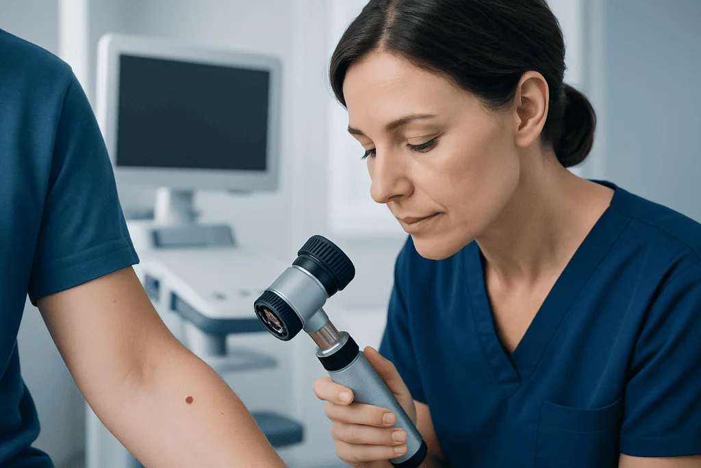
Cherry angiomas are typically small, ranging from pinpoint size to several millimeters across. Their color may vary from bright red to dark red or bluish-red, especially if they are older or have been irritated. Some remain flat, while others become dome-shaped over time.
They are usually painless and do not itch. Many are discovered by chance during bathing, dressing, or looking in the mirror. Because they contain blood vessels, they may bleed more easily than ordinary skin spots if scratched, shaved, or rubbed by clothing.
Red moles can sometimes resemble other skin lesions. For example, spider angiomas, petechiae, inflamed spots, or vascular birthmarks may look similar at first glance. A dermatologist may use a dermatoscope during a dermatology evaluation to look at the lesion in more detail and distinguish a benign angioma from other conditions.
- Usually round or oval
- Bright red, dark red, or purple
- Flat or slightly raised
- Common on the trunk and upper limbs
- May bleed if injured
What causes red moles and who gets them?

The exact cause of cherry angiomas is not fully understood. They seem to arise when very small blood vessels in the skin grow in a clustered way. For many people, they simply appear with age and do not reflect an underlying disease.
Genetics may play a role, since some families appear more prone to developing them. Hormonal changes, pregnancy, and certain environmental exposures have also been discussed in medical literature, but these factors do not explain every case. In most healthy adults, a few red moles are considered a normal skin finding.
They can appear at any age but are more common after the third decade of life. People with lighter or darker skin tones can both develop them. Importantly, ordinary red moles are not caused by poor hygiene and are not contagious.
Rarely, a sudden eruption of many vascular lesions may be associated with medication effects or other medical issues, but this is uncommon. If someone develops many new red spots quickly or has other symptoms, a clinician may evaluate whether another skin or internal condition needs to be ruled out, including disorders that can mimic angiomas such as hemangioma.
How doctors tell red moles from other skin changes
Diagnosis usually begins with a clinical skin examination. A doctor will ask when the spot first appeared, whether it has changed, and whether it bleeds, itches, or becomes painful. They will also look at the rest of the skin to see if there are similar lesions elsewhere.
In many cases, an experienced clinician can identify a cherry angioma by appearance alone. Dermoscopy, a close-up examination tool, can improve accuracy by showing the vascular pattern of the lesion. This is especially helpful when a red mole is dark, crusted, or unusually shaped.
If the diagnosis is uncertain, the doctor may recommend removing a small sample or the whole lesion for laboratory testing. A biopsy is particularly important if the lesion changes quickly, has irregular borders, develops a scab that does not heal, or looks different from the person’s other skin spots. In some cases, clinicians may use skin biopsy to confirm whether a lesion is a benign angioma or another condition.
Possible look-alikes include pyogenic granuloma, spider angioma, angiokeratoma, inflamed seborrheic keratosis, and some forms of skin cancer. This is why self-diagnosis based on photographs alone can be unreliable.
Do red moles need treatment?
Most red moles do not need treatment. If they are clearly benign and not bothersome, they can simply be observed. Many people choose no treatment at all once they understand that the lesion is harmless.
Treatment may be considered if a red mole bleeds often, is repeatedly irritated by shaving or clothing, or causes cosmetic concern. The choice depends on the lesion’s size, location, and appearance, as well as patient preference. A doctor can explain which method is most suitable and what kind of healing to expect.
Common options include laser therapy, electrocautery, cryotherapy, or minor shave removal. These methods aim to remove the visible lesion and usually involve little downtime, although any skin procedure can leave temporary redness or, less commonly, a small scar or pigment change. When appropriate, some patients may be offered laser treatment for selected vascular skin lesions.
Home removal should be avoided. Trying to cut, burn, or tie off a red mole can lead to bleeding, infection, scarring, and delayed diagnosis if the lesion was not actually a cherry angioma.
Skin self-checks, prevention, and everyday care
There is no proven way to prevent ordinary cherry angiomas. Because they are largely related to aging and individual susceptibility, they may appear even in people with excellent skin-care habits. Still, good skin awareness can help people notice changes early and seek advice when needed.
A practical approach is to check the skin regularly in good light, including the back, scalp, and areas that are easy to overlook. Looking for new lesions is useful, but paying attention to change is even more important. A spot that suddenly enlarges, darkens, forms a crust, or begins bleeding repeatedly should not be ignored.
General skin protection is also worthwhile. Sun protection does not specifically prevent cherry angiomas, but it supports overall skin health and may reduce confusion caused by multiple sun-related spots. A broad-spectrum sunscreen, protective clothing, and avoiding intense midday sun are sensible measures. If there is a family history of suspicious lesions, a clinician may recommend periodic skin cancer screening.
- Avoid picking or scratching red moles
- Take care when shaving around raised spots
- Use sunscreen as part of routine skin health
- Photograph unusual lesions to track change over time
- Ask a doctor before using over-the-counter removal products
When to seek medical care
Most red moles are harmless, but medical review is appropriate if a spot is new and unusual, changes quickly, or does not match the person’s other skin marks. Repeated bleeding without clear injury is another reason to arrange an assessment. This is especially true if the lesion grows rapidly or becomes tender.
Prompt evaluation is also important if a red lesion develops an irregular border, mixed colors, persistent crusting, or an open sore that does not heal. These features do not automatically mean cancer, but they deserve professional attention. People with a personal history of skin cancer, weakened immunity, or significant sun damage should be particularly cautious.
Anyone who is unsure what a skin lesion is should not rely only on internet images or home treatments. A qualified doctor can usually clarify whether the spot is a benign cherry angioma or another condition and recommend observation, removal, or further testing if needed.
For people seeking specialist assessment, Acibadem International’s multidisciplinary teams and JCI-accredited hospitals evaluate and treat skin lesions for international patients in a structured, evidence-based way.
Frequently asked questions
Are red moles dangerous?
Most red moles are not dangerous. They are usually cherry angiomas, which are benign growths of tiny blood vessels. A doctor should assess any lesion that changes, bleeds often, or looks unusual.
Are red moles the same as cherry angiomas?
In everyday language, many people use the term red moles to mean cherry angiomas. Medically, cherry angiomas are vascular lesions rather than true pigmented moles. The distinction matters because different skin lesions can require different evaluation.
Why do red moles appear with age?
Red moles often become more common as people get older, likely because of age-related changes in the skin’s small blood vessels. Genetics may also contribute. In most cases, they are a normal and benign finding.
Can a red mole turn into cancer?
A typical cherry angioma is benign and does not usually become cancer. The main issue is that some other skin lesions can resemble a red mole. If there is uncertainty or the spot changes, a doctor may examine it closely or biopsy it.
Should a bleeding red mole be removed?
A red mole that bleeds after repeated friction or shaving may be considered for removal, especially if it is troublesome. However, bleeding can also be a sign that the lesion should be checked to confirm the diagnosis. A clinician can advise whether observation or treatment is best.
Can red moles be removed for cosmetic reasons?
Yes, benign red moles can often be removed if they cause cosmetic concern or frequent irritation. Common methods include laser treatment, electrocautery, cryotherapy, or minor surgical removal. The right option depends on the lesion’s size, depth, and location.
This article is for general information only and is not a substitute for professional medical advice. Please consult a qualified doctor about your individual situation.
Explore treatments in Turkey — costs, top hospitals & a free quote
JCI-accredited · board-certified surgeons · reply within 24h
Add us as a Preferred Source to see more of our trusted health content across Google Search, AI Overviews and Discover.

More from the Health Library

Pores: An Evidence-Based Guide for Patients

Non Blanchable: A Complete Medical Overview

Elevated Liver Enzymes: An Evidence-Based Guide for Patients

Thoracotomy: A Complete Medical Overview

Fatal Mld: An Evidence-Based Guide for Patients