

Retinal Detachment: Flashes, Floaters, and Emergency Warning Signs

Retinal detachment is a medical emergency that usually needs prompt assessment by an ophthalmologist. Warning signs include sudden flashes of light, a burst of new floaters, blurred vision, or a dark curtain or shadow.
Key Takeaways
- Retinal detachment is a medical emergency that usually needs prompt assessment by an ophthalmologist.
- Warning signs include sudden flashes of light, a burst of new floaters, blurred vision, or a dark curtain or shadow.
- Risk is higher in people with severe nearsightedness, previous eye surgery, eye injury, diabetes-related retinal disease, or a past retinal detachment in the other eye.
- Diagnosis is usually made with a dilated eye examination and retinal imaging when needed.
- Treatment may include laser or freezing for a tear, or surgery such as pneumatic retinopexy, scleral buckle, or vitrectomy for detachment.
Retinal detachment is a serious eye condition in which the light-sensitive retina separates from its normal position. Sudden flashes, new floaters, or a curtain-like shadow in vision should be checked urgently because early treatment can help protect sight.
Overview
Retinal detachment occurs when the retina, the thin layer of nerve tissue lining the back of the eye, pulls away from the tissue that normally supports it. The retina receives light and sends visual signals to the brain, so it needs to remain attached and healthy to function well. When it separates, retinal cells may not receive enough oxygen and nutrients, and vision can be affected.
The condition is usually painless. This is why visual symptoms such as flashes, floaters, or a shadow in the field of vision are especially important. A person may feel otherwise well but still have an urgent eye problem. Prompt evaluation can identify whether symptoms are due to a common age-related change, a retinal tear, or a retinal detachment.
Not every floater or flash means the retina has detached. Many people develop floaters from changes in the vitreous, the clear gel inside the eye. However, because a retinal tear can lead to detachment, sudden or worsening symptoms should not be ignored. Same-day or urgent ophthalmology assessment is the safest approach when warning signs appear.
Symptoms and Emergency Warning Signs
Retinal detachment symptoms often begin suddenly and may affect one eye. The person may notice brief flashes of light, especially in side vision, or a shower of new floaters that look like dots, cobwebs, threads, or specks. Vision may become blurry, wavy, or less clear than usual.
A classic warning sign is a dark shadow, veil, or curtain moving across part of the visual field. This can start at the side, top, or bottom of vision and may gradually spread. Some people describe missing areas of vision rather than darkness. If the central retina, called the macula, becomes involved, central vision may become distorted or significantly reduced.
Symptoms that need urgent eye care include:
- Sudden flashes of light, especially with new floaters
- A rapid increase in floaters or a “shower” of spots
- A curtain, veil, or shadow over vision
- Sudden loss of side vision or central vision
- New blurred vision after an eye injury or eye surgery
Retinal detachment is usually not painful, and redness may be absent. Waiting for symptoms to improve on their own can delay treatment. Anyone with these signs should contact an eye specialist or emergency service promptly, particularly if symptoms are new, increasing, or limited to one eye.
Causes and Risk Factors

The most common type is rhegmatogenous retinal detachment. It begins with a break or tear in the retina. Fluid from the vitreous can pass through the tear and collect under the retina, lifting it away from the back wall of the eye. Retinal tears often develop when the vitreous gel shrinks and pulls on the retina, a process that becomes more common with age.
Other types are less common but important. Tractional retinal detachment occurs when scar tissue pulls the retina away from its normal position, often in advanced diabetic eye disease or other conditions that cause retinal scarring. Exudative retinal detachment happens when fluid builds under the retina without a tear, sometimes due to inflammation, vascular disease, tumors, or other eye disorders.
Risk factors include severe nearsightedness, previous cataract or other eye surgery, a history of retinal tear or detachment, family history, eye trauma, and certain inherited retinal conditions. People with diabetes, especially if diabetic retinopathy is present, may also be at increased risk of traction-related retinal problems. A detachment in one eye increases the need for careful monitoring of the other eye.
Age-related posterior vitreous detachment is a frequent trigger for flashes and floaters. In many cases it is not dangerous, but it can sometimes create a retinal tear. This is why an eye examination is needed to distinguish a harmless vitreous change from a sight-threatening retinal problem.
Diagnosis
Diagnosis begins with a detailed symptom history and a comprehensive eye examination by an ophthalmologist. The doctor will ask when symptoms started, whether they are changing, and whether there has been eye injury, previous surgery, high myopia, diabetes, or a previous retinal problem. Visual acuity and eye pressure may be checked as part of the assessment.
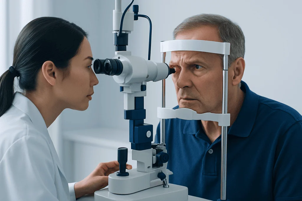
A dilated retinal examination is central to diagnosis. Eye drops are used to widen the pupil so the doctor can examine the retina, including the far peripheral areas where tears often occur. Special lenses and a bright light allow the ophthalmologist to look for retinal holes, tears, bleeding, traction, or areas where the retina has lifted.
If the view of the retina is blocked by bleeding, cataract, or other opacity, ultrasound imaging of the eye may be used. Optical coherence tomography may help evaluate the macula and fluid under or within the retina. Wide-field retinal photography can document findings and support follow-up, but it does not replace a careful dilated examination when detachment is suspected.
Because timing matters, people with warning symptoms should not wait for a routine eye appointment weeks later. Urgent assessment helps determine whether observation, laser treatment, or surgery is needed. The eye doctor will also explain whether activity should be limited until treatment is completed.
Treatment Options
Treatment depends on whether there is a retinal tear without detachment, a small localized detachment, or a more extensive detachment. If a retinal tear is found early, the doctor may use laser photocoagulation or cryopexy, a freezing treatment, to create a scar around the tear. This helps seal the retina and reduce the chance of fluid passing underneath it.
When the retina has detached, surgery is usually required. Pneumatic retinopexy involves placing a gas bubble inside the eye to press the retina back into position, often combined with laser or freezing treatment. The patient may need to keep the head in a specific position for a period of time so the bubble supports the correct area.
Scleral buckle surgery places a soft band or sponge around the outside of the eye to reduce pulling on the retina and support reattachment. Vitrectomy removes the vitreous gel that is pulling on the retina and replaces it with gas, air, or silicone oil to help hold the retina in place while healing occurs. Sometimes more than one technique is used, depending on the location and complexity of the detachment.
Recovery varies according to the type of detachment, whether the macula was involved, and the treatment performed. Vision often improves gradually, but the final result can take time. Follow-up visits are essential to monitor healing, eye pressure, and any need for additional treatment. Patients should follow their surgeon’s instructions about positioning, eye drops, activity, and air travel restrictions if a gas bubble is used.
Prevention and Self-Care
Not all retinal detachments can be prevented, but early detection of retinal tears can reduce risk. People with high myopia, previous retinal detachment, significant eye trauma, or known peripheral retinal changes may need periodic dilated retinal examinations. Those with diabetes should keep regular eye screening appointments and manage blood sugar, blood pressure, and cholesterol with their healthcare team.
Protective eyewear can lower the risk of eye injuries during sports, home repairs, and work that involves flying particles or impact. After cataract surgery or other eye procedures, patients should attend follow-up appointments and report new floaters, flashes, or vision changes promptly. A person who has had detachment in one eye should ask the ophthalmologist how often the other eye should be examined.
Self-care does not treat a detached retina, and there are no eye exercises, supplements, or home remedies that can reattach it. The most important step is recognizing warning symptoms and seeking timely medical care. If vision is suddenly affected, the person should avoid driving and arrange safe transport to an eye emergency service or specialist clinic.
General eye health habits still matter. Regular eye exams, good control of chronic diseases, avoiding smoking, and using appropriate eye protection can support long-term vision health. However, sudden flashes, new floaters, or a curtain-like shadow should always be treated as a reason for urgent evaluation.
When to See a Doctor
A person should seek urgent eye care the same day if they develop sudden flashes, a sudden increase in floaters, a dark curtain or shadow, or sudden loss of any part of vision. These symptoms are especially important if they occur after an eye injury, after eye surgery, or in someone with high myopia, diabetes-related eye disease, or a previous retinal tear.
It is also important to be checked if floaters are accompanied by blurred vision, bleeding-like spots, or reduced side vision. Even if symptoms seem mild, a retinal tear can be present before a full detachment develops. Early treatment of a tear is often simpler than treatment after a larger detachment has occurred.
At Acibadem International, multidisciplinary eye specialists and JCI-accredited hospitals diagnose and treat retinal conditions for international patients, including urgent retinal assessment and surgical care when appropriate. Patients should follow the advice of a qualified ophthalmologist, who can recommend the safest plan based on examination findings and individual risk factors.
Frequently asked questions
Is retinal detachment painful?
Retinal detachment is usually painless. This can make it easy to underestimate, because the main warning signs are visual changes such as flashes, floaters, shadows, or blurred vision. Any sudden change in vision should be assessed urgently by an eye specialist.
Are floaters always a sign of retinal detachment?
No. Floaters are common and often come from age-related changes in the vitreous gel inside the eye. However, a sudden shower of new floaters, especially with flashes or a shadow in vision, can signal a retinal tear or detachment and needs prompt examination.
How quickly should someone seek care for flashes and floaters?
New flashes, a sudden increase in floaters, or any curtain-like shadow should be checked urgently, ideally the same day. If vision is decreasing or part of the visual field is missing, emergency eye care is recommended. Timely diagnosis helps determine whether treatment is needed to protect vision.
Can a detached retina heal without surgery?
A true retinal detachment usually does not heal on its own and often requires a procedure or surgery. Small retinal tears may be treated with laser or freezing before a larger detachment develops. The correct treatment depends on the examination findings and should be guided by an ophthalmologist.
What is the difference between a retinal tear and retinal detachment?
A retinal tear is a break in the retina, often caused by pulling from the vitreous gel. Retinal detachment occurs when fluid passes under the retina and lifts it away from its supporting tissue. Treating a tear early can sometimes prevent progression to detachment.
What happens after retinal detachment surgery?
After surgery, patients usually need close follow-up and may use prescribed eye drops. Some procedures require specific head positioning, and gas bubbles in the eye can restrict flying or certain anesthesia until the bubble resolves. Recovery and visual improvement vary, so the surgeon’s instructions are essential.
References
- American Academy of Ophthalmology
- National Eye Institute
- Mayo Clinic
- Royal National Institute of Blind People
- Merck Manual Professional Edition
This article is for general information only and is not a substitute for professional medical advice. Please consult a qualified doctor about your individual situation.
Explore treatments in Turkey — costs, top hospitals & a free quote
JCI-accredited · board-certified surgeons · reply within 24h
Add us as a Preferred Source to see more of our trusted health content across Google Search, AI Overviews and Discover.








