

Thoracoscopy: Candidacy, Procedure Steps, and Recovery Timeline

Thoracoscopy uses a thin camera inserted through a small incision to examine the pleura, lungs, and other structures in the chest. Medical thoracoscopy is commonly used for diagnosis and pleural procedures, while video-assisted thoracoscopic surgery (VATS) can also treat lung and chest conditions.
Key Takeaways
- Thoracoscopy uses a thin camera inserted through a small incision to examine the pleura, lungs, and other structures in the chest.
- Medical thoracoscopy is commonly used for diagnosis and pleural procedures, while video-assisted thoracoscopic surgery (VATS) can also treat lung and chest conditions.
- Candidates are assessed individually based on the reason for the procedure, imaging results, lung function, overall health, and ability to tolerate anesthesia or sedation.
- Many people are up and walking on the day of or day after the procedure, but full recovery can take days to several weeks depending on the extent of treatment.
- Possible risks include pain, bleeding, infection, air leakage from the lung, and anesthesia-related complications, although serious complications are uncommon.
- New or worsening breathlessness, chest pain, fever, or concerning wound changes after thoracoscopy should be assessed promptly.
Thoracoscopy is a minimally invasive procedure that allows a specialist to view the space around the lungs and, when needed, perform biopsies or treatment through small chest incisions. It may be used to investigate unexplained fluid around the lung, evaluate abnormal tissue, or treat certain chest conditions; the procedure and recovery time depend on whether it is performed medically or as surgery.
Thoracoscopy: an overview
Thoracoscopy is a procedure that lets a chest specialist look inside the thorax, or chest cavity, using a narrow tube with a camera called a thoracoscope. The camera is passed through one or more small incisions between the ribs. It provides detailed views of the pleura, which is the thin lining around the lungs and inside the chest wall, as well as selected areas of the lung and diaphragm.
The term may describe two related approaches. Medical thoracoscopy, also called pleuroscopy, is generally performed by a respiratory physician to examine pleural disease, obtain tissue samples, drain fluid, or carry out pleurodesis. Video-assisted thoracoscopic surgery (VATS) is performed by a thoracic surgeon under general anesthesia and can be used for more extensive diagnostic or surgical treatment.
Thoracoscopy is less invasive than an open thoracotomy, which requires a larger incision and spreading of the ribs. However, it is still an important chest procedure and requires careful planning. The exact purpose, type of anesthesia, expected hospital stay, and recovery plan should be discussed with the treating team before the procedure.
Why thoracoscopy may be recommended
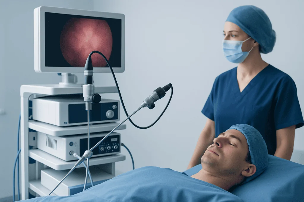
A doctor may recommend thoracoscopy when chest imaging, fluid analysis, or symptoms do not provide a clear diagnosis. One of its most common uses is evaluating a pleural effusion, meaning an abnormal collection of fluid around a lung. During the procedure, the doctor can inspect the pleural surfaces directly and collect targeted biopsies, which may be more informative than fluid testing alone.
Thoracoscopy can help investigate pleural thickening, nodules, persistent air leaks, recurrent collapsed lung, or abnormalities seen on a chest X-ray or CT scan. It may be used when doctors need to distinguish between infection, inflammation, benign disease, and cancer. Tissue obtained during thoracoscopy is examined by a pathology laboratory and can guide the next steps in care.
Depending on the approach, thoracoscopy may also provide treatment. Examples include draining fluid, breaking down selected adhesions, placing a chest drain, treating recurrent pneumothorax, removing a small lung lesion, or performing pleurodesis. Pleurodesis creates controlled irritation between the pleural layers so they adhere, helping reduce recurrent fluid buildup or repeated lung collapse in appropriate patients.
Thoracoscopy does not automatically mean cancer is present. It is a diagnostic and treatment tool used for a wide range of chest conditions. The doctor will explain what is being investigated and what information the procedure may or may not provide.
Who may be a candidate for thoracoscopy
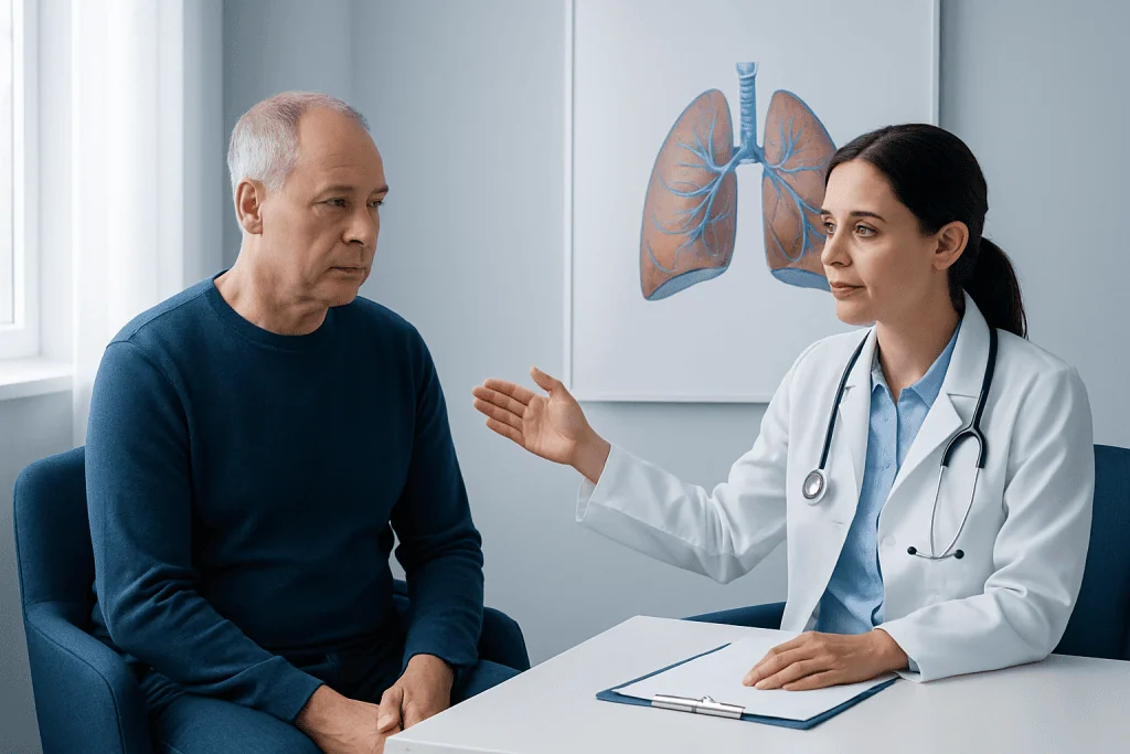
Suitability for thoracoscopy is individualized. A person may be considered when there is a clear clinical question that cannot be answered adequately by less invasive tests, such as imaging, blood tests, sputum testing, or needle biopsy. The expected benefit of obtaining a diagnosis or providing treatment is weighed against the possible risks of a chest procedure.
Before recommending thoracoscopy, the medical team commonly reviews CT imaging and other test results, current symptoms, medical history, medications, allergies, and prior chest surgery. They may also assess oxygen levels, heart and lung function, and blood clotting. These checks help determine whether local anesthesia with sedation or general anesthesia is more suitable.
Some people need additional preparation or may not be suitable for a particular thoracoscopic approach. Factors that can affect candidacy include severe breathing impairment, unstable heart disease, uncontrolled bleeding risk, certain extensive pleural adhesions, active infection at the incision site, or inability to safely receive anesthesia. These factors do not always rule out care; they may mean another diagnostic method or a modified plan is safer.
People taking anticoagulants, antiplatelet medicines, diabetes medications, or supplements should tell their care team well in advance. The team will provide personalized instructions about fasting and whether any medicines should be paused or adjusted. Medicines should not be stopped independently.
What happens during the procedure
Thoracoscopy begins with preparation in a procedure room or operating theatre. The patient’s identity, planned procedure, imaging, and consent are confirmed. Monitoring equipment tracks breathing, oxygen level, blood pressure, and heart rhythm. Medical thoracoscopy may use local anesthetic around the incision together with sedation, while VATS usually requires general anesthesia and a breathing tube.
The specialist makes a small incision, usually on the side of the chest being examined. A thoracoscope is introduced between the ribs, allowing the team to inspect the pleural cavity on a monitor. If fluid is present, it may be drained and sent for laboratory analysis. The doctor can take biopsies from areas that appear inflamed, thickened, or otherwise unusual.
If treatment is planned, additional small incisions may be made for surgical instruments. During VATS, the surgeon may repair an air leak, remove a wedge of lung tissue, remove part of a lung when clinically indicated, treat a pneumothorax, or complete another planned chest procedure. The precise steps depend on the diagnosis and surgical goal.
At the end, a chest tube may be placed temporarily to remove air or fluid and help the lung stay expanded. The incisions are closed with stitches, adhesive strips, or surgical glue and covered with dressings. The procedure length varies considerably, from a relatively short diagnostic examination to several hours for more complex surgery.
Recovery timeline and aftercare
Recovery begins in a monitored area as sedation or anesthesia wears off. It is common to have soreness around the incision, temporary fatigue, a mild sore throat after general anesthesia, or discomfort when taking deep breaths. Pain relief is important because comfortable breathing, coughing, and early movement can help reduce the risk of chest complications.
A chest X-ray is often performed after thoracoscopy to confirm that the lung is expanded and the chest tube, if present, is positioned appropriately. Some patients having uncomplicated medical thoracoscopy may go home the same day or after an overnight stay. After VATS or a more extensive procedure, hospitalization may last several days, especially if a chest drain needs to remain in place.
Many people can stand and walk with assistance on the day of surgery or the following day. Gentle activity is generally encouraged, while heavy lifting, strenuous exercise, swimming, and driving should wait until the treating team says they are safe. The timing varies with the procedure, pain medicines used, wound healing, and a person’s overall condition.
After a simple diagnostic thoracoscopy, some people feel ready for usual light activities within several days to two weeks. Recovery after VATS may take a few weeks, and recovery after more extensive lung surgery can take longer. Follow-up visits review wound healing, pathology results, chest imaging when needed, and the next stage of treatment. Acibadem International’s multidisciplinary specialists and JCI-accredited hospitals can assess and treat thoracic conditions for international patients.
Benefits and possible risks
The main benefit of thoracoscopy is that it combines direct visualization of the chest with the ability to take targeted samples or provide selected treatment through small incisions. Compared with open chest surgery, minimally invasive techniques may involve less tissue disruption, smaller scars, less postoperative pain for some patients, and a shorter recovery. Results can clarify a diagnosis and help the care team recommend appropriate treatment.
All procedures carry risks. Common short-term effects include incision pain, bruising, tiredness, and discomfort from a chest tube. Other potential complications include bleeding, infection, a persistent air leak, fluid buildup, lung collapse, irregular heart rhythm, blood clots, or reactions to sedatives or anesthesia. Rarely, an injury to nearby structures or a need to convert VATS to open surgery may occur if this is necessary for safety or treatment.
The likelihood of complications depends on the procedure’s purpose and complexity, existing lung and heart disease, smoking status, medications, and overall health. The specialist will discuss the risks that are most relevant to the individual situation and explain how they are reduced, including careful imaging review, infection-prevention measures, pain management, breathing exercises, and early mobility.
Thoracoscopy may not always produce a definitive diagnosis. For example, a biopsy can occasionally be inconclusive, or the findings may indicate that another test is needed. This does not mean the procedure was unsuccessful; direct findings and sample results can still provide valuable information for further decision-making.
When to seek medical care
Before a planned thoracoscopy, medical advice is important if a person develops a new respiratory infection, fever, worsening cough, chest pain, significant breathlessness, or any change in health that could affect anesthesia. The procedural team should also know about recent hospital visits, new medicines, pregnancy, or changes in anticoagulant use.
After discharge, patients should follow their written aftercare instructions and contact their medical team for increasing wound redness, warmth, swelling, drainage, persistent vomiting, pain that is not controlled by the prescribed plan, or a fever. A small amount of tenderness and tiredness can be expected, but symptoms should gradually improve rather than worsen.
Urgent medical assessment is needed for sudden or worsening shortness of breath, severe or new chest pain, coughing up a substantial amount of blood, fainting, confusion, blue or gray lips, or signs of a severe allergic reaction. These symptoms can have several causes and should not be managed at home.
People with ongoing symptoms that led to thoracoscopy, such as recurrent fluid around the lung, persistent cough, unexplained weight loss, or repeated pneumothorax, should keep all follow-up appointments. Timely review of pathology and imaging results helps ensure that any required care is planned without unnecessary delay.
Frequently asked questions
Is thoracoscopy the same as VATS?
Thoracoscopy is a broad term for looking inside the chest with a camera. Medical thoracoscopy is often used for pleural diagnosis and treatment under local anesthesia and sedation, while VATS is a surgical form performed under general anesthesia. VATS can allow more extensive procedures through small incisions.
How painful is thoracoscopy?
Pain levels vary according to the type of thoracoscopy and whether a chest tube is needed. Most people have soreness around the incisions and may feel discomfort with deep breathing or coughing for a short time. The care team provides a pain-control plan to support movement and comfortable breathing.
How long does a thoracoscopy take?
A straightforward diagnostic thoracoscopy may take less time than a surgical VATS procedure, but the total time also includes anesthesia preparation and recovery monitoring. More complex procedures can take several hours. The surgeon or respiratory specialist can provide the most accurate estimate for the planned procedure.
Will a chest tube be needed after thoracoscopy?
A chest tube is commonly used after VATS and may also be used after medical thoracoscopy. It removes air and fluid from the chest and helps the lung remain expanded. It is removed when the medical team confirms that drainage and lung expansion are satisfactory.
When can a person return to work after thoracoscopy?
Return-to-work timing depends on the procedure, occupation, recovery progress, and whether the work involves lifting or physical exertion. Some people return to desk-based work within days to a couple of weeks after a simple procedure, while recovery after surgery may take longer. The treating clinician should clear the return to work and activity.
What do thoracoscopy biopsy results show?
A biopsy can show whether pleural or lung tissue has signs of infection, inflammation, scarring, benign disease, or cancer. Results are interpreted together with symptoms, imaging, fluid tests, and medical history. Timing varies because laboratory processing and specialized testing may be needed.
References
- American Thoracic Society
- European Respiratory Society
- National Heart, Lung, and Blood Institute
- Society of Thoracic Surgeons
- National Cancer Institute
This article is for general information only and is not a substitute for professional medical advice. Please consult a qualified doctor about your individual situation.
Explore treatments in Turkey — costs, top hospitals & a free quote
JCI-accredited · board-certified surgeons · reply within 24h
Add us as a Preferred Source to see more of our trusted health content across Google Search, AI Overviews and Discover.

More from the Health Library



Related Specialists
Dr. Yunus Çelik
Anesthesiology
Prof. Dr. Emel Güngör
Dermatology
Prof. Dr. Ayşe Korkmaz Toygar
Neonatal Intensive Care Unit
Prof. Dr. Ayşe Nurdan Tözün
Internal Medicine