Transthoracic Echocardiogram: What Patients Need to Know

A transthoracic echocardiogram uses sound waves to create real-time images of the heart from outside the chest. It can help assess heart size, pumping strength, valve function, and patterns of blood flow.
Key Takeaways
- A transthoracic echocardiogram uses sound waves to create real-time images of the heart from outside the chest.
- It can help assess heart size, pumping strength, valve function, and patterns of blood flow.
- The test is usually safe, painless, and does not involve radiation.
- Preparation is typically minimal, and most people can return to normal activities right away.
- Results are interpreted alongside symptoms, physical examination, and sometimes other heart tests.
A transthoracic echocardiogram is a painless, noninvasive ultrasound of the heart performed through the chest. It helps doctors see how the heart muscle, valves, chambers, and blood flow are working and is often one of the first tests used to evaluate heart symptoms.
Overview: what a transthoracic echocardiogram shows
A transthoracic echocardiogram is a standard ultrasound test of the heart. A trained healthcare professional places a small handheld device called a transducer on the chest, and sound waves are used to create moving pictures of the heart. These images help doctors evaluate how the heart looks and how well it works.
This test is often called a “heart ultrasound” or “transthoracic echo.” “Transthoracic” means the images are taken through the chest wall. Unlike more invasive heart tests, it does not require instruments to be placed inside the body, and it does not use ionizing radiation.
A transthoracic echocardiogram can show the heart’s chambers, valves, major blood vessels near the heart, and the motion of the heart muscle as it pumps. It can also estimate how blood flows through the heart. For this reason, it is commonly used when a person has symptoms such as shortness of breath, chest discomfort, palpitations, fainting, leg swelling, or a newly detected heart murmur.
Why doctors order this test
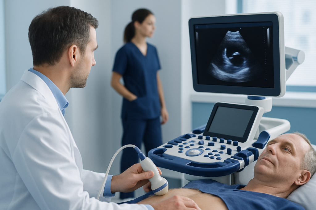
Doctors use a transthoracic echocardiogram to answer specific questions about heart structure and function. It can help explain symptoms, follow a known heart condition, or provide more detail after an abnormal physical exam, electrocardiogram, or chest imaging result.
The test is especially useful for looking at the heart valves, the thickness and movement of the heart muscle, and the size of the chambers. It can help identify changes linked to heart failure, problems with the heart valves, fluid around the heart, and signs of pressure changes in the lungs or right side of the heart.
Common reasons for ordering a transthoracic echocardiogram include:
- Evaluation of a heart murmur
- Assessment of shortness of breath or swelling
- Checking heart pumping function after a heart event or during treatment
- Investigating suspected valve disease, cardiomyopathy, or congenital heart abnormalities
- Monitoring known conditions over time
Because it is noninvasive and widely available, it is often the first imaging test used before other options such as transesophageal echo, cardiac CT, or cardiac MRI are considered.
What happens before, during, and after the exam
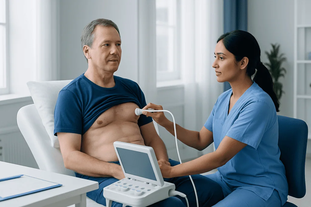
Preparation for a transthoracic echocardiogram is usually simple. Most people do not need to fast or stop normal daily activities unless their doctor gives specific instructions. It is often helpful to wear a two-piece outfit, since clothing from the waist up may need to be removed and a gown provided.
During the exam, the person lies on an examination table, often slightly turned onto the left side. A technician or doctor applies gel to the chest to help the transducer make good contact with the skin. The transducer is moved to several positions on the chest to obtain views from different angles. At times, the patient may be asked to hold their breath briefly or change position.
The test is usually painless, though mild pressure from the transducer can sometimes feel uncomfortable over the ribs. A transthoracic echocardiogram commonly takes about 30 to 60 minutes, depending on the reason for the study and how easily images can be obtained.
After the exam, the gel is wiped off and normal activities can generally be resumed right away. A cardiologist or another qualified physician reviews the images and prepares a report, which is then discussed with the patient in the context of their symptoms and overall medical history.
What the images can reveal
A transthoracic echocardiogram provides more than a single picture. It shows the heart beating in real time, allowing doctors to observe the opening and closing of the valves and the contraction of the heart muscle. It can also estimate ejection fraction, a measurement often used to describe how strongly the left ventricle pumps blood.
Doppler ultrasound, which is commonly included in the exam, helps evaluate the direction and speed of blood flow. This can be important when looking for narrowing or leakage of the valves, abnormal pressure gradients, or unusual communication between chambers. In this way, a transthoracic echocardiogram often plays a central role in assessing heart valve diseases.
The test may help detect or support evaluation of:
- Enlarged heart chambers
- Weak or stiff heart muscle
- Valve narrowing or valve leakage
- Fluid around the heart
- Blood clots or masses in selected cases
- Congenital structural differences
- Evidence suggesting <a href="https://acibademinternational.com/diseases/pulmonary-hypertension/”>pulmonary hypertension or strain on the right heart
In some situations, image quality may be limited by body shape, chest wall structure, lung disease, or other factors. If the images do not answer the clinical question, a doctor may recommend further testing, such as a stress echo or cardiac MRI.
Benefits, safety, and limitations
One of the main advantages of a transthoracic echocardiogram is that it provides important information without surgery, needles into the heart, or radiation. It is considered very safe for most people, including many who need repeat studies over time. This makes it useful both for diagnosis and for monitoring changes during follow-up care.
The test can be done in outpatient clinics, hospitals, and emergency settings. It gives rapid information and is often well tolerated by children, adults, and older patients. In many cases, it helps guide whether medications, closer monitoring, or additional imaging are needed.
Still, it has limits. Ultrasound images depend on how clearly sound waves can pass through the chest. Some areas of the heart may be difficult to visualize, and small abnormalities can occasionally be missed. Also, while a transthoracic echocardiogram can strongly suggest a diagnosis, it does not replace clinical judgment or every other form of heart testing.
If doctors need a closer look at certain structures, they may recommend a transesophageal echocardiogram, which places the ultrasound probe in the esophagus. If exercise-related symptoms are the concern, a stress echocardiography study may offer additional information about how the heart performs under exertion.
How results are interpreted and what comes next
Results from a transthoracic echocardiogram are interpreted by looking at multiple measurements together rather than relying on one number alone. The report may include information about chamber size, wall motion, valve structure, ejection fraction, blood flow patterns, and estimated pressures. A finding described as “normal” generally means the heart’s major structures and pumping function appear within expected ranges for that person.
Abnormal results do not always mean a serious problem, and the meaning depends on the full clinical picture. Mild valve leakage, for example, may be common and may only require observation, while other findings may need treatment or closer follow-up. Doctors use the test alongside symptoms, examination, blood tests, and sometimes additional studies such as electrocardiography, Holter monitoring, or cardiology evaluation.
Possible next steps vary. Some people simply need reassurance or repeat imaging after a period of time. Others may be advised to start medication, manage risk factors such as blood pressure, or see a specialist for more detailed assessment. The goal is not only to name a condition, but also to understand how the heart is functioning in everyday life.
For international patients who need coordinated assessment, Acibadem International’s multidisciplinary specialists and JCI-accredited hospitals diagnose and treat heart conditions using evidence-based imaging and follow-up care plans.
When to seek medical care
A transthoracic echocardiogram is usually arranged because a clinician wants more information about a possible heart issue, but symptoms should still guide when to seek care. Prompt medical attention is important if a person has chest pain, severe shortness of breath, fainting, sudden weakness, new confusion, or a rapid or irregular heartbeat that does not settle. These symptoms need urgent evaluation rather than waiting for a scheduled imaging test.
Non-urgent medical review is also appropriate when symptoms develop gradually, such as reduced exercise tolerance, ankle swelling, unexplained fatigue, or repeated dizziness. A doctor can decide whether a transthoracic echocardiogram is the right test and whether other causes outside the heart should also be considered.
People with known heart disease should ask for review if symptoms change or treatment no longer seems to be working as expected. Early assessment can help clarify whether the condition is stable or whether management needs to be adjusted.
Frequently asked questions
What is the difference between a transthoracic echocardiogram and other types of echocardiograms?
A transthoracic echocardiogram is performed through the chest wall using an external ultrasound probe. Other types include transesophageal echocardiography, which uses a probe in the esophagus for closer views, and stress echocardiography, which examines heart function during exercise or medication-induced stress.
Does a transthoracic echocardiogram hurt?
The test is usually painless. Some people notice mild pressure from the probe on the chest, but it should not cause significant discomfort and does not involve incisions or radiation.
How should a patient prepare for a transthoracic echocardiogram?
Most people do not need special preparation. They can usually eat, drink, and take regular medications as normal unless their doctor gives different instructions for a specific reason.
How long does a transthoracic echocardiogram take?
Many exams take around 30 to 60 minutes. The exact length depends on why the test is being done and how many image views are needed to answer the clinical question clearly.
Can a transthoracic echocardiogram detect heart failure?
It can provide important information that helps diagnose and monitor heart failure. The test can show how well the heart pumps, whether the chambers are enlarged, and whether valve problems or other structural issues may be contributing.
Are there any risks after the test?
A standard transthoracic echocardiogram is considered very safe, and most people can return to normal activities immediately. Because it is noninvasive and does not use radiation, there are no usual recovery restrictions after the exam.
References
- American Heart Association
- American Society of Echocardiography
- National Heart, Lung, and Blood Institute
- European Society of Cardiology
- MedlinePlus
This article is for general information only and is not a substitute for professional medical advice. Please consult a qualified doctor about your individual situation.
Explore treatments in Turkey — costs, top hospitals & a free quote
JCI-accredited · board-certified surgeons · reply within 24h
Add us as a Preferred Source to see more of our trusted health content across Google Search, AI Overviews and Discover.

More from the Health Library

Stages of Passing a Kidney Stone — Explained by Medical Evidence, Not Myths

BMI Women: What Patients Need to Know

Sulfites — Explained by Medical Evidence, Not Myths

Pelvic Phleboliths: A Complete Medical Overview

Stretching of Trapezius: What Patients Need to Know