Treatment for Acoustic Neuroma: How It Works, Results and What to Expect

Acoustic neuroma, also called vestibular schwannoma, is usually a noncancerous and slow-growing tumor. Not every acoustic neuroma needs immediate treatment; observation can be appropriate for selected small or stable tumors.
Key Takeaways
- Acoustic neuroma, also called vestibular schwannoma, is usually a noncancerous and slow-growing tumor.
- Not every acoustic neuroma needs immediate treatment; observation can be appropriate for selected small or stable tumors.
- Stereotactic radiosurgery aims to stop or slow growth without an open operation, while microsurgery removes the tumor.
- Hearing preservation cannot be guaranteed with any approach, but early specialist assessment helps guide planning.
- Care commonly involves ear, skull-base surgery, neurosurgery, radiation oncology, audiology and rehabilitation specialists.
Treatment for acoustic neuroma is individualized and may involve careful MRI monitoring, focused radiation treatment or microsurgery. The best approach depends on tumor growth, hearing and balance symptoms, pressure on nearby brain structures, age, general health and personal priorities.
Overview: how treatment for acoustic neuroma is chosen
Treatment for acoustic neuroma may involve active monitoring, stereotactic radiosurgery or microsurgical removal. These options are selected after a detailed assessment of the tumor and the person’s symptoms, with the goal of protecting neurological function, managing tumor growth and supporting quality of life.
An acoustic neuroma is more accurately called a vestibular schwannoma. It develops from Schwann cells around the vestibular nerve, which carries balance signals from the inner ear to the brain. Although it is usually benign, it can affect hearing, balance, facial nerve function and, if it becomes large, structures near the brainstem.
There is no single “best” treatment for every person. A small tumor that is not growing may be followed safely, while a growing tumor or one causing significant symptoms may require active treatment. Decisions are usually made with a multidisciplinary skull-base team and should reflect the individual’s health needs and preferences.
How treatment options work
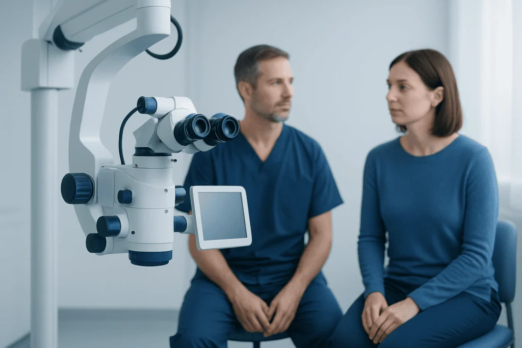
Active surveillance, sometimes called watchful waiting, means monitoring the tumor with scheduled MRI scans, hearing tests and clinical reviews. It does not mean that care is being delayed or ignored. It is often considered for small tumors, minimal symptoms, older adults, people with medical conditions that make procedures less suitable, or tumors that appear stable over time.
Stereotactic radiosurgery delivers a carefully planned, high dose of radiation to the tumor while limiting exposure to nearby tissue. Despite its name, it is not open surgery. The purpose is generally to control tumor growth rather than remove the tumor immediately; the tumor may remain the same size or gradually shrink over months to years. Gamma Knife radiosurgery is one form of focused radiosurgical treatment that may be considered in appropriate cases.
Microsurgery is an operation to remove all or part of the tumor through the skull base. It may be recommended when a tumor is large, growing, compressing the brainstem, causing hydrocephalus, or when removal is preferred after an individualized discussion. Surgical planning focuses on safe tumor removal and preservation of facial nerve function; hearing preservation may be possible in selected situations but cannot be promised.
Occasionally, treatment is deliberately staged. For example, surgeons may remove most of a large tumor to reduce pressure and then monitor or treat a small residual portion later. This approach may help balance tumor control with protection of important nerves in selected patients.
Candidacy and the assessment before treatment

Before recommending treatment, specialists review MRI images to measure the tumor and determine whether it is touching or compressing nearby structures. They also assess symptoms such as one-sided hearing loss, tinnitus, dizziness, unsteadiness, facial numbness and headaches. Formal audiology testing shows the level and usefulness of hearing in each ear.
Important considerations include whether the tumor has grown on serial scans, its size and location, hearing status, age, general health, prior treatment and the person’s daily activities. A person’s priorities matter as well. For example, some people place particular importance on avoiding an operation, while others prefer a treatment intended to remove the tumor.
Further tests may include balance assessment, facial nerve examination and, when needed, evaluation of swallowing, eye protection or other neurological functions. People with tumors on both sides or a family history suggesting neurofibromatosis type 2 may need genetic counseling and more specialized follow-up.
A second opinion can be helpful when options are closely balanced. The discussion should include what each approach can realistically achieve, how future monitoring will work and what treatment may be needed if the tumor changes later.
What happens during radiosurgery and microsurgery
For stereotactic radiosurgery, the care team first creates a precise treatment plan using high-resolution MRI and sometimes CT imaging. A head frame or fitted mask helps keep the head still, depending on the system used. The treatment itself is painless, and most people return home the same day or shortly afterward. The radiation effect is gradual, so follow-up scans remain essential.
Microsurgery is performed under general anesthesia by a skull-base surgical team. The surgical route is selected according to tumor size, location, hearing level and the goal of treatment. Common approaches include retrosigmoid, translabyrinthine and middle fossa approaches. The translabyrinthine route requires sacrificing hearing in the operated ear, while other approaches may be considered when hearing preservation is a realistic goal.
During surgery, clinicians commonly use specialized monitoring of the facial nerve and other neurological functions. Hospital stay varies with the procedure, tumor characteristics and individual recovery. Pathology testing confirms the diagnosis after tissue is removed.
Both radiosurgery and microsurgery require long-term imaging follow-up. Even after complete surgical removal, periodic MRI may be advised because recurrence is uncommon but possible. After radiosurgery, imaging helps distinguish temporary treatment-related swelling from continued growth.
Expected benefits, recovery and possible risks
The main potential benefit of active treatment is durable tumor control. Surgery can reduce or remove tumor mass immediately, which is especially important when there is pressure on the brainstem. Radiosurgery is less invasive and often has a shorter initial recovery, but its effects on growth are assessed over time rather than immediately.
Recovery after radiosurgery is usually brief. Some people feel tired or develop a temporary headache, scalp tenderness or balance symptoms. Recovery after microsurgery is more variable and may take weeks to months. Early tiredness, headache, dizziness and imbalance are common, and vestibular rehabilitation can help the brain adapt to changes in balance signals.
Potential risks depend on the tumor and treatment approach. They can include reduced or lost hearing in the affected ear, tinnitus, persistent balance problems, facial weakness or numbness, dry eye, taste changes, cerebrospinal fluid leak, infection, stroke and, rarely, more serious complications. Focused radiation also carries a small possibility of delayed nerve effects or later tumor growth, which is why follow-up is important.
Hearing outcomes are difficult to predict. Hearing may already decline because of the tumor itself, and no treatment can guarantee preservation. A hearing specialist can discuss options such as conventional hearing aids, CROS systems, bone-conduction devices or cochlear implantation when appropriate.
Living well during monitoring or recovery
People under observation should attend planned MRI and hearing appointments even when symptoms are stable. They should report meaningful changes, such as worsening hearing, increasing imbalance, new facial weakness or persistent numbness. Keeping a brief symptom record can help make follow-up visits more productive.
After treatment, gradual activity and a structured rehabilitation plan can support recovery. Balance exercises prescribed by a vestibular physiotherapist may improve steadiness and confidence with walking. Rest, hydration and practical support at home can also be valuable during the early recovery period.
Hearing changes and dizziness can affect work, driving, communication and emotional wellbeing. Audiology support, rehabilitation and counseling may help. It is reasonable to ask the care team about work restrictions, travel, exercise and when it is safe to resume specific activities.
Acibadem International’s multidisciplinary specialists and JCI-accredited hospitals evaluate and treat acoustic neuroma for international patients, coordinating neurosurgical, neuro-otology, radiation oncology and rehabilitation care when needed.
When to seek medical care
Medical assessment is appropriate for gradual hearing loss in one ear, persistent one-sided tinnitus, ongoing imbalance, recurrent dizziness or facial numbness. These symptoms can have many causes, and acoustic neuroma is uncommon, but early evaluation with an ear specialist and hearing test can clarify the cause.
Prompt medical advice is important if symptoms worsen noticeably, if new facial weakness develops, or if balance problems create a risk of falls. People already diagnosed with an acoustic neuroma should contact their treating team if they develop new or changing neurological symptoms between planned visits.
Urgent evaluation is needed for sudden severe headache, confusion, fainting, new weakness affecting an arm or leg, trouble speaking, or severe sudden loss of coordination. These symptoms are not specific to acoustic neuroma and may signal another condition requiring immediate care.
Frequently asked questions
Can an acoustic neuroma be treated without surgery?
Yes. Small or stable tumors may be monitored with MRI scans and hearing tests. Focused radiation treatment, such as stereotactic radiosurgery, is another nonsurgical option that aims to control growth in selected people.
Is stereotactic radiosurgery a cure for acoustic neuroma?
Stereotactic radiosurgery is designed primarily to stop or slow tumor growth, rather than remove the tumor right away. Many tumors remain stable or shrink gradually, but continued MRI follow-up is needed to confirm control over time.
When is surgery recommended for acoustic neuroma?
Surgery may be recommended for a large or growing tumor, pressure on the brainstem, hydrocephalus, or symptoms that require more immediate decompression. It may also be chosen based on individual circumstances and preferences after discussion with a specialist team.
Will hearing return after acoustic neuroma treatment?
Hearing that has been affected by the tumor does not always return after treatment. Depending on the tumor, hearing level and chosen approach, care may focus on preserving remaining hearing or supporting communication with hearing rehabilitation options.
How long is recovery after acoustic neuroma surgery?
Initial hospital recovery often takes several days, but fatigue, imbalance and adjustment to hearing changes may continue for weeks or months. Recovery varies according to tumor size, surgical approach, general health and whether rehabilitation is needed.
Does acoustic neuroma always grow?
No. Some acoustic neuromas grow very slowly or remain stable for years. This is why observation with regular MRI scans is a suitable initial approach for some people.
References
- National Institute of Neurological Disorders and Stroke
- American Academy of Otolaryngology–Head and Neck Surgery
- Congress of Neurological Surgeons
- National Cancer Institute
This article is for general information only and is not a substitute for professional medical advice. Please consult a qualified doctor about your individual situation.
Oncology care in Turkey — second opinion and treatment plan
JCI-accredited · board-certified surgeons · reply within 24h
Add us as a Preferred Source to see more of our trusted health content across Google Search, AI Overviews and Discover.

More from the Health Library



Related Specialists
Dr. Özge Karaosmanoğlu
Gynecology & Obstetrics
Dt. Cüneyt Işıker
Oral & Dental Health
Dr. Tuna Yildirim
General Surgery
Assoc. Prof. Dr. Ahmet Ifran
Hematology