Understanding Carcinoma Skin Types: A Complete Patient Guide

The main carcinoma skin types are basal cell carcinoma and squamous cell carcinoma. These skin cancers often appear as new, changing, nonhealing, scaly, or pearly skin lesions.
Key Takeaways
- The main carcinoma skin types are basal cell carcinoma and squamous cell carcinoma.
- These skin cancers often appear as new, changing, nonhealing, scaly, or pearly skin lesions.
- Long-term ultraviolet exposure is the leading risk factor, but skin type, age, and immune status also matter.
- Diagnosis usually requires a skin examination and biopsy.
- Treatment depends on the type, size, depth, and location of the lesion.
- Early medical evaluation improves outcomes and may reduce the need for more extensive treatment.
Carcinoma skin types mainly refer to basal cell carcinoma and squamous cell carcinoma, the two most common forms of nonmelanoma skin cancer. They usually develop on sun-exposed skin, often grow slowly, and are often highly treatable when recognized and treated early.
Overview: What are carcinoma skin types?
Carcinoma skin types are the main forms of skin cancer that begin in the skin’s epithelial cells. For most patients, this term refers to basal cell carcinoma and squamous cell carcinoma. Both are often grouped as nonmelanoma skin cancers, and both are commonly linked to cumulative ultraviolet exposure from sunlight or tanning devices.
Basal cell carcinoma starts in basal cells, which are found in the lower part of the epidermis. It is the most common skin cancer and tends to grow slowly. Squamous cell carcinoma begins in squamous cells closer to the skin surface. It can also often be treated successfully, but some cases behave more aggressively than basal cell carcinoma and may spread if left untreated.
Patients sometimes use the term “carcinoma skin types” when trying to understand how these cancers differ from melanoma. Melanoma is a separate form of skin cancer that develops from pigment-producing cells and is managed differently. Knowing the type matters because appearance, risk, treatment choices, and follow-up plans can vary.
A useful way to think about carcinoma skin types is that they are usually visible cancers of the skin that can often be found early. That makes regular skin awareness important. A new bump, sore, patch, or lesion that changes over time deserves medical attention, especially if it does not heal.
How basal cell carcinoma and squamous cell carcinoma differ
Basal cell carcinoma often appears as a pearly or shiny bump, a pink growth, a sore that crusts and reopens, or a flat scar-like area. It is especially common on the face, ears, scalp, neck, shoulders, and back. Although it rarely spreads to distant parts of the body, it can gradually invade nearby tissue if not treated.
Squamous cell carcinoma often looks different. It may appear as a rough, scaly patch, a thickened or wart-like growth, a firm red bump, or an ulcer that does not heal. It commonly develops on sun-exposed areas such as the face, lips, ears, hands, forearms, and scalp, but it can also occur in scars, chronic wounds, or areas of long-term inflammation.
Another key difference is behavior. Basal cell carcinoma usually grows slowly and tends to stay local. Squamous cell carcinoma can also be localized, but some tumors have a higher chance of growing deeper or spreading to nearby lymph nodes, particularly when they are large, recurrent, poorly defined, or located on high-risk areas such as the lip or ear.
Doctors may also discuss less common skin carcinomas, but for most patients, understanding these two main categories provides the clearest foundation. If a diagnosis is confirmed, the next step is to assess the size, depth, borders, and location of the lesion so treatment can be matched to the individual case.
Symptoms and warning signs to watch for

The symptoms of carcinoma skin types are often subtle at first. Many lesions are painless, which is one reason they may be overlooked. A spot that seems minor but does not go away can be more important than a dramatic symptom that appears suddenly.
Common warning signs include a new skin growth, a sore that bleeds or does not heal, a spot that crusts repeatedly, or a lesion that slowly enlarges over weeks or months. Patients may also notice changes in texture, such as scaling, roughness, tenderness, or skin thickening.
Features that can be seen with common carcinoma skin types include:
- A pearly, translucent, or shiny bump
- A flat patch that looks pale, yellowish, or scar-like
- A rough, red, or scaly patch
- A wart-like or crusted growth
- An open sore that bleeds, oozes, or heals and returns
- A painful or tender lesion, especially if it becomes deeper
These findings do not always mean cancer. Benign growths, eczema, psoriasis, infections, and actinic keratoses can look similar. Even so, any persistent or changing lesion should be checked by a qualified clinician rather than self-diagnosed.
Causes and risk factors
The leading cause of most carcinoma skin types is ultraviolet radiation exposure over time. Sunlight is the main source, but indoor tanning devices also increase risk. Damage accumulates gradually, so skin cancer may develop many years after repeated exposure, especially on areas that receive the most sun.
Risk also depends on individual factors. People with fair skin, light eyes, blond or red hair, or a tendency to burn easily often have a higher risk. Increasing age, a personal history of skin cancer, and a family history of certain skin cancers can also raise the likelihood of developing these conditions.
Additional risk factors include a weakened immune system, prior radiation therapy, chronic skin injury or inflammation, exposure to certain chemicals such as arsenic, and precancerous skin changes. Some squamous cell carcinomas arise from actinic keratoses, which are rough sun-damaged patches that can develop into cancer over time if not monitored or treated.
Importantly, darker skin tones are not fully protected. Skin carcinoma can occur in any skin color, though patterns and body locations may differ. Because early lesions may be harder to notice or may appear in less expected areas, everyone benefits from routine skin awareness and prompt assessment of suspicious changes.
How doctors diagnose carcinoma skin types
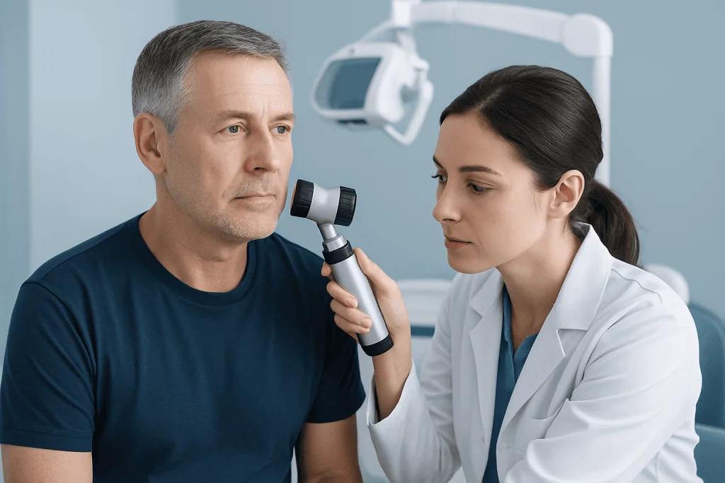
Diagnosis usually begins with a focused history and skin examination. A doctor will ask how long the lesion has been present, whether it has changed, and if there are symptoms such as bleeding, pain, itching, or repeated crusting. They will also look for other suspicious spots and ask about sun exposure, prior skin cancers, immune status, and family history.
If a lesion looks suspicious, the usual next step is a skin biopsy. During a biopsy, part or all of the lesion is removed and sent to a pathology laboratory. This is the only reliable way to confirm whether a lesion is basal cell carcinoma, squamous cell carcinoma, or another condition.
Depending on the lesion, the biopsy may be shave, punch, or excisional. If cancer is confirmed, the pathology report helps guide treatment by describing the tumor type and features such as depth or how aggressive the cells appear under the microscope. In selected cases, imaging or lymph node evaluation may be considered, especially for higher-risk squamous cell carcinoma.
Patients may also hear about related conditions such as skin cancer in broader terms. While the umbrella term is useful, treatment decisions are best made once the exact pathology is known. This helps the care team choose the most effective and tissue-sparing option when possible.
Treatment options and what care may involve
Treatment for carcinoma skin types depends on several factors: the cancer type, its size and depth, where it is located, whether it is a first occurrence or a recurrence, and the patient’s overall health. Many cases can be treated with a minor procedure, especially when found early.
Surgery is one of the most common approaches. Standard excision removes the lesion along with a margin of healthy tissue. For some tumors in cosmetically or functionally important areas, such as the face, doctors may recommend Mohs surgery, which removes the cancer layer by layer while preserving as much healthy skin as possible. This technique is often used for high-risk, recurrent, or poorly defined tumors.
Other treatments may include curettage and electrodesiccation for selected superficial lesions, cryotherapy in limited settings, topical therapies for certain early cancers, or radiation therapy when surgery is not suitable. More advanced or high-risk cases may require a multidisciplinary plan that can include radiation oncology or, less commonly, systemic treatment through medical oncology.
Follow-up is an important part of care because patients who have had one skin cancer have a higher chance of developing another. Near the end of the treatment pathway, some patients may also need repair or reconstruction, particularly after removal of a larger facial lesion, and this can involve plastic and reconstructive surgery. Acibadem International’s multidisciplinary specialists in JCI-accredited hospitals diagnose and treat skin cancers for international patients when such care is needed.
Prevention and self-care after diagnosis
Prevention focuses largely on reducing ultraviolet exposure. This includes seeking shade, wearing protective clothing and a wide-brimmed hat, and using broad-spectrum sunscreen on exposed skin. Sunscreen should be part of a broader sun-protection routine rather than the only measure.
Avoiding tanning beds is also important. Indoor tanning exposes the skin to concentrated ultraviolet radiation and increases long-term skin cancer risk. For patients who work outdoors or spend significant time outside, planning around peak sunlight hours and using physical barriers can make a meaningful difference.
After treatment, self-care includes regular skin checks at home and keeping scheduled follow-up visits. Patients can monitor for any new lesion, a change in a scar, or a spot that bleeds or crusts repeatedly. Taking simple photographs over time may help track whether an area is changing.
Healthy skin habits also support recovery. These include following wound-care instructions after procedures, avoiding picking at healing areas, and informing the doctor about any delayed healing or signs of infection. Prevention does not eliminate risk completely, but it can reduce future damage and improve the chances of catching problems early.
When to seek medical care
Patients should seek medical care if they notice a new skin lesion that persists, a spot that grows, or a sore that does not heal within a few weeks. A lesion that bleeds easily, crusts repeatedly, or becomes painful should also be evaluated. Even a small change can be worth checking if it continues over time.
Prompt assessment is especially important for lesions on the face, lips, ears, scalp, hands, or genitals, and for changes arising in scars or chronic wounds. People with a weakened immune system or a personal history of skin cancer should have a lower threshold for arranging an appointment.
Urgent care may be needed if a lesion is rapidly enlarging, causing significant pain, or associated with a firm nearby lump that could suggest lymph node involvement. Although many skin lesions are benign, it is safer to let a clinician decide rather than wait for more obvious symptoms.
Regular skin examinations by a dermatologist or other qualified doctor may be advised for people at increased risk. Early evaluation often leads to simpler treatment and better preservation of healthy skin, especially for visible or sensitive body areas.
Frequently asked questions
What are the main carcinoma skin types?
The main carcinoma skin types are basal cell carcinoma and squamous cell carcinoma. Both are common forms of nonmelanoma skin cancer and often develop on sun-exposed skin. They differ in appearance, behavior, and sometimes in treatment approach.
Is basal cell carcinoma less serious than squamous cell carcinoma?
Basal cell carcinoma usually grows more slowly and is less likely to spread to other parts of the body. Squamous cell carcinoma can sometimes behave more aggressively, especially if it is large, deep, recurrent, or located in a higher-risk area. Both still need proper medical evaluation and treatment.
Can skin carcinoma look like a harmless spot?
Yes. Some skin carcinomas begin as subtle changes, such as a small shiny bump, a rough patch, or a sore that seems to heal and then returns. Because they can resemble benign conditions, a persistent or changing lesion should be assessed by a doctor.
How is skin carcinoma confirmed?
A doctor usually confirms the diagnosis with a skin biopsy. This means removing part or all of the suspicious lesion and examining it under a microscope. The biopsy shows the exact cancer type and helps guide treatment.
Are carcinoma skin types curable?
Many cases are highly treatable, especially when diagnosed early. Outcomes depend on the type of carcinoma, where it is located, how deep it is, and whether it has come back before. Following the treatment plan and attending follow-up visits are important parts of care.
Can skin carcinoma come back after treatment?
Yes, recurrence is possible, and people who have had one skin cancer are also at higher risk of developing another. That is why doctors often recommend regular follow-up and self-examination of the skin. Reporting new or changing lesions early can help detect recurrence promptly.
References
- American Academy of Dermatology
- National Cancer Institute
- American Cancer Society
- World Health Organization
- National Comprehensive Cancer Network
This article is for general information only and is not a substitute for professional medical advice. Please consult a qualified doctor about your individual situation.
Oncology care in Turkey — second opinion and treatment plan
JCI-accredited · board-certified surgeons · reply within 24h
Add us as a Preferred Source to see more of our trusted health content across Google Search, AI Overviews and Discover.