Understanding Leiomyosarcoma: A Complete Patient Guide
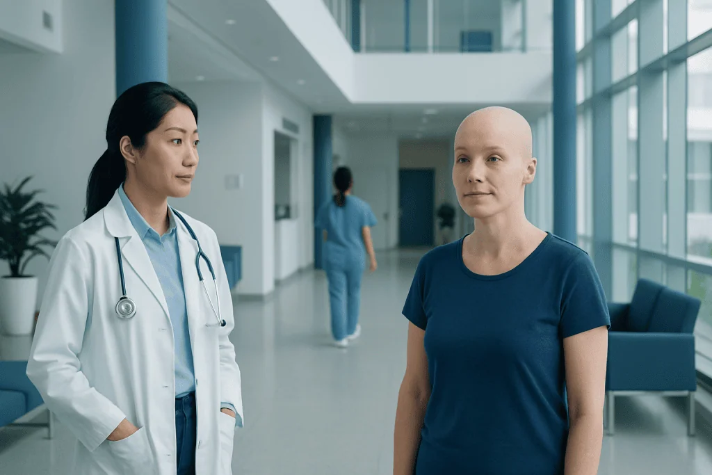
Leiomyosarcoma is a rare malignant tumor that develops from smooth muscle cells. It may grow in the uterus, abdomen, retroperitoneum, blood vessels, or other soft tissues.
Key Takeaways
- Leiomyosarcoma is a rare malignant tumor that develops from smooth muscle cells.
- It may grow in the uterus, abdomen, retroperitoneum, blood vessels, or other soft tissues.
- Symptoms are often nonspecific at first and depend on where the tumor is located.
- Diagnosis usually involves imaging and a biopsy reviewed by specialists experienced in sarcomas.
- Treatment often combines surgery with selected use of chemotherapy, radiation therapy, or other systemic treatments.
- Early specialist assessment is important for persistent unexplained symptoms or a growing mass.
Leiomyosarcoma is a rare cancer that starts in smooth muscle, the tissue found in organs such as the uterus, intestines, and blood vessels. It can occur in different parts of the body, so symptoms, treatment, and outlook vary depending on the tumor’s location, size, and stage.
Overview: what leiomyosarcoma means
Leiomyosarcoma is a rare type of cancer that begins in smooth muscle cells. Smooth muscle is the involuntary muscle found in many parts of the body, including the uterus, digestive tract, bladder, and the walls of blood vessels. Because these cells are present in several organs, leiomyosarcoma can develop in different locations and may behave differently from one person to another.
This cancer belongs to the broader group of soft tissue sarcomas. Unlike the much more common benign leiomyoma, such as a typical uterine fibroid, leiomyosarcoma is malignant and can invade nearby tissues or spread to other organs. Common sites of spread include the lungs, liver, and abdominal cavity, although patterns vary by the primary tumor site.
Leiomyosarcoma is not one single disease with one single presentation. Doctors usually describe it by where it starts, such as uterine leiomyosarcoma or leiomyosarcoma of the retroperitoneum, abdomen, or blood vessel wall. This location matters because it affects symptoms, imaging choices, surgery planning, and follow-up.
Because it is uncommon, evaluation is often best guided by a multidisciplinary team with experience in sarcoma care. In some patients, the condition may be considered within the wider context of sarcoma management, where pathology review and careful treatment planning play an important role.
Where it can occur and how symptoms may appear
The symptoms of leiomyosarcoma depend mainly on where the tumor is located. In the early stages, some people have no symptoms at all, especially if the tumor grows deep in the abdomen or retroperitoneum. In others, the first sign may be a painless lump, abdominal fullness, or symptoms caused by pressure on nearby organs.
When leiomyosarcoma develops in the uterus, it may cause abnormal uterine bleeding, pelvic pain, pressure, or a rapidly enlarging uterine mass. However, these symptoms can overlap with more common noncancerous conditions, which is one reason the diagnosis is not usually made from symptoms alone.
Leiomyosarcoma in the abdomen or retroperitoneum may lead to abdominal pain, bloating, constipation, early fullness after eating, urinary symptoms, or unexplained weight loss. Tumors involving blood vessels or other soft tissues may cause swelling, discomfort, or reduced function in the affected area.
- A new or enlarging lump under the skin or deep in soft tissue
- Persistent pelvic or abdominal pain or pressure
- Abnormal vaginal bleeding, especially after menopause
- Bloating, early satiety, or change in bowel habits
- Unexplained fatigue or unintended weight loss
- Shortness of breath or cough if the disease has spread to the lungs
Causes and risk factors

For most patients, there is no clear single cause of leiomyosarcoma. It develops when smooth muscle cells acquire genetic changes that allow them to grow uncontrollably. These changes are usually not something a person caused, and in most cases they are not inherited in a straightforward way.
Known risk factors for soft tissue sarcomas in general may include prior radiation exposure, certain inherited cancer syndromes, and long-term lymphatic or tissue changes, but many people with leiomyosarcoma have none of these factors. Most cases appear sporadically, meaning they arise without an obvious trigger.
It is also important to address a common concern: leiomyosarcoma is different from a benign leiomyoma. In the uterus, ordinary fibroids are very common and leiomyosarcoma is rare. Current understanding suggests that leiomyosarcoma usually does not develop from a typical fibroid transforming into cancer.
Researchers continue to study the molecular features of this disease to better understand why it starts and why some tumors behave more aggressively than others. These findings may help guide future treatment choices, especially in advanced disease.
How doctors diagnose leiomyosarcoma
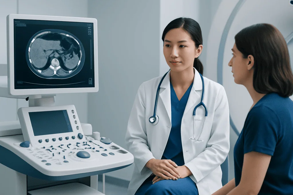
Diagnosing leiomyosarcoma usually begins with a medical history, physical examination, and imaging tests. The goal is to define the tumor’s size, location, relationship to nearby structures, and whether there are signs of spread. Depending on the body area involved, imaging may include ultrasound, CT, MRI, or PET/CT.
A biopsy is typically needed to confirm the diagnosis before treatment planning, especially for tumors outside the uterus. During a biopsy, a sample of tissue is removed and examined under a microscope by a pathologist. Because sarcomas are uncommon and can resemble other tumors, expert pathology review is often very valuable.
Pathology helps determine whether the tumor is truly leiomyosarcoma and may also describe grade, cell appearance, and other features linked to behavior. After diagnosis, staging tests are used to see whether the cancer remains localized or has spread. Chest imaging is often included because the lungs are a common site of metastasis in soft tissue sarcomas.
Imaging and biopsy often work together in a structured diagnostic pathway. Depending on the case, doctors may use MRI to define local tumor extent or PET/CT as part of broader cancer assessment, while tissue sampling may be guided by biopsy techniques chosen for the tumor’s location.
Treatment options and what care may involve
Treatment for leiomyosarcoma is individualized. The main factors include where the cancer started, whether it is localized or metastatic, the tumor’s size and grade, the patient’s overall health, and whether complete surgical removal is possible. Care is often coordinated by a team that may include surgical oncologists, gynecologic oncologists, medical oncologists, radiation oncologists, radiologists, and pathologists.
Surgery is often the main treatment when the disease is localized and removable. The aim is to remove the tumor completely with an adequate margin of healthy tissue when possible. In uterine leiomyosarcoma, surgery may involve removal of the uterus, while surgery for tumors in the abdomen or soft tissues may be more complex because of nearby organs and blood vessels.
Chemotherapy may be recommended before surgery, after surgery, or for advanced disease that cannot be fully removed. Radiation therapy can also be considered in selected cases, especially to help reduce the risk of local recurrence depending on tumor site and surgical margins. Not every patient needs both chemotherapy and radiation, and treatment plans differ from one case to another.
For advanced or recurrent disease, treatment may focus on controlling growth, relieving symptoms, and maintaining quality of life. Depending on the situation, options may include chemotherapy, targeted approaches in selected settings, symptom-directed supportive care, or enrollment in clinical trials when appropriate. At the end of the treatment pathway, some international patients may seek coordinated evaluation at centers such as Acibadem International, where multidisciplinary specialists in JCI-accredited hospitals diagnose and treat complex cancers including leiomyosarcoma.
Outlook, follow-up, and living with the diagnosis
The outlook for leiomyosarcoma varies widely. Important influences include the primary site, tumor size, grade, stage at diagnosis, whether it can be completely removed, and how the tumor responds to treatment. Some patients are treated successfully for localized disease, while others need long-term care for recurrence or metastasis.
Follow-up is an important part of care because leiomyosarcoma can return after treatment. Doctors usually recommend scheduled visits with physical examinations and imaging tailored to the original tumor site and the risk of recurrence. Chest imaging is often included in surveillance for soft tissue sarcomas.
Living with a rare cancer can also bring emotional and practical challenges. Patients may benefit from clear communication with their care team, written treatment summaries, support from family or cancer counseling services, and help with managing fatigue, pain, nutrition, and physical recovery.
Questions about pathology review, treatment goals, side effects, fertility considerations, and follow-up planning are all reasonable and important. Because the disease is rare, many patients also find reassurance in a second opinion from a team experienced in oncology and sarcoma treatment.
When to seek medical care
A person should seek medical care if they notice a persistent or enlarging lump, unexplained pelvic or abdominal pain, abnormal vaginal bleeding, ongoing bloating, or a feeling of pressure that does not improve. These symptoms are often caused by conditions other than cancer, but they should still be evaluated if they persist or worsen.
Prompt assessment is especially important for postmenopausal bleeding, rapidly increasing abdominal girth, unexplained weight loss, or shortness of breath. A doctor can decide which tests are needed and whether referral to a sarcoma or gynecologic oncology team is appropriate.
After a diagnosis, patients should contact their care team sooner if they develop new severe pain, fever, heavy bleeding, sudden swelling, breathing difficulties, or treatment side effects that interfere with eating, hydration, or daily activities. Early communication can help problems be managed more safely and comfortably.
Self-care cannot prevent all cases of leiomyosarcoma, but general measures can support overall health during evaluation and treatment. These include attending follow-up appointments, reporting new symptoms promptly, eating as well as possible, staying physically active within medical advice, and asking for support when needed.
Frequently asked questions
Is leiomyosarcoma the same as a fibroid?
No. A fibroid, also called a leiomyoma, is a benign growth of smooth muscle, while leiomyosarcoma is a malignant tumor. Although symptoms may overlap, they are biologically different conditions and require different management.
How rare is leiomyosarcoma?
Leiomyosarcoma is considered a rare cancer. It is one of several subtypes of soft tissue sarcoma and is seen much less often than common cancers such as breast, colon, or lung cancer.
Can leiomyosarcoma be cured?
Some patients with localized leiomyosarcoma can be treated successfully, especially when the tumor can be completely removed. The chance of long-term control depends on the tumor’s site, stage, grade, and response to treatment.
What tests are used to confirm leiomyosarcoma?
Doctors usually use imaging such as ultrasound, CT, or MRI to locate and assess the tumor, but a biopsy is generally needed to confirm the diagnosis. The tissue sample is examined by a pathologist, often with expert review when sarcoma is suspected.
Does leiomyosarcoma spread quickly?
Its behavior can vary. Some tumors grow and spread more aggressively than others, which is why grade, stage, and location are important. Doctors use these details to estimate risk and guide treatment and follow-up.
What specialist should a patient see for leiomyosarcoma?
Care is usually best coordinated by a multidisciplinary cancer team with experience in sarcomas. Depending on the tumor site, this may include a surgical oncologist, gynecologic oncologist, medical oncologist, radiation oncologist, and specialist pathologist.
References
- National Cancer Institute
- American Cancer Society
- National Comprehensive Cancer Network
- World Health Organization
- European Society for Medical Oncology
This article is for general information only and is not a substitute for professional medical advice. Please consult a qualified doctor about your individual situation.
Persistent digestive symptoms? Get evaluated in Turkey
JCI-accredited · board-certified surgeons · reply within 24h
Add us as a Preferred Source to see more of our trusted health content across Google Search, AI Overviews and Discover.

More from the Health Library



Related Specialists

Assoc. Prof. Dr. Fatih Aygün
Pediatric Intensive Care Unit
Dr. Ahsen Ercan
Physical Medicine & Rehabilitation
Prof. Dr. Ethem Kumbay
Cardiology
Prof. Dr. Erkan Özcan
Psychiatry