Urachal Remnant Ultrasound: Preparation, Procedure and Results

Ultrasound is often the first imaging test for a suspected urachal remnant because it does not use radiation. A full bladder may improve views of the bladder dome and nearby urachal tissue, so preparation instructions should be followed.
Key Takeaways
- Ultrasound is often the first imaging test for a suspected urachal remnant because it does not use radiation.
- A full bladder may improve views of the bladder dome and nearby urachal tissue, so preparation instructions should be followed.
- Ultrasound can suggest a urachal cyst or infection, but CT, MRI, or other tests may be needed to confirm the diagnosis or plan treatment.
- Not every urachal remnant requires surgery; decisions depend on symptoms, infection, imaging findings, and clinical assessment.
- Fever, worsening lower abdominal pain, a tender lump, or discharge from the umbilicus should be assessed promptly.
A urachal remnant ultrasound uses sound waves to examine the area between the bladder and the umbilicus (belly button). It is usually a simple, noninvasive first test when a clinician suspects a urachal cyst, sinus, diverticulum, or other remaining part of the fetal urachus.
Overview: what is a urachal remnant ultrasound?
A urachal remnant ultrasound is an imaging examination of tissue extending from the top of the bladder toward the umbilicus. Before birth, the urachus is a temporary channel that connects the developing bladder to the umbilical cord. It usually closes before birth and becomes a fibrous cord called the median umbilical ligament. If a portion remains open or forms a fluid-filled space, it is called a urachal remnant.
Several forms are possible, including a urachal cyst, urachal sinus opening toward the umbilicus, a diverticulum connected to the bladder, or a fully patent urachus. Many remnants cause no symptoms and are found incidentally. Others may become infected, cause umbilical drainage, or lead to discomfort in the lower middle abdomen.
Ultrasound is commonly used as an initial test because it is painless, widely available, and does not expose the patient to ionizing radiation. It can help clinicians determine whether there is a cystic structure, fluid collection, inflammation, or another explanation for symptoms near the bladder or navel.
Who may need this examination and how to prepare
A clinician may request a urachal remnant ultrasound for persistent or recurrent umbilical discharge, redness or tenderness around the navel, lower abdominal pain, a midline lump, urinary symptoms, or repeated infections in this region. The test may be used in children or adults. It can also help assess a finding noticed during another scan or examination.
Preparation is usually straightforward. The imaging department may ask the patient to arrive with a comfortably full bladder, since this can improve visualization of the bladder dome and the region immediately above it. Instructions vary by age and by facility, so patients should follow the guidance provided when the appointment is scheduled.
Usually, there is no need to fast, stop regular medicines, or arrange recovery time after an ultrasound. Patients should tell the imaging team about current symptoms, prior abdominal or pelvic surgery, recent imaging, pregnancy, and any known urinary or umbilical drainage. These details help the sonographer target the examination appropriately.
How the ultrasound works: procedure step by step

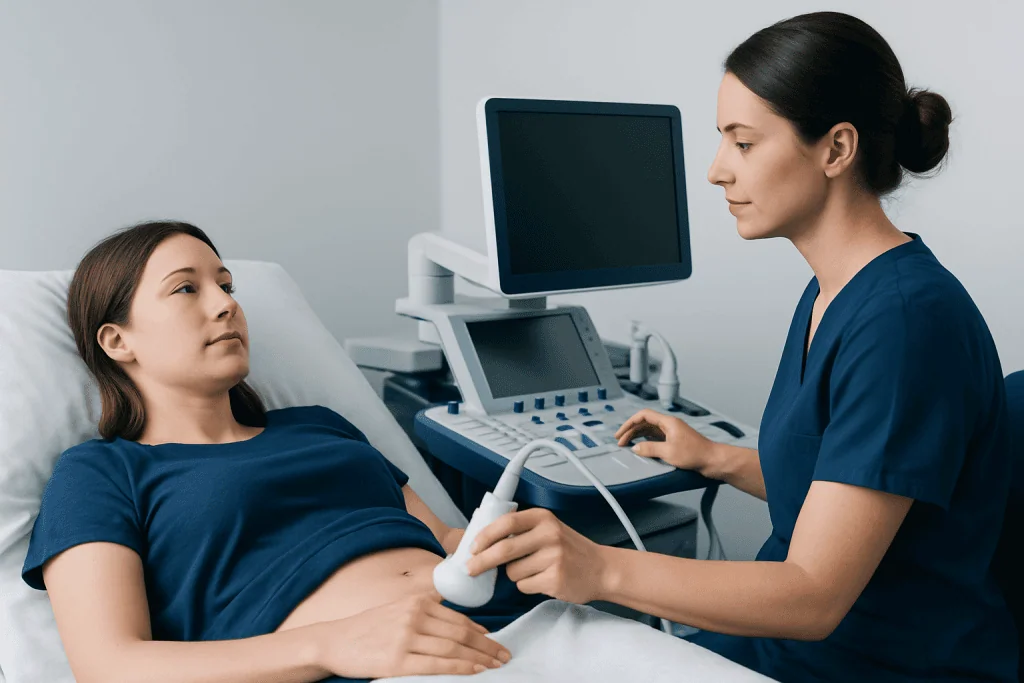
Ultrasound creates images using high-frequency sound waves. A sonographer applies a small amount of gel to the lower abdomen and moves a handheld probe over the skin. The probe sends sound waves into the body and receives returning echoes, which are converted into live images on a monitor.
For a urachal assessment, the scan generally focuses on the midline between the umbilicus and the upper part of the bladder. Images may be taken from several angles. The sonographer may apply gentle pressure or ask the patient to change position, hold their breath briefly, or empty the bladder if comparison images are useful.
The examination commonly takes about 20 to 40 minutes, although timing depends on the reason for the scan and how clearly the area can be seen. It does not involve needles, contrast dye, or radiation. The images are interpreted by a radiologist, who sends a report to the referring clinician.
Afterward, the patient can usually return to normal activities immediately. The referring clinician discusses the report in the context of symptoms, examination findings, urine tests, and any further testing that may be appropriate.
Can an ultrasound detect an urachal remnant?
Yes. An ultrasound can often detect an urachal remnant, particularly when it forms a fluid-filled cyst or visible tract in the typical midline location between the bladder and the umbilicus. A urachal cyst may appear as a small, well-defined fluid-containing structure above the bladder. If infection is present, the collection may look more complex, with internal echoes, debris, or surrounding inflammatory change.
However, ultrasound does not identify every remnant with equal certainty. Very small remnants, deep structures, bowel gas, body habitus, incomplete bladder filling, or significant inflammation can limit the view. Ultrasound findings can also overlap with other conditions, such as an umbilical hernia, abscess, bowel-related condition, or a different pelvic cyst.
When the scan is unclear or suggests an infection or complication, CT or MRI may give a more detailed view. Additional tests are selected individually, balancing the clinical question, age, symptoms, and the need to avoid unnecessary radiation. In some cases, a urine test or specialist review is also part of the assessment.
How to check for urachal remnant?
Checking for a urachal remnant begins with a clinical assessment. A doctor asks about symptoms such as umbilical fluid or pus, lower abdominal pain, fever, urinary discomfort, blood in the urine, or repeated infections. They may examine the umbilicus and lower abdomen for tenderness, redness, swelling, a mass, or active drainage.
Imaging then helps clarify the anatomy. Ultrasound is frequently the first examination. CT can provide detailed information about an infected collection, communication with nearby structures, and surrounding inflammation. MRI may be considered when detailed soft-tissue imaging is needed or when avoiding radiation is especially important. A specialist may occasionally recommend bladder evaluation, such as cystoscopy, if a connection to the bladder or another bladder concern is suspected.
Blood tests and urine testing may be used when infection or urinary involvement is possible. It is important not to try to probe, squeeze, or insert objects into a draining umbilicus at home. This can irritate the area and may worsen infection. A qualified clinician should interpret symptoms and imaging together before confirming a diagnosis.
What are the CT findings of an infected urachal cyst?
On CT, an infected urachal cyst commonly appears as a midline fluid collection between the umbilicus and the dome of the bladder. Instead of looking like simple clear fluid, it may have a thicker or irregular wall, internal debris, and surrounding fat stranding, which indicates inflammation in nearby tissues. The cyst may also show enhancement of its wall after contrast is given.
Depending on severity, CT may reveal an abscess, gas within the collection, extension of inflammation into the abdominal wall, or a tract toward the umbilicus or bladder. CT can also help distinguish a urachal problem from other causes of lower abdominal pain or umbilical symptoms and can guide decisions about drainage or surgery.
These findings are not interpreted in isolation. Infection can resemble other inflammatory or cystic conditions, and imaging appearances must be considered alongside fever, pain, blood tests, and physical examination. If infection is suspected, timely medical evaluation is important because treatment may involve antibiotics, drainage, surgery, or a combination of approaches.
Results, treatment options and recovery
Ultrasound results may be reported as normal, suggestive of a urachal remnant, consistent with a simple cyst, or concerning for infection or another condition. A result does not automatically mean treatment is needed. The clinician considers whether the finding explains symptoms, whether there is infection, and whether further imaging is necessary.
Does an urachal remnant need to be removed? Not always. An incidental, symptom-free remnant may be monitored in selected situations, particularly after specialist review. Recurrent infection, persistent umbilical drainage, pain, an enlarging or complex mass, or uncertainty about the diagnosis may lead a urologist or surgeon to recommend removal. An acute infection often needs treatment first, and the timing of surgery is individualized.
When excision is recommended, the aim is to remove the remnant and reduce the chance of recurrent symptoms or infection. Procedures may be performed through minimally invasive or open surgical approaches, depending on the anatomy, inflammation, and the patient’s overall health. Patients can discuss laparoscopic surgery with their surgical team if this approach is suitable.
Recovery varies with the treatment used. After ultrasound alone, there is no recovery period. Following drainage or surgery, patients may have short-term discomfort, activity restrictions, wound-care instructions, and follow-up appointments. The care team explains expected recovery, warning signs, and when usual work, school, exercise, and bathing can safely resume.
Benefits, limitations and when to seek medical care
The main benefits of urachal remnant ultrasound are that it is noninvasive, does not use radiation, and can provide useful real-time information about the bladder dome and adjacent midline tissues. It is particularly helpful for identifying a fluid-containing lesion and for deciding whether more detailed imaging or specialist input is needed. Its limitations include reduced clarity in some body types and the possibility that a small or complicated remnant may not be fully characterized.
There are very few risks from a standard ultrasound. Some people experience mild pressure from the probe over a tender area, but the examination is generally well tolerated. The gel may feel cool and can be wiped away immediately after the scan.
Medical care should be sought promptly for fever, increasing lower abdominal pain, redness or swelling around the umbilicus, pus or foul-smelling drainage from the navel, vomiting, difficulty passing urine, or blood in the urine. Emergency assessment is appropriate for severe pain, high fever, confusion, fainting, or rapidly worsening symptoms.
Urologists, radiologists, pediatric specialists, and general surgeons may work together when a urachal condition is suspected. Acibadem International’s multidisciplinary specialists and JCI-accredited hospitals assess and treat urologic conditions for international patients, with care plans based on the individual’s symptoms and imaging findings.
Frequently asked questions
Is a urachal remnant ultrasound painful?
No. A standard ultrasound does not involve needles or radiation and is usually painless. The probe may cause mild pressure, particularly if the lower abdomen is already tender, but the examination can be adjusted for comfort.
Do patients need a full bladder for a urachal remnant ultrasound?
Often, yes. A comfortably full bladder can help show the bladder dome and nearby urachal region more clearly. The imaging center should provide specific instructions, as preparation may vary by age and clinical situation.
Can an ultrasound tell whether a urachal cyst is infected?
Ultrasound may show features that raise concern for infection, such as a complex fluid collection, internal debris, thick walls, or surrounding inflammation. It cannot always confirm infection by itself, so symptoms, examination, blood tests, and sometimes CT are also considered.
What happens if the ultrasound result is unclear?
The clinician may recommend CT or MRI for a more detailed view of the area. Urine tests, blood tests, or referral to a urologist or surgeon may also be appropriate depending on the symptoms and imaging findings.
Can a urachal remnant cause belly button discharge?
Yes. A urachal sinus or patent urachus can be associated with drainage from the umbilicus, especially if there is inflammation or infection. Umbilical discharge can have several causes, so it should be evaluated rather than self-treated.
Is surgery always necessary for a urachal cyst?
No. The need for surgery depends on whether the cyst is causing symptoms, has become infected, recurs, appears complex, or cannot be confidently distinguished from other conditions. A urologist or surgeon can explain the advantages and limitations of monitoring versus removal in an individual case.
References
- National Institute of Diabetes and Digestive and Kidney Diseases
- Radiological Society of North America
- American Urological Association
- Merck Manual Professional Edition
This article is for general information only and is not a substitute for professional medical advice. Please consult a qualified doctor about your individual situation.
Ultrasound in Turkey — costs, top hospitals & a free quote
JCI-accredited · board-certified surgeons · reply within 24h
Add us as a Preferred Source to see more of our trusted health content across Google Search, AI Overviews and Discover.

Check-up & Preventive Medicine
Comprehensive health screenings designed for early detection and prevention.
2 specialists in this unit