Vision Loss in One Eye: How Neuro-Ophthalmologists Find the Cause

Vision loss in one eye is a symptom, not a diagnosis, and it has many possible causes. The pattern of vision change, pain, timing, and associated symptoms helps guide diagnosis.
Key Takeaways
- Vision loss in one eye is a symptom, not a diagnosis, and it has many possible causes.
- The pattern of vision change, pain, timing, and associated symptoms helps guide diagnosis.
- Some causes are urgent, including retinal detachment, stroke-related reduced blood flow, and giant cell arteritis.
- A neuro-ophthalmologist evaluates both the visual system and the nervous system to locate the problem.
- Prompt medical assessment can protect vision and may also identify broader health risks such as vascular disease.
Vision loss in one eye can happen suddenly or gradually and may start in the eye, optic nerve, blood vessels, or brain. Neuro-ophthalmologists help identify the cause by combining a detailed history, a focused eye and nerve examination, and targeted testing.
Overview
Vision loss in one eye means reduced sight affecting only one eye, either partially or completely. It may appear as blurring, dimming, a dark curtain, missing areas in the field of vision, faded colors, or complete loss of sight. In some people it happens within seconds or minutes, while in others it develops over hours, days, or longer.
This symptom can arise from problems in different parts of the visual pathway. The cause may be in the front of the eye, such as the cornea or lens; in the retina at the back of the eye; in the optic nerve that carries visual signals to the brain; or more rarely in parts of the brain that process vision. Because these structures work together, careful evaluation is needed to determine exactly where the problem begins.
Neuro-ophthalmologists are specialists who assess vision problems that overlap eye disease and nervous system disease. They are especially helpful when symptoms suggest optic nerve inflammation, reduced blood flow to the eye or nerve, double vision, unexplained visual field loss, or a connection to neurological conditions. Their role is not only to name the condition, but also to identify whether it is urgent and whether it reflects a broader health issue.
Symptoms and patterns doctors look for
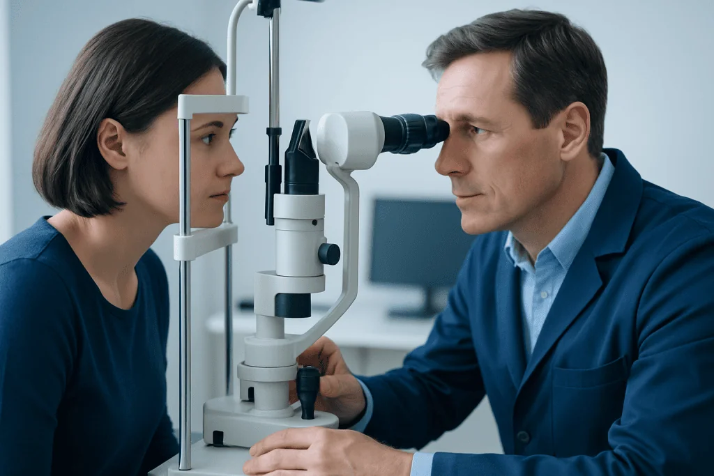
The way vision changes often provides important clues. Sudden, painless dimming or blackout in one eye may suggest a vascular event, sometimes called amaurosis fugax when it is temporary. Flashes of light, new floaters, or a curtain-like shadow may point to a retinal tear or retinal detachment. Pain with eye movement, reduced color vision, and blurred central vision can be seen with optic neuritis.
Doctors also ask whether the vision loss is constant or comes and goes. Temporary episodes lasting seconds to minutes can occur with reduced blood flow, migraine-related visual symptoms, or pressure-related changes. More gradual decline may occur with inflammation, swelling, glaucoma, cataract, retinal disease, or compressive problems affecting the optic nerve.
Associated symptoms matter as well. Headache, scalp tenderness, jaw pain when chewing, weakness, numbness, speech changes, imbalance, eye pain, or double vision can all change the level of concern and narrow the possibilities. A neuro-ophthalmologist may also ask about recent infection, autoimmune disease, diabetes, high blood pressure, smoking history, and previous neurological symptoms because these details can guide the next steps.
- Blurred or dim vision in one eye
- Blind spots or loss of peripheral vision
- Flashes, floaters, or a curtain over vision
- Pain with eye movement or around the eye
- Color vision that seems washed out
- Temporary episodes of vision loss
Common causes and risk factors
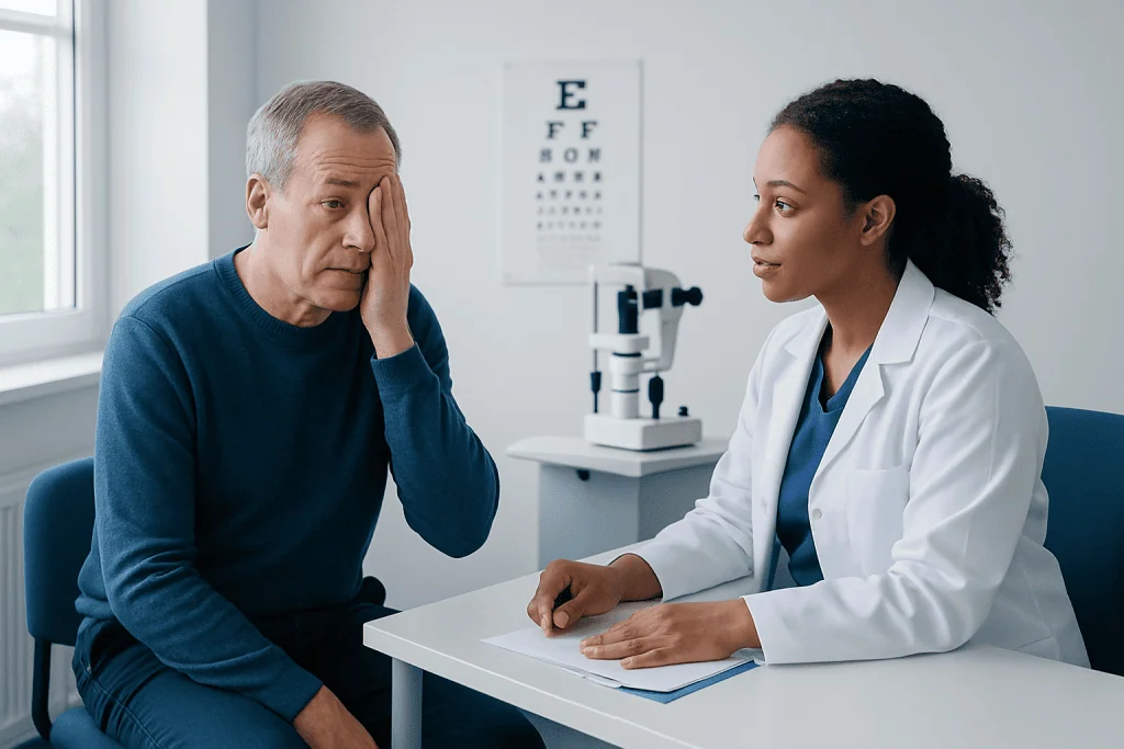
Causes can be grouped by where the problem starts. In the eye itself, common causes include retinal detachment, retinal artery or vein blockage, bleeding in the eye, severe dry eye, corneal disease, or advanced glaucoma. In the optic nerve, possible causes include optic neuritis, reduced blood supply to the nerve, swelling of the optic nerve, toxic or nutritional injury, and compression from nearby structures. In a smaller number of cases, lesions in the brain’s visual pathways can reduce vision in a way that first seems to affect one eye.
Vascular causes are especially important because they may reflect general circulation problems. A temporary or permanent drop in blood flow can be associated with high blood pressure, diabetes, high cholesterol, smoking, heart rhythm problems, carotid artery disease, or clotting disorders. In older adults, giant cell arteritis is a key cause that needs urgent recognition because it can threaten vision in both eyes if not treated quickly.
Inflammatory and immune-related causes are also considered. Optic neuritis may occur on its own or in association with conditions such as multiple sclerosis or other inflammatory disorders. Infections, certain medications, trauma, and tumors can affect the optic nerve or retina as well. The doctor considers age, speed of onset, pain, medical history, and examination findings to build a focused list of likely causes rather than assuming all vision loss has the same explanation.
How neuro-ophthalmologists make the diagnosis
Diagnosis begins with a detailed history. The specialist asks exactly when the symptom began, whether it was sudden or progressive, whether there is pain, and what part of vision is affected. They also review prior eye disease, recent illnesses, vascular risk factors, autoimmune symptoms, medication use, and neurological symptoms. This history often points toward whether the problem is more likely retinal, optic nerve, vascular, or neurological.
The examination is equally important. It typically includes visual acuity, color vision, pupil responses, eye movements, visual field testing, and a close look at the front and back of the eye after the pupils are dilated. The pupils can reveal whether one optic nerve is transmitting visual signals less effectively. The appearance of the optic nerve and retina may show swelling, pallor, bleeding, detachment, or vessel blockage.
Additional tests are chosen based on the suspected cause. These may include optical coherence tomography to image the retina and optic nerve, formal visual field testing, blood tests for inflammation or infection, and vascular evaluation. If the exam suggests a neurological or structural cause, imaging such as MRI scanning may be recommended to look at the optic nerve, brain, and surrounding tissues. Some patients may also need comprehensive eye examination by retinal or glaucoma specialists, or neurological evaluation when the findings point beyond the eye.
Treatment depends on the cause
There is no single treatment for vision loss in one eye because therapy must match the cause. Some conditions need urgent care to protect sight or reduce the risk of stroke and other complications. For example, retinal detachment often requires prompt retinal treatment, giant cell arteritis requires immediate medical therapy, and retinal artery blockage may prompt emergency evaluation for vascular risk. When inflammation of the optic nerve is suspected, treatment and follow-up depend on the severity, imaging findings, and overall neurological context.
Not every case requires surgery or hospital treatment. Some causes are managed with medication, close observation, vascular risk reduction, or referral to another specialist such as a retina specialist, neurologist, or rheumatologist. Treatment may also include controlling blood pressure, blood sugar, cholesterol, and smoking exposure when circulation problems are involved.
Recovery varies by diagnosis. Some people experience significant improvement, while others may have lasting visual changes despite appropriate care. The most helpful step is early assessment, because delays can reduce treatment options in time-sensitive conditions. When specialized care is needed, Acibadem International’s multidisciplinary specialists in JCI-accredited hospitals evaluate and treat neuro-ophthalmic conditions for international patients.
Self-care, prevention, and reducing risk
People cannot prevent every cause of vision loss in one eye, but they can lower risk by protecting both eye health and general vascular health. Regular eye examinations are especially important for those with diabetes, high blood pressure, high cholesterol, glaucoma risk, or previous retinal disease. Protective eyewear can help reduce eye injuries during sports, home repairs, or certain jobs.
Healthy daily habits support the blood vessels and nerves that vision depends on. Not smoking, staying physically active, following a balanced diet, taking prescribed medications, and keeping chronic conditions well controlled all help lower the chance of some vascular and nerve-related eye problems. Patients with migraine, autoimmune disease, or neurological disorders should also keep regular follow-up appointments and report any new visual symptoms promptly.
After an episode of one-eye vision loss, self-care should focus on safety and medical follow-up rather than self-diagnosis. It is wise to avoid driving until the cause is understood and a clinician advises that it is safe. Keeping a record of when symptoms started, how long they lasted, whether there was pain, and any triggers or associated symptoms can be very useful at the appointment.
When to seek urgent medical attention
Some forms of vision loss in one eye should be treated as urgent. Sudden loss of vision, a curtain or shadow over part of the visual field, new flashes and floaters, severe eye pain, or vision loss with weakness, numbness, trouble speaking, or severe headache needs immediate medical attention. These symptoms can signal retinal detachment, stroke-related reduced blood flow, acute glaucoma, or other time-sensitive conditions.
Older adults should seek urgent care if one-eye vision loss occurs with headache, scalp tenderness, fever, fatigue, or jaw pain while chewing. These symptoms may suggest giant cell arteritis, an inflammatory blood vessel condition that can threaten vision quickly. Prompt treatment can help protect the other eye and reduce complications.
Even when the change is temporary and vision returns, it still deserves medical assessment. A brief episode can be a warning sign of a circulation problem or another disorder that may recur. If a person is unsure whether symptoms are serious, it is safest to contact an eye specialist or emergency service without delay.
Frequently asked questions
Is vision loss in one eye always an emergency?
Not every case is an emergency, but sudden vision loss in one eye should always be assessed promptly. Some causes are time-sensitive and can threaten vision or signal a stroke-related circulation problem.
What does a neuro-ophthalmologist do differently from a general eye doctor?
A neuro-ophthalmologist evaluates how the eyes, optic nerves, brain, and nervous system work together. This specialist is especially helpful when vision loss may come from the optic nerve, blood flow problems, inflammation, or a neurological condition.
Can temporary vision loss in one eye still be serious?
Yes. Even if vision returns within minutes, temporary loss can be a warning sign of reduced blood flow or another underlying condition that needs medical attention. It should not be ignored, especially in older adults or people with vascular risk factors.
Will vision come back after loss in one eye?
Recovery depends on the cause, how quickly treatment starts, and how much tissue was affected. Some conditions improve substantially, while others can leave lasting changes despite appropriate treatment.
What tests might be needed?
Doctors may use visual acuity testing, pupil assessment, dilated eye examination, visual field testing, retinal imaging, blood tests, and sometimes MRI or vascular studies. The exact tests depend on the symptom pattern and examination findings.
What symptoms should never be ignored?
A sudden curtain over vision, flashes and floaters, severe eye pain, new weakness or speech trouble, or vision loss with headache and jaw pain all need urgent assessment. These features can point to retinal, vascular, or neurological emergencies.
References
- American Academy of Ophthalmology
- North American Neuro-Ophthalmology Society
- National Eye Institute
- American Stroke Association
- National Institute of Neurological Disorders and Stroke
This article is for general information only and is not a substitute for professional medical advice. Please consult a qualified doctor about your individual situation.
Explore treatments in Turkey — costs, top hospitals & a free quote
JCI-accredited · board-certified surgeons · reply within 24h
Add us as a Preferred Source to see more of our trusted health content across Google Search, AI Overviews and Discover.
Check your numbers in seconds
BMI, calories, due date, blood pressure and 30+ more clinical calculators — free, instant, doctor-reviewed ranges.