When Do You Need a Cerebral Angiogram? Diagnosis, Safety, and What to Expect

A cerebral angiogram provides detailed images of blood vessels in the brain and neck. It is commonly used to investigate aneurysms, narrowing, blockages, vascular malformations, and some stroke-related problems.
Key Takeaways
- A cerebral angiogram provides detailed images of blood vessels in the brain and neck.
- It is commonly used to investigate aneurysms, narrowing, blockages, vascular malformations, and some stroke-related problems.
- The procedure is generally safe when performed by experienced teams, though it carries small risks such as bleeding, allergic reaction, or stroke.
- Most people go home the same day or after a short observation period, depending on why the test was done.
- Preparation may include fasting, blood tests, medication review, and arranging for someone to help after the procedure.
A cerebral angiogram is a specialized imaging test that shows blood flow in the brain and neck in very fine detail. It is often used when doctors need clearer answers than standard scans can provide or when they are planning minimally invasive treatment.
Overview: What Is a Cerebral Angiogram?
A cerebral angiogram, also called cerebral angiography or a brain angiogram, is a test that creates highly detailed images of the blood vessels that supply the brain, head, and sometimes the neck. During the procedure, a doctor inserts a thin tube called a catheter into a blood vessel, usually in the wrist or groin, and guides it toward the arteries being examined. A special contrast dye is then injected while X-ray images are taken.
This test helps doctors see the shape of blood vessels, how blood flows through them, and whether there are areas of narrowing, blockage, bulging, or abnormal connections. Although noninvasive scans such as CT angiography and MR angiography are very useful, a cerebral angiogram can provide more precise detail when a diagnosis is uncertain or when treatment planning requires a closer look.
In many cases, cerebral angiography is both a diagnostic and a procedural tool. If the angiogram identifies a problem that can be treated through the catheter, some patients may go on to have minimally invasive care during the same session or in a planned follow-up procedure. This makes it a central part of modern interventional neurology and neurovascular care.
When Is a Cerebral Angiogram Needed?
Doctors may recommend a cerebral angiogram when symptoms, examination findings, or other scans suggest a problem in the brain’s blood vessels. It is often used when more detailed imaging is needed to explain symptoms such as sudden severe headache, stroke-like symptoms, weakness, numbness, vision changes, dizziness, or unexplained neurological events.
Common reasons for cerebral angiography include evaluating a suspected brain aneurysm, checking narrowing or blockage in arteries, investigating abnormal blood vessel tangles called arteriovenous malformations, and assessing bleeding in or around the brain. It may also be used to better understand conditions linked to transient ischemic attack or stroke, including stroke and other neurovascular disorders.
In some people, the goal is to plan treatment rather than make the first diagnosis. For example, a cerebral angiogram can help map the exact size and shape of an aneurysm before aneurysm treatment or show the location of a vessel blockage before stroke treatment. It may also be used before or during embolization for certain vascular malformations or bleeding problems.
What Conditions Can It Help Diagnose?

Cerebral angiography is especially useful for conditions that affect blood vessels in the brain and neck. One of the best-known examples is a brain aneurysm, where a weak area in an artery wall bulges outward. The test can show the aneurysm’s neck, shape, and relationship to nearby branches, which helps guide management decisions.
It can also identify arterial narrowing from atherosclerosis, blood vessel blockage, vasospasm after certain types of bleeding, and dissections, which are tears in the vessel wall. In patients with suspected brain aneurysm, a cerebral angiogram may clarify findings that are not fully defined on CT or MRI-based tests.
Other important uses include diagnosing arteriovenous malformations, dural arteriovenous fistulas, and some tumors with unusual blood supply. Because the images are dynamic, meaning they show blood flowing through the vessels over time, doctors can understand not only anatomy but also circulation patterns. This can be important for choosing the safest and most effective next step.
How the Procedure Is Performed and What to Expect
Before the procedure, the care team reviews the person’s medical history, allergies, medications, and kidney function. Blood thinners, diabetes medicines, or other drugs may need special instructions. Patients are often asked not to eat or drink for several hours beforehand, and they usually sign a consent form after the procedure, benefits, and risks are explained.
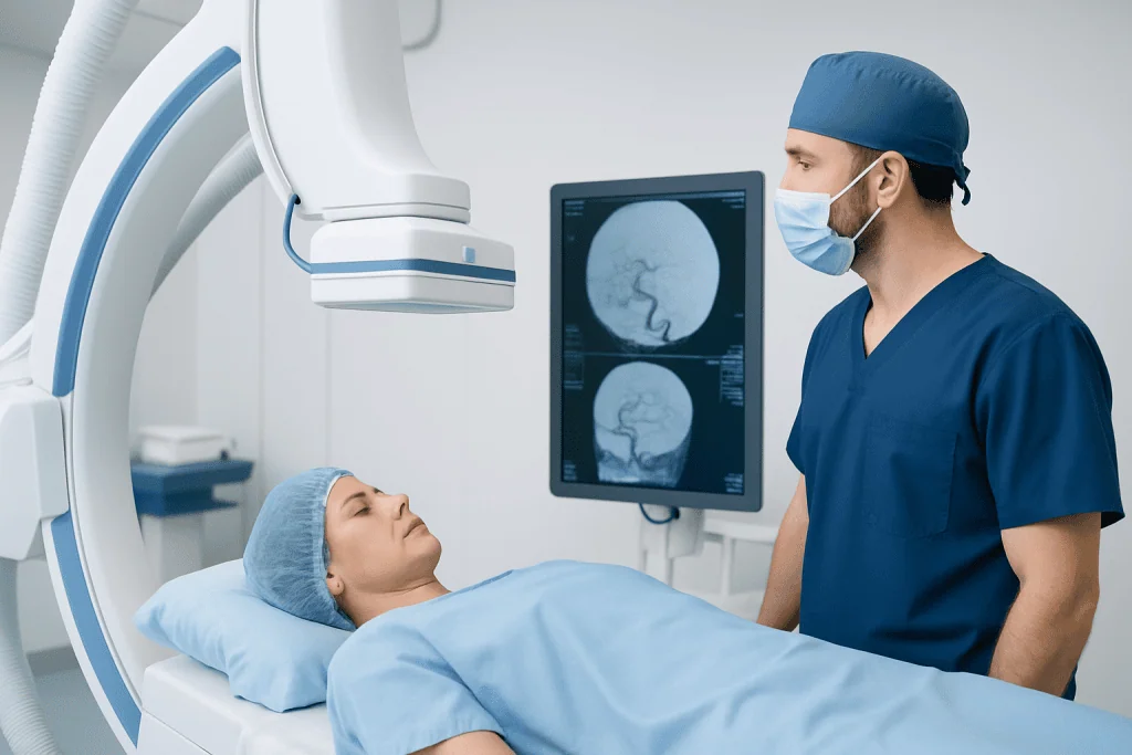
During the angiogram, the patient lies on an X-ray table while the skin over the access site is cleaned and numbed. A catheter is inserted into an artery, often in the wrist or groin, and guided carefully to the vessels of interest. Contrast dye is injected, and rapid X-ray images are taken. Some people notice a brief warm feeling or a metallic taste when the dye is delivered. Sedation may be used, but many cerebral angiograms are done with the patient awake and comfortable.
After the images are completed, the catheter is removed and pressure or a closure device is used to reduce bleeding at the access site. The patient is then observed for a period of time while the team checks blood pressure, neurological status, and the puncture site. Depending on the reason for the test and the person’s overall condition, discharge may occur the same day or after overnight observation.
- Wear comfortable clothing and leave valuables at home.
- Bring a list of medications and allergies.
- Arrange a ride home if sedation is planned or advised.
- Follow instructions about eating, drinking, and medicines carefully.
Safety and Possible Risks
A cerebral angiogram is generally considered safe, especially when it is performed by experienced specialists in an appropriate hospital setting. However, because it involves a catheter, contrast dye, and blood vessels supplying the brain, it is more invasive than CT or MRI-based angiography. For that reason, doctors recommend it when the expected benefit outweighs the risk.
Possible complications include bruising or bleeding at the catheter site, temporary discomfort, allergic reaction to contrast dye, and kidney strain in susceptible patients. A small number of people may develop blood vessel injury, clot formation, or stroke-related complications. The care team works carefully to reduce these risks by reviewing medical history, monitoring the patient closely, and using meticulous technique.
Patients can help improve safety by telling the team about any prior contrast allergy, kidney disease, pregnancy, bleeding disorders, or use of blood thinners. It is also important to report new symptoms after the procedure, such as increasing swelling at the puncture site, severe headache, chest pain, weakness, numbness, trouble speaking, or worsening confusion. Most people complete the test without major problems, but prompt communication matters if something does not feel right.
Recovery, Results, and Next Steps
Recovery after a cerebral angiogram is usually straightforward. Mild soreness, bruising, or tenderness at the wrist or groin can happen for a few days. Patients are commonly advised to rest, drink fluids if appropriate, avoid heavy lifting for a short period, and keep the access site clean and dry according to the care team’s instructions.
Results may sometimes be discussed soon after the procedure, especially if the findings are clear or urgent. In other cases, a specialist reviews the images in detail and then explains what they show. The angiogram may confirm that blood vessels are normal, or it may reveal aneurysms, narrowing, malformations, bleeding sources, or other circulation problems that need monitoring or treatment.
If a problem is found, next steps depend on the diagnosis, symptoms, and overall health of the patient. Options may include medication, follow-up imaging, lifestyle measures, surgery, or catheter-based therapy. In specialized centers, multidisciplinary teams can combine expertise from neurology, neurosurgery, neuroradiology, and vascular care. Acibadem International’s multidisciplinary specialists in JCI-accredited hospitals diagnose and treat neurovascular conditions for international patients when advanced evaluation is needed.
When to Call a Doctor After the Test
Most people recover without difficulty, but some symptoms should not be ignored. Medical advice should be sought promptly if there is persistent bleeding from the catheter site, a rapidly growing lump, severe pain, coldness or color change in the limb used for access, or fever.
Urgent evaluation is especially important if neurological symptoms appear after the test. These may include sudden weakness, facial drooping, speech difficulty, severe headache, vision loss, numbness, loss of balance, or confusion. Although these problems are uncommon, they require immediate attention because they can signal a serious complication.
It is also sensible to contact the care team if nausea, rash, reduced urination, or unusual fatigue develops after contrast exposure, particularly in patients with known kidney issues or past dye reactions. Clear discharge instructions help patients know what is expected and what deserves a quick call or emergency care.
Frequently asked questions
Is a cerebral angiogram the same as a CT angiogram or MR angiogram?
No. A cerebral angiogram usually refers to catheter angiography, in which a thin tube is placed into an artery and contrast dye is injected directly for detailed X-ray imaging. CT angiogram and MR angiogram are less invasive and often used first, but catheter angiography can provide more precise information when needed.
Why would a doctor order a cerebral angiogram after other scans?
A doctor may recommend it when CT, MRI, or ultrasound does not fully explain the problem or when treatment planning requires a more exact view of the blood vessels. It can show very small abnormalities and detailed blood flow patterns that other tests may miss.
Is a cerebral angiogram painful?
Most people do not describe the procedure as painful, though they may feel a needle stick, pressure at the access site, or a brief warm sensation when contrast is injected. The area is numbed, and sedation may be used in some cases to improve comfort.
How long does recovery take after a cerebral angiogram?
Many patients return home the same day after a period of monitoring, while others stay longer depending on the reason for the test and their health status. Mild soreness or bruising can last a few days, but normal light activities often resume fairly soon if the doctor approves.
What are the main risks of a cerebral angiogram?
The main risks include bleeding or bruising at the catheter site, contrast allergy, kidney strain, blood vessel injury, and a small risk of stroke-related complications. These risks are carefully weighed against the benefit of obtaining detailed information that may be important for diagnosis or treatment.
Can a cerebral angiogram be used to treat a problem at the same time?
Sometimes, yes. In certain situations, the angiogram is part of a planned interventional procedure, such as treating an aneurysm or closing an abnormal blood vessel. Whether treatment can happen at the same time depends on the diagnosis, preparation, and the specialist's judgment.
References
- American Heart Association
- National Institute of Neurological Disorders and Stroke
- Radiological Society of North America
- Society of NeuroInterventional Surgery
- Mayo Clinic
This article is for general information only and is not a substitute for professional medical advice. Please consult a qualified doctor about your individual situation.
Explore treatments in Turkey — costs, top hospitals & a free quote
JCI-accredited · board-certified surgeons · reply within 24h
Add us as a Preferred Source to see more of our trusted health content across Google Search, AI Overviews and Discover.