When Is a Cerebral Angiogram Needed After Brain MRI or CT?
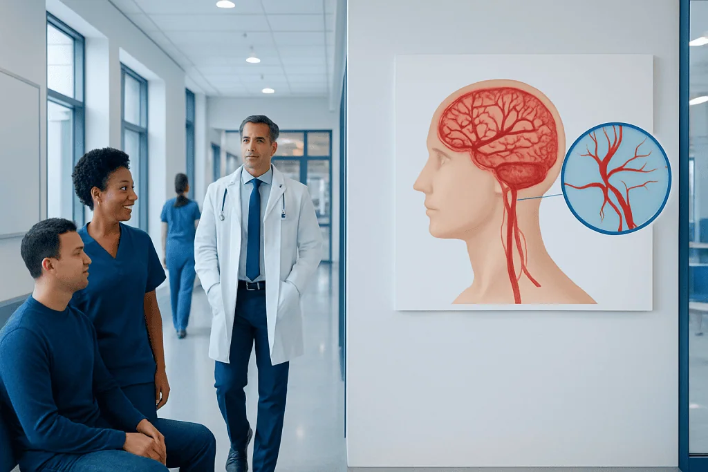
MRI and CT often provide important first answers, but they may not always show brain blood vessels in enough detail. A cerebral angiogram is most often used to evaluate aneurysms, arteriovenous malformations, narrowing, blockage, or bleeding sources.
Key Takeaways
- MRI and CT often provide important first answers, but they may not always show brain blood vessels in enough detail.
- A cerebral angiogram is most often used to evaluate aneurysms, arteriovenous malformations, narrowing, blockage, or bleeding sources.
- This test is an invasive imaging procedure, but it can provide highly detailed, real-time pictures of the brain’s circulation.
- Doctors recommend a cerebral angiogram when the result is likely to change diagnosis, treatment planning, or urgent care decisions.
- The benefits and risks depend on the person’s symptoms, imaging results, and overall health.
A cerebral angiogram may be recommended after a brain MRI or CT when doctors need a more precise look at the blood vessels in the brain. It is usually used to clarify uncertain findings, confirm a suspected vascular problem, or help plan treatment.
Overview: What a Cerebral Angiogram Does
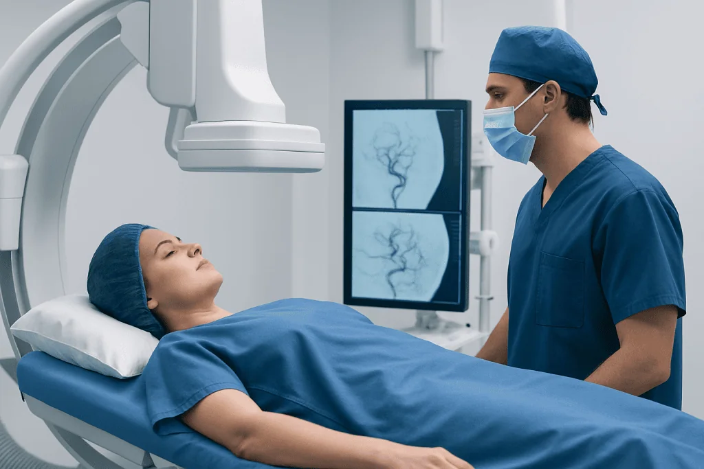
A cerebral angiogram, also called cerebral angiography, is a specialized imaging test that shows the blood vessels in the brain in very fine detail. During the procedure, a doctor injects contrast dye into an artery and takes X-ray images as the dye travels through the brain’s circulation. This allows the medical team to see the size, shape, and flow patterns of blood vessels much more clearly than with many routine scans.
Brain MRI and CT scans are often the first tests used when someone has headaches, stroke-like symptoms, bleeding, seizures, or other neurological concerns. These tests can identify many problems quickly and noninvasively. However, MRI or CT may sometimes suggest a blood vessel abnormality without fully defining it, or the findings may not clearly explain the symptoms.
In these situations, a cerebral angiogram can provide the extra detail needed to make a confident diagnosis. It may help determine whether a person has an aneurysm, an arteriovenous malformation, narrowing of an artery, a blockage, or another vascular condition. In some cases, it can also be used at the same time as treatment, especially in interventional neuroradiology.
When Is a Cerebral Angiogram Needed After Brain MRI or CT?
A cerebral angiogram is usually considered when MRI or CT results are incomplete, uncertain, or show signs of a vascular problem that needs closer evaluation. Doctors generally do not order this test routinely after every brain scan. Instead, they recommend it when the additional detail is likely to affect important medical decisions.
Common reasons include a suspected or known brain aneurysm, unexplained brain bleeding, possible arteriovenous malformation, narrowing or spasm of arteries, or concern about a blockage related to stroke. It may also be used when symptoms strongly suggest a vascular disorder, but noninvasive imaging has not provided a clear answer.
In practical terms, doctors may advise a cerebral angiogram if:
- MRI or CT shows a possible aneurysm that needs confirmation or better measurement
- There is bleeding around the brain and the source is not clear
- A vascular malformation is suspected and treatment planning is needed
- Stroke imaging suggests an unusual blockage, vessel narrowing, or inflammation
- There is concern for conditions such as carotid artery disease or complex intracranial vessel disease that needs precise mapping
The decision is individualized. The care team weighs the quality of the previous scans, the urgency of symptoms, and whether the angiogram could change the next step in care.
Symptoms and Imaging Findings That May Lead to Angiography

Certain symptoms raise concern for a problem in the brain’s blood vessels and may prompt more advanced imaging. These include a sudden severe headache, stroke-like weakness or numbness, trouble speaking, visual changes, unexplained seizures, or sudden confusion. A cerebral angiogram may be considered if MRI or CT suggests that one of these symptoms could be linked to an abnormal blood vessel.
For example, a CT scan might show bleeding in or around the brain, but not clearly identify the exact source. An MRI may suggest an area of abnormal blood flow, but it may not define the vessel anatomy well enough for safe treatment planning. In these settings, angiography can show whether the bleeding comes from an aneurysm or whether an abnormal tangle of vessels is present.
Doctors may also recommend angiography after noninvasive vascular imaging, such as CT angiography or MR angiography, if those tests show a possible abnormality that requires confirmation. This can happen when a small aneurysm is suspected, when vessel narrowing appears complex, or when findings are affected by movement, artifacts, or limited image resolution.
Conditions a Cerebral Angiogram Can Help Diagnose
One of the main reasons for cerebral angiography is to evaluate a brain aneurysm. An aneurysm is a weakened, bulging area in a blood vessel wall. MRI or CT may suggest one, but an angiogram can often show its exact shape, neck, relationship to nearby vessels, and whether it looks suitable for procedures such as brain aneurysm embolization.
Another important use is the assessment of arteriovenous malformations and other abnormal vessel connections. These are complex conditions involving unusual links between arteries and veins. Because treatment depends heavily on the detailed vessel pattern, cerebral angiography remains a key test when planning procedures such as AVM embolization.
The test may also help in evaluating stroke-related vessel problems, including severe narrowing, blockage, dissection, or vasospasm. In some cases, it is used when other scans do not fully explain symptoms of transient ischemic attack or stroke, or when a person may need an endovascular procedure such as mechanical thrombectomy.
Other possible indications include suspected vasculitis affecting the brain, unusual tumors with a rich blood supply, and selected cases of head or neck vascular disease. Depending on the findings, a doctor may also compare them with related conditions such as brain aneurysm or arteriovenous malformation.
How Doctors Decide Between MRI, CT, CTA, MRA, and Angiography
Modern brain imaging includes several different tools, and each has strengths. Standard CT is fast and very useful for detecting acute bleeding or major structural problems. MRI provides detailed images of the brain tissue and can reveal stroke, inflammation, tumors, and many other abnormalities. CTA and MRA are specialized forms of CT or MRI that focus more directly on the blood vessels.
Even so, noninvasive tests have limits. Small aneurysms may be difficult to characterize. Overlapping vessels can make complex malformations harder to understand. Some flow-related abnormalities are better seen in real time, and this is where catheter-based angiography is especially valuable. It remains the reference standard in many situations because it offers very high spatial detail and dynamic views of blood flow.
Doctors choose the next test based on the clinical question. If MRI or CT has already answered the question clearly, an angiogram may not be necessary. But if there is doubt, if treatment is being considered, or if the findings could carry serious consequences, the extra precision of angiography may be worth the added invasiveness.
What to Expect During the Procedure and What the Risks Are
A cerebral angiogram is usually performed in a hospital by an interventional neuroradiologist or another specially trained physician. A thin catheter is inserted through an artery, often in the wrist or groin, and guided toward the arteries supplying the brain. Contrast dye is injected while a series of X-ray images is taken. The procedure team monitors the patient closely throughout.
Patients are typically asked not to eat or drink for a certain period beforehand, and blood tests may be checked in advance. The care team reviews allergies, kidney function, medications, and any history of bleeding problems. Most people receive local anesthesia and medicine to help them relax, though the exact approach depends on the situation and the hospital’s practice.
Like any invasive procedure, cerebral angiography carries risks. These may include bruising or bleeding at the catheter site, allergic reaction to contrast, kidney strain from contrast dye, blood vessel injury, or, rarely, stroke. Although serious complications are uncommon, the medical team recommends the procedure only when the expected diagnostic or treatment benefit outweighs the risk.
After the test, the patient is observed for a period of time and advised to rest, drink fluids if appropriate, and watch for any concerning symptoms such as worsening pain, swelling, weakness, or speech changes. The doctor then explains the results and whether further treatment, follow-up imaging, or monitoring is needed.
Treatment Planning, Follow-Up, and Self-Care
The value of a cerebral angiogram often lies not only in diagnosis, but also in guiding what happens next. If the test confirms an aneurysm, vascular malformation, or vessel blockage, the care team can discuss treatment choices more accurately. These may include monitoring, medicines, surgery, or minimally invasive endovascular procedures depending on the condition, symptoms, and risk profile.
Some angiograms are purely diagnostic, while others may be followed by treatment planning for a separate date. In urgent cases, diagnosis and treatment can sometimes happen in the same session. This is especially relevant when a dangerous bleeding source is found or when a blocked vessel needs rapid intervention.
Self-care after the procedure usually includes protecting the catheter site, avoiding heavy activity for a short period, and following instructions about medications and hydration. People with diabetes, kidney disease, or blood thinners may need more specific advice from their doctor. Keeping follow-up appointments is important so results can be reviewed in context.
Near the end of the care pathway, some patients seek evaluation at centers with neuroradiology, neurology, and neurosurgery working together. Acibadem International’s multidisciplinary specialists and JCI-accredited hospitals diagnose and treat complex brain vascular conditions for international patients when this level of coordinated care is needed.
When to See a Doctor Urgently
Anyone with sudden stroke-like symptoms should seek urgent medical care. Warning signs include facial drooping, weakness on one side, numbness, trouble speaking, vision loss, severe dizziness, or sudden confusion. A sudden, extremely severe headache, especially if it is different from previous headaches, also needs immediate evaluation.
People should also contact a doctor promptly if a recent MRI or CT showed a possible vascular abnormality and they have not yet discussed the findings with a specialist. While many imaging abnormalities do not require emergency treatment, they do need proper interpretation so the next step can be planned safely.
After a cerebral angiogram, urgent care is needed if there is increasing swelling or bleeding at the catheter site, chest pain, shortness of breath, fever, severe headache, or new neurological symptoms such as weakness or difficulty speaking. Quick medical attention helps doctors assess whether these symptoms are part of normal recovery or a sign of a complication.
Frequently asked questions
Is a cerebral angiogram always needed if MRI or CT shows something abnormal?
No. Many abnormalities seen on MRI or CT can be evaluated further with follow-up scans or noninvasive vascular imaging such as CTA or MRA. A cerebral angiogram is usually reserved for cases where doctors need more exact vessel detail to confirm a diagnosis or plan treatment.
Why would a doctor order an angiogram after an MRI already looked at the brain?
MRI is excellent for viewing brain tissue, but it may not always show the blood vessels with enough precision. If there is concern about an aneurysm, vascular malformation, bleeding source, or complex vessel narrowing, angiography can provide a more detailed and dynamic view.
Is a cerebral angiogram the same as CT angiography or MR angiography?
No. CT angiography and MR angiography are noninvasive imaging methods that show blood vessels using CT or MRI technology. A cerebral angiogram is a catheter-based procedure that is more invasive but often gives the most detailed information.
How long does a cerebral angiogram take?
The procedure itself often takes about one to a few hours, depending on why it is being done and how complex the blood vessel anatomy is. Additional time is usually needed before and after the test for preparation, monitoring, and recovery.
Is a cerebral angiogram painful?
Most people feel pressure or a brief sting when the area is numbed, and they may notice warmth when the contrast dye is injected. Significant pain is not expected, and the team works to keep the person comfortable throughout the procedure.
What are the main risks of a cerebral angiogram?
The main risks include bleeding or bruising where the catheter enters the body, reaction to contrast dye, kidney strain, and, rarely, stroke or blood vessel injury. Doctors recommend the test only when its expected benefit is greater than these risks.
References
- World Health Organization
- American Heart Association
- National Institute of Neurological Disorders and Stroke
- Radiological Society of North America
- Society of NeuroInterventional Surgery
This article is for general information only and is not a substitute for professional medical advice. Please consult a qualified doctor about your individual situation.
Explore treatments in Turkey — costs, top hospitals & a free quote
JCI-accredited · board-certified surgeons · reply within 24h
Add us as a Preferred Source to see more of our trusted health content across Google Search, AI Overviews and Discover.