Zygomatic Bone: A Complete Medical Overview
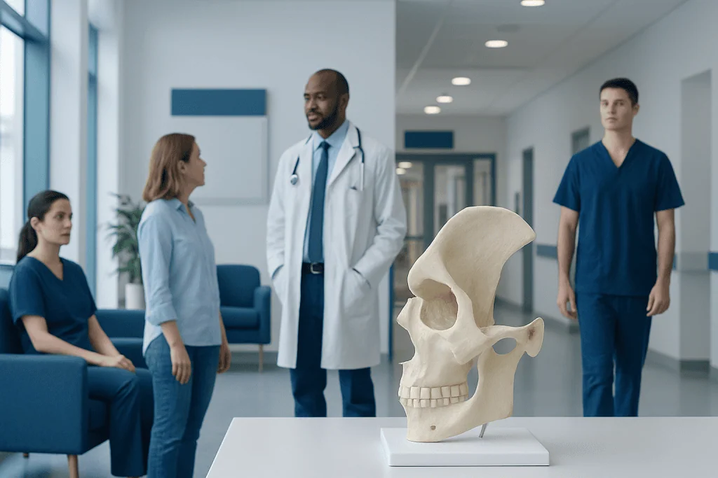
The zygomatic bone is the cheekbone and a key part of the midface and eye socket. It helps protect the eye, supports facial structure, and contributes to chewing muscle attachment.
Key Takeaways
- The zygomatic bone is the cheekbone and a key part of the midface and eye socket.
- It helps protect the eye, supports facial structure, and contributes to chewing muscle attachment.
- The most common medical problem involving the zygomatic bone is fracture after facial trauma.
- Symptoms can include cheek flattening, swelling, bruising, numbness, double vision, or trouble opening the mouth.
- Diagnosis usually involves a physical examination and imaging such as CT scans.
- Treatment depends on severity and may range from observation to surgical repair.
The zygomatic bone is the cheekbone, an important facial bone that helps form the cheek, supports the eye socket, and connects with nearby bones for facial shape and function. Understanding its anatomy, common injuries, symptoms, and treatment options can help patients recognize when medical evaluation is needed.
Overview of the Zygomatic Bone
The zygomatic bone, commonly called the cheekbone, is a paired bone on each side of the face. It helps create the prominence of the cheeks and forms part of the outer wall and floor of the eye socket. In simple terms, it is both a structural and protective bone, giving the midface its contour while helping support nearby tissues.
This bone connects with several other facial bones, including the maxilla, frontal bone, temporal bone, and sphenoid bone. Because of these connections, the zygomatic bone acts like an important meeting point within the facial skeleton. Its position means that injury to the cheek can affect not only appearance but also vision, jaw movement, and sensation.
Although the zygomatic bone is often discussed in anatomy, it is also clinically important. Doctors pay close attention to it after facial trauma because fractures can change the shape of the face, disturb the eye socket, or compress nearby nerves. A clear understanding of this bone helps explain why cheek injuries sometimes need prompt specialist assessment.
Anatomy and Function
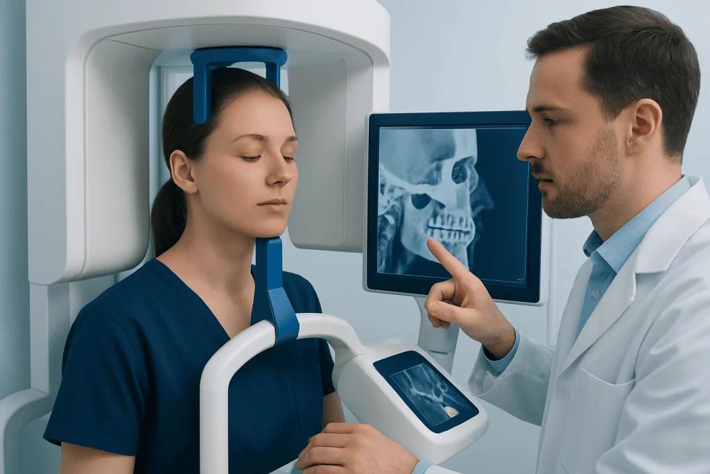
The zygomatic bone has several surfaces and processes that join with neighboring bones. Together these structures form the zygomatic arch region, the lateral part of the orbit, and the front of the cheek. This arrangement gives the face width and definition while helping maintain the shape of the midface.
Functionally, the zygomatic bone does more than create appearance. It helps protect the eye by contributing to the orbital rim and socket. It also provides attachment points for muscles and connective tissues involved in facial expression and chewing, especially in the region where the jaw muscles pass beneath the cheekbone arch.
A nerve called the zygomaticofacial nerve passes through this area and provides sensation to part of the cheek. This is one reason why an injury to the zygomatic bone can cause numbness or tingling. Because the bone sits close to the maxillary sinus and the orbit, problems in this area may overlap with symptoms seen in maxillary sinusitis or orbital injuries, though the causes are different.
Common Problems That Affect the Zygomatic Bone
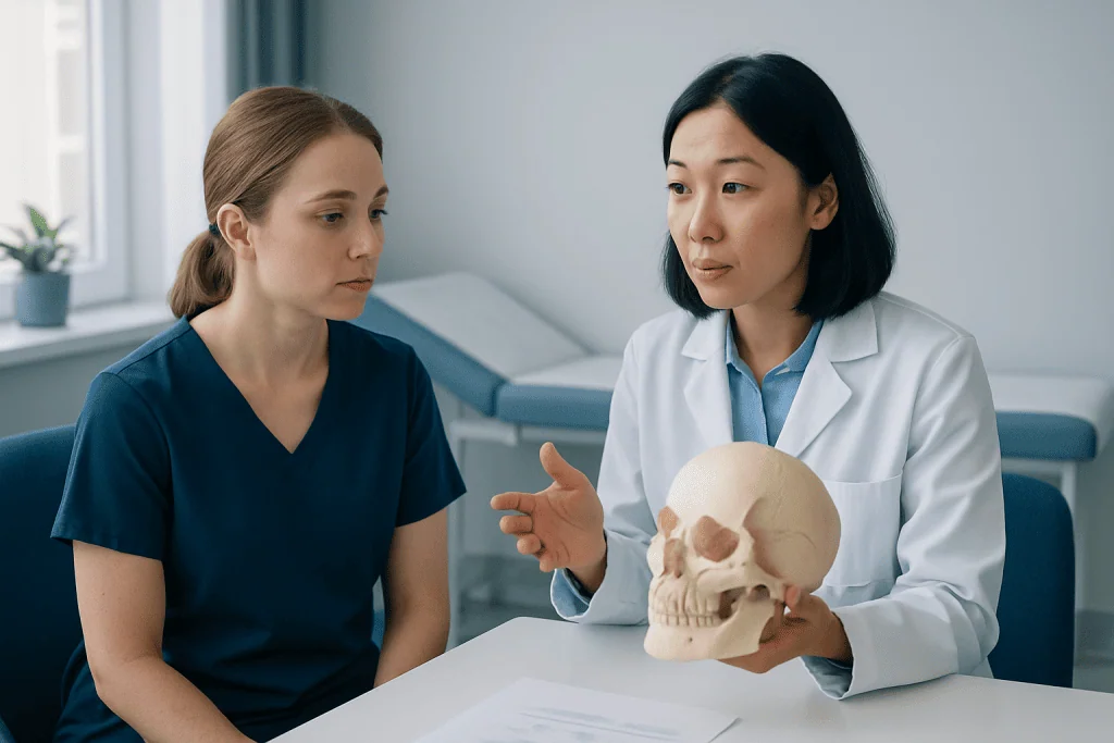
The most common condition involving the zygomatic bone is fracture, usually caused by direct trauma. Falls, sports injuries, vehicle accidents, and physical impact to the face are frequent reasons. Doctors may describe these injuries as zygomatic fractures, zygomaticomaxillary complex fractures, or cheekbone fractures depending on the exact pattern.
Not every problem is traumatic. Less commonly, the zygomatic bone may be affected by congenital differences, infection spreading from nearby tissues, benign bone lesions, or tumors involving the facial bones. These are much less common than fracture, but they may still lead to swelling, pain, or visible changes in facial contour.
Some people notice ongoing cheek discomfort after previous trauma, facial surgery, or dental procedures near the upper jaw. In certain cases, symptoms may reflect associated problems rather than the bone itself, such as nerve irritation or nearby joint issues. A clinician may consider related facial conditions, including temporomandibular joint disorders, when symptoms include jaw pain or difficulty opening the mouth.
Symptoms of Injury or Disease
Symptoms vary depending on the cause, but trauma to the zygomatic bone often leads to pain, swelling, and bruising over the cheek and around the eye. Some people notice that the cheek looks flatter than usual or that one side of the face appears asymmetrical. Tenderness is common, especially when touching the cheekbone area.
Because the zygomatic bone forms part of the orbit, eye-related symptoms can occur. These may include double vision, pain with eye movement, reduced eye movement, or a feeling that the eye is sunken. Any change in vision after facial trauma deserves timely medical assessment.
Numbness of the cheek, upper lip, side of the nose, or upper teeth can happen if nearby sensory nerves are affected. Trouble opening the mouth or pain when chewing may suggest involvement of the zygomatic arch or surrounding muscles. In non-traumatic conditions, symptoms may develop more gradually and include swelling, facial fullness, or persistent discomfort.
- Cheek pain, swelling, or bruising
- Flattening or asymmetry of the face
- Numbness in the cheek or upper lip
- Double vision or eye movement problems
- Jaw pain or difficulty opening the mouth
- Persistent facial tenderness after injury
How Doctors Diagnose Zygomatic Bone Problems
Diagnosis starts with a careful history and physical examination. The doctor will ask how the injury happened, when symptoms began, and whether there are changes in vision, bite, sensation, or jaw movement. During the exam, they may gently feel the facial bones for tenderness, step-offs, or movement that suggests a fracture.
The eyes are also assessed because midface injuries can affect the orbit. A clinician may check eye movement, vision, pupil response, and the position of the eyeball. They may also look inside the mouth and assess how widely the jaw opens, since cheekbone injuries can influence the mechanics of chewing.
Imaging is usually needed when fracture is suspected. CT scanning is often the most useful test because it shows the facial bones in detail and helps define whether the fracture is displaced or affecting the orbit. X-rays may be used in some settings, but CT is generally more informative for treatment planning. When facial trauma is complex, evaluation may involve ENT, maxillofacial, plastic surgery, ophthalmology, or neurosurgery teams.
Treatment Options
Treatment for a zygomatic bone problem depends on the cause, the severity of symptoms, and whether the bone has shifted out of place. Mild, non-displaced fractures may be treated conservatively with observation, pain control, ice, rest, and follow-up imaging or examinations. Patients are often advised to avoid further facial trauma and follow instructions about eating, activity, and nose blowing if the sinus or orbit is involved.
More significant fractures may need surgical repair, especially if there is facial asymmetry, double vision, limited jaw opening, sensory loss, or disruption of the orbital rim or floor. Surgical treatment aims to restore normal bone position and stabilize the fracture, often using small plates and screws. When the eye socket is affected, surgeons may also need to address orbital support through orbital fracture surgery.
Some patients require multidisciplinary treatment because facial trauma can involve several structures at once. Depending on the injury pattern, doctors may recommend maxillofacial surgery or plastic and reconstructive surgery to improve function and facial symmetry. Near the end of the care pathway, rehabilitation may include swelling management, soft-diet guidance, and monitoring of sensation, bite, and eye symptoms. Acibadem International’s multidisciplinary specialists and JCI-accredited hospitals diagnose and treat facial bone conditions for international patients when this level of care is needed.
Prevention and Self-Care
Because most zygomatic bone injuries are caused by trauma, prevention mainly focuses on safety. Seat belt use, protective sports gear, helmets where appropriate, and fall prevention strategies can lower the risk of facial injury. People in contact sports or high-risk work settings should use recommended face protection whenever possible.
After a diagnosed cheekbone injury, self-care should follow the treating clinician’s advice. This may include using cold compresses early on, sleeping with the head elevated, avoiding pressure on the injured side, and eating softer foods if chewing is uncomfortable. Patients should not attempt to press or reshape the bone themselves.
Recovery is usually smoother when follow-up instructions are taken seriously. If surgery has been performed, patients may be asked to keep incisions clean, avoid strenuous activity for a period, and report any new visual symptoms, fever, increasing swelling, or worsening pain. Self-care is supportive, but it does not replace professional assessment for suspected fractures.
When to Seek Medical Care
Medical care should be sought after any significant blow to the cheek or eye area, especially if there is visible facial deformity, severe swelling, persistent pain, or numbness. Even when the skin looks normal, an underlying fracture may still be present. A prompt evaluation can help prevent missed injuries affecting the orbit, nerves, or jaw function.
Urgent assessment is especially important if there is double vision, blurred vision, inability to move the eye normally, eye pain, heavy bleeding, or difficulty opening the mouth. These symptoms may signal a more complex facial injury that needs imaging and specialist care. Any loss of consciousness, severe headache, confusion, or suspected head injury should also be treated as urgent.
People should also see a doctor for slowly developing cheek swelling, unexplained facial asymmetry, or persistent tenderness that does not improve. While many causes are minor, ongoing symptoms deserve evaluation to rule out fracture healing problems, nerve involvement, infection, or less common bone conditions.
Frequently asked questions
What is the zygomatic bone?
The zygomatic bone is the cheekbone on each side of the face. It helps form the cheek, supports part of the eye socket, and connects with several other facial bones.
Can a zygomatic bone fracture heal without surgery?
Yes, some mild fractures that are not displaced may heal with observation and supportive care. Surgery is more likely when there is bone displacement, facial asymmetry, eye symptoms, numbness, or problems with jaw movement.
What are the signs of a broken cheekbone?
Common signs include cheek pain, swelling, bruising, flattening of the cheek, numbness, and difficulty opening the mouth. Some people also develop double vision or pain around the eye if the orbit is involved.
How is a zygomatic bone injury diagnosed?
Doctors diagnose it through a physical examination and imaging, most often a CT scan. The assessment usually includes checks for vision changes, facial symmetry, sensation, and jaw movement.
Is the zygomatic bone important for the eye?
Yes, it forms part of the outer wall and floor of the eye socket. That is why injuries to this bone can sometimes affect eye position, eye movement, or vision.
How long does recovery take after a cheekbone injury?
Recovery time depends on the severity of the injury and whether surgery is needed. Minor injuries may improve over weeks, while more complex fractures can take longer and require follow-up to monitor healing and function.
References
- American Academy of Otolaryngology–Head and Neck Surgery
- American Association of Oral and Maxillofacial Surgeons
- American Academy of Ophthalmology
- National Library of Medicine
- Merck Manual
This article is for general information only and is not a substitute for professional medical advice. Please consult a qualified doctor about your individual situation.
Explore treatments in Turkey — costs, top hospitals & a free quote
JCI-accredited · board-certified surgeons · reply within 24h
Add us as a Preferred Source to see more of our trusted health content across Google Search, AI Overviews and Discover.

More from the Health Library

WBC: A Complete Medical Overview

Life Expectancy After Mini Stroke: What Patients Need to Know

Lactate Dehydrogenase Low — Explained by Medical Evidence, Not Myths

Zepbound Storage — Explained by Medical Evidence, Not Myths
Lateral Collateral Ligament Sprain: An Evidence-Based Guide for Patients

Masseter Botox: A Complete Medical Overview
Orthopedic Surgery & Traumatology Specialists at Acibadem

Prof. Dr. Aziz Kaya Alturfan
Orthopedic Surgery & Traumatology
Dr. Baran Şen
Orthopedic Surgery & Traumatology
Prof. Dr. Barış Kocaoğlu
Orthopedic Surgery & Traumatology
Dr. Bengü Kasım
Physical Medicine & Rehabilitation