

Quick answer
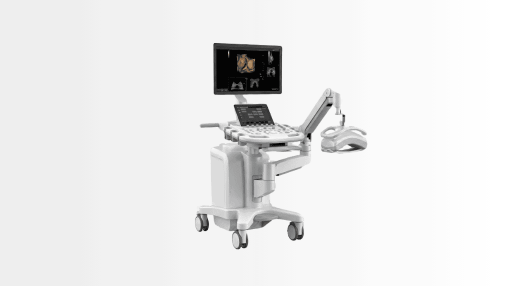
4D breast ultrasonography is an imaging technology that creates real-time volumetric views of breast tissue using sound waves, without ionising radiation. At Acibadem in Turkey, it may be used to assess areas of concern, help characterize breast lesions, and guide decisions about further imaging or diagnostic procedures.
4D Breast Ultrasonography
4D Breast Ultrasonography is an advanced imaging technology that uses sound waves to create detailed, real-time volumetric views of breast tissue. Unlike X-ray-based imaging, ultrasound does not use ionising radiation. It can help specialists examine an area of concern from different angles, assess breast tissue in greater detail and support informed decisions about follow-up care.
At Acibadem International, 4D breast ultrasonography may be used as part of a personalised breast assessment plan. Our radiology and breast care teams combine imaging findings with your medical history, symptoms, previous reports and, when appropriate, other diagnostic tests. The goal is to provide clear information while avoiding unnecessary invasive procedures whenever possible.
This technology can be particularly helpful when specialists need a closer look at a breast lump, an imaging finding or an area that is difficult to assess with standard two-dimensional ultrasound alone. It is not a replacement for every breast imaging test. Your doctor will recommend the most suitable approach according to your age, breast density, symptoms, risk factors and previous imaging results.
What Is 4D Breast Ultrasonography?
4D breast ultrasonography is a type of ultrasound examination designed to provide three-dimensional images of breast tissue in real time. The term “4D” refers to 3D imaging that can be viewed dynamically as the specialist scans the breast. This allows the radiologist to assess the structure, shape and position of an area of interest while observing the images live.
Traditional ultrasound produces two-dimensional images, similar to individual slices through tissue. With 4D imaging, the system gathers a larger volume of information. The radiologist can review the area in multiple planes and reconstruct the images to better understand its features. This can improve visualisation of certain findings and support more precise communication between imaging, surgery and breast care teams.
Because the examination uses ultrasound waves rather than radiation, it may be considered when a radiation-free imaging method is appropriate. However, the best breast imaging plan differs from one patient to another. Your specialist will explain whether 4D breast ultrasonography should be used alone, alongside mammography, with magnetic resonance imaging, or as part of another diagnostic pathway.
How 4D Breast Ultrasonography Works
During the examination, a trained specialist places a small handheld device called a transducer on the skin of the breast. The transducer sends high-frequency sound waves into the tissue. These sound waves travel through the body and reflect back differently depending on the type of tissue they encounter.
The ultrasound system converts these returning signals into images. Advanced software then helps create detailed volumetric views that can be examined in real time. The radiologist may view the same area in different directions, measure structures accurately and compare findings with previous studies where available.
A clear gel is applied to the skin before scanning. The gel helps the transducer maintain good contact with the skin and improves the quality of the images. The gel may feel cool, but the examination is generally well tolerated and does not require injections, incisions or recovery time.
In some situations, Doppler ultrasound may also be used. Doppler technology assesses blood flow in and around a finding. This information can be useful when characterising certain lesions, although it is always interpreted alongside the complete clinical and imaging picture.
Clinical Applications
4D breast ultrasonography can support the assessment of a wide range of breast concerns. It may be used when a patient notices a change, when another imaging examination identifies an area that requires clarification, or when additional detail is needed before planning the next steps.
| Clinical applications | How 4D breast ultrasonography may help |
|---|---|
| Assessment of a palpable lump | Helps evaluate a breast lump or localised area of thickening and determine whether it appears cystic, solid or requires further assessment. |
| Characterisation of lesions | Provides volumetric views that may help specialists assess lesion shape, margins, orientation and internal features. |
| Dense breast tissue assessment | May offer additional information in patients with dense breast tissue, where some findings can be more difficult to evaluate with other imaging methods alone. |
| Follow-up of known findings | Can support imaging follow-up of selected benign-appearing findings or areas previously identified by a specialist. |
| Evaluation of breast symptoms | May be used to investigate symptoms such as focal pain, a newly noticed area of concern, skin changes or nipple-related concerns when clinically appropriate. |
| Assessment after previous imaging | Can help clarify findings seen on mammography, standard ultrasound or other imaging examinations. |
| Pre-procedure planning | May assist the clinical team in reviewing the location and dimensions of an area before a biopsy, surgery or another recommended diagnostic procedure. |
| Post-treatment or post-procedure review | May contribute to the assessment of selected changes after surgery, treatment or an intervention, based on your doctor’s recommendation. |
Although 4D breast ultrasonography can provide valuable information, imaging results must always be interpreted by an experienced specialist. Some findings may need further imaging, short-term follow-up or tissue sampling through a biopsy. An ultrasound examination can guide decision-making, but it does not by itself confirm or exclude every breast condition.
Conditions It May Help Specialists Assess
4D breast ultrasonography may be considered in the assessment of both benign and suspicious breast findings. Many breast changes are non-cancerous, such as simple cysts, fibroadenomas, areas of fibrocystic change or inflammation. Imaging can help specialists understand the nature of a finding and decide whether observation, further imaging or a biopsy is appropriate.
The technology may also support the assessment of breast lesions that need closer evaluation for possible malignancy. Features such as irregular borders, changes in tissue architecture or particular patterns of blood flow may guide the radiologist’s recommendations. No single imaging feature should be interpreted in isolation, and your care team will discuss the meaning of your results in the context of your overall health.
For patients with a personal or family history of breast disease, prior breast surgery, breast implants or previous imaging abnormalities, the examination plan may need to be tailored. Acibadem International coordinates specialist review so that your assessment is based on your individual clinical needs rather than a one-size-fits-all pathway.
Benefits and Advantages for Patients
One of the main advantages of 4D breast ultrasonography is its ability to provide detailed, real-time images without ionising radiation. This can be reassuring for patients who require additional assessment or follow-up and want to understand why a particular imaging method has been recommended.
The examination is usually comfortable and non-invasive. There are no needles, contrast injections or radiation exposure as part of a routine ultrasound scan. Most patients can return to their normal daily activities immediately after the appointment.
Volumetric imaging can also support more confident clinical decisions. By examining an area from multiple perspectives, specialists may obtain clearer information about its dimensions, location and characteristics. This can be valuable when planning further tests, monitoring an existing finding or coordinating care with breast surgeons and other specialists.
Support for coordinated careFindings can be reviewed by radiology and breast care specialists when multidisciplinary input is needed.
| Patient benefits at a glance | What this can mean for you |
|---|---|
| Radiation-free imaging | Uses sound waves rather than ionising radiation to create breast images. |
| Real-time assessment | The specialist can examine the area as images are produced and focus on the specific region of concern. |
| Volumetric detail | 3D imaging views may help clarify the shape, size and position of a finding. |
| Comfortable examination | Usually performed externally with ultrasound gel and a handheld transducer, without injections or incisions. |
| Personalised care planning | Results can be considered alongside your symptoms, history and other imaging to guide the next steps. |
| No routine recovery period | Patients can generally continue with travel plans, work or usual activities after the examination. |
What to Expect During Your 4D Breast Ultrasound
Before your appointment, the Acibadem International patient team can help coordinate your visit, collect available medical records and arrange language support where needed. Sharing previous mammograms, ultrasound reports, pathology results or surgical records before travel can help the specialist compare current findings with earlier examinations.
At your consultation, the radiologist or breast specialist will review your medical history and discuss any symptoms you have noticed. You may be asked about previous breast procedures, medications, family history and prior imaging. This information helps the team determine whether 4D breast ultrasonography is the right examination for your situation.
For the scan, you will usually lie comfortably on an examination bed, often with one arm positioned above your head. The specialist applies ultrasound gel to the breast and moves the transducer gently across the skin. You may feel light pressure in certain areas, especially if there is tenderness, but the examination is not usually painful.
The scan itself commonly takes a relatively short time, although the duration can vary depending on whether one or both breasts need assessment, whether the examination is focused on a particular area and whether previous images need to be reviewed. If the specialist identifies an area that needs additional attention, more images or measurements may be taken during the same visit.
After the scan, the images are reviewed and a report is prepared. Your results may be discussed with you directly or shared through your coordinating team, depending on your care plan. If further imaging, follow-up or a biopsy is recommended, your international patient coordinator can help arrange the next stage of care with the appropriate department.
Preparing for the Examination
In most cases, no special preparation is required for 4D breast ultrasonography. You can normally eat, drink and take regular medications as advised by your doctor. Wearing a comfortable two-piece outfit may make changing easier on the day of your appointment.
If possible, avoid applying heavy lotions, powders or oils to the breast area on the day of the scan, as they can interfere with the contact between the transducer and the skin. Bring any previous breast imaging, reports and relevant medical documentation. If you have had breast surgery, a biopsy, implants or treatment for breast cancer, please let your care team know in advance.
Patients travelling from abroad are encouraged to send available records before arrival. This helps Acibadem International coordinate the most appropriate specialist appointments and reduce unnecessary repetition of tests where clinically possible.
Safety and Quality of Care
Ultrasound imaging has been used in clinical practice for many years and does not involve ionising radiation. When performed by trained professionals and interpreted by experienced radiologists, it is a well-established tool for breast assessment. As with any diagnostic examination, the value of the test depends on selecting it appropriately and interpreting the findings in the full clinical context.
Acibadem International supports care in JCI-accredited hospital settings, where clinical pathways, patient safety processes and quality standards are designed to support reliable care. Breast imaging is performed by qualified professionals using advanced equipment and structured reporting practices. When a finding requires further discussion, radiology teams can work closely with general surgery and breast care specialists.
Your doctor will explain any case-specific considerations, including whether another imaging method may be needed. A normal ultrasound does not eliminate the need for recommended screening or follow-up, and an abnormal result does not automatically mean cancer. The purpose of careful imaging assessment is to ensure that each patient receives the most appropriate next step.
Why 4D Breast Ultrasonography Matters for International Patients
Travelling for breast assessment can feel stressful, particularly when you are waiting for answers. International patients often need more than an imaging appointment: they need clear communication, coordinated scheduling and access to specialist guidance in a language they understand.
At Acibadem International, our international patient services team can help organise the process from the first medical review through to appointments, interpretation support and follow-up planning. Where appropriate, your reports can be reviewed before travel so that the clinical team can advise whether 4D breast ultrasonography may be useful within your diagnostic plan.
If additional care is needed, we can help coordinate consultations with radiology, general surgery and other relevant specialists. This integrated approach can be especially important when time is limited or when patients wish to combine imaging, specialist review and recommended next steps within a carefully planned visit.
After your assessment, your team can provide the medical documentation needed for continuity of care at home. If further follow-up is recommended, the plan can be explained clearly so that you understand what should happen next, whether in Türkiye or with your local doctor.
Frequently Asked Questions
Is 4D breast ultrasonography safe?
Yes. It uses ultrasound waves and does not involve ionising radiation. The examination is routinely performed by trained specialists who follow established safety and quality standards. Your doctor will explain whether it is suitable for your individual situation.
Is 4D breast ultrasonography painful?
The examination is generally comfortable. You may feel gentle pressure from the transducer, particularly over a tender area, but there are usually no injections, incisions or invasive steps involved.
Can 4D breast ultrasonography detect breast cancer?
It can help specialists assess suspicious findings and guide decisions about further tests. However, no imaging examination alone can diagnose every breast condition with complete certainty. If an area has concerning features, your specialist may recommend additional imaging or a biopsy.
Does it replace mammography?
Not always. Mammography, ultrasound and magnetic resonance imaging provide different types of information. Your specialist will recommend the most appropriate combination of tests based on your age, symptoms, breast density, medical history and previous imaging results.
Will my insurance cover the examination?
Coverage depends on your individual insurance policy and the clinical reason for the examination. You can share your insurance information with the international patient team, who can help check available options and explain the planned process before treatment.
How do I get started?
You can submit your available medical reports through a consultation request. A specialist can review the information provided and advise whether 4D breast ultrasonography may be appropriate as part of your personalised breast assessment plan.
Related Medical Units
Available at these Hospitals


















Speak with our medical team
Find out how this technology could support your diagnosis or treatment.