

Quick answer
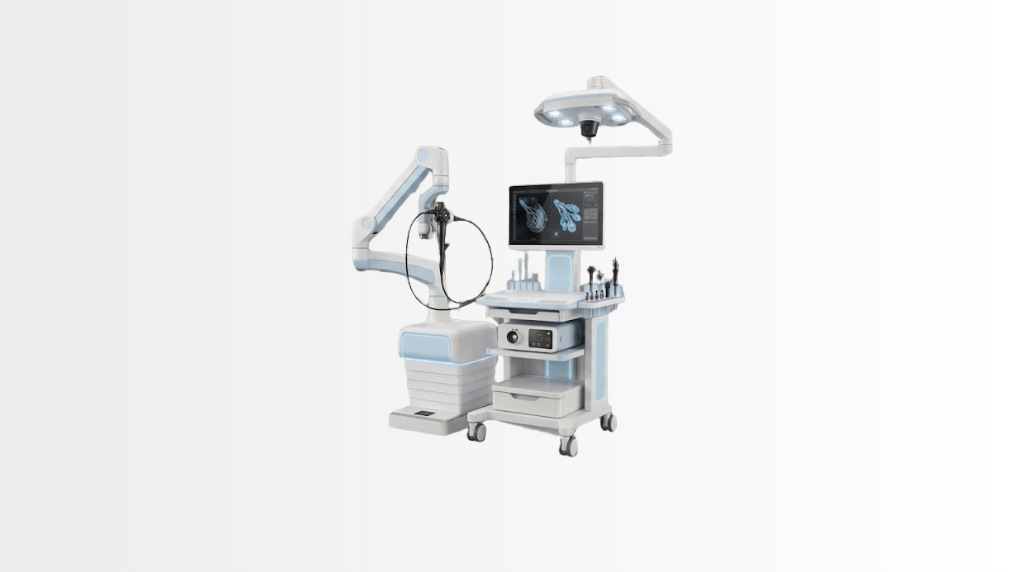
Ductoscopy is a minimally invasive procedure that uses a very thin endoscope to examine the inside of breast milk ducts through the nipple, often when persistent or suspicious nipple discharge is present. At Acibadem in Turkey, breast specialists use ductoscopy to assess duct-related abnormalities and guide further diagnostic or treatment planning when needed.
Ductoscopy: A Micro-Endoscopic Examination of the Breast Milk Ducts
Ductoscopy is a minimally invasive diagnostic procedure that allows breast specialists to look directly inside the milk ducts of the breast. Using a very fine endoscope, the specialist can examine the duct system through the nipple and assess possible causes of nipple discharge.
It is particularly helpful when nipple discharge is persistent, occurs from a single duct, is clear or bloody, or is associated with other clinical findings that require closer investigation. Ductoscopy can provide detailed, real-time images from inside the duct, helping the care team understand whether further tests, monitoring or treatment may be needed.
At Acibadem International, ductoscopy supports a precise and personalised breast assessment. Our General Surgery and breast care teams combine advanced diagnostic technology with clinical examination, breast imaging and pathology services when appropriate. The aim is to reach a clear diagnosis while avoiding unnecessary invasive procedures whenever possible.
What Is Ductoscopy?
Ductoscopy is also known as breast duct endoscopy or mammary ductoscopy. It uses a micro-endoscope, a very thin flexible camera designed to enter the opening of a breast milk duct at the nipple.
Milk ducts are small channels within the breast that carry milk toward the nipple. Although they are most active during breastfeeding, they remain present throughout adult life. In some cases, a duct may become affected by inflammation, a benign growth, a narrowing or another abnormality that can lead to nipple discharge.
Traditional breast imaging examinations, such as mammography, ultrasound and magnetic resonance imaging, remain important tools in breast assessment. However, they do not always show the inside surface of an individual milk duct. Ductoscopy can add a different level of detail by enabling direct visual inspection of the duct lining.
The procedure is not a routine screening test for all patients. It is considered when a breast specialist believes direct assessment of the duct may help clarify an unresolved clinical concern. Your suitability depends on your symptoms, imaging results, medical history and physical examination.
How Ductoscopy Works
Ductoscopy begins with identifying the duct from which discharge is coming, if discharge is present. The specialist then gently introduces a tiny endoscope through the natural opening of that duct in the nipple.
The micro-endoscope contains a camera and light source. It transmits magnified images to a monitor, allowing the specialist to assess the inner walls of the duct in real time. Depending on the clinical situation, the team may document areas of inflammation, narrowing, debris, changes in the duct lining or a lesion that needs further evaluation.
In selected cases, ductoscopy can help guide the planning of a targeted surgical procedure. For example, if a suspicious duct-related lesion is identified, direct visualisation may help the surgeon focus treatment on the relevant duct rather than removing more tissue than necessary. Whether this is appropriate will always be decided individually.
Ductoscopy is usually performed as an outpatient assessment. It may be carried out with local anaesthesia or another comfort measure depending on the patient’s needs, the planned procedure and the specialist’s recommendation.
What the Specialist May Look For
The purpose of ductoscopy is not simply to find an abnormality, but to understand its location, appearance and possible clinical significance. Findings are interpreted together with breast imaging, laboratory tests, pathology results and the patient’s symptoms.
- Intraductal papillomas, which are often benign growths within a milk duct
- Inflammation or irritation of the duct lining
- Duct narrowing, widening or obstruction
- Changes associated with duct ectasia, where ducts become widened
- Blood, debris or other material within the duct
- Lesions that may require biopsy, surgery or additional imaging
- Findings that help distinguish a duct-related issue from other causes of nipple discharge
Clinical Applications
Ductoscopy is most often used in the assessment of nipple discharge, especially when symptoms are persistent or concerning and a focused examination of the duct system may add useful information. It may be considered alongside ultrasound, mammography, breast MRI, cytology, biopsy or surgical assessment.
Not all nipple discharge indicates a serious condition. Discharge can occur for many reasons, including hormonal changes, medication use, pregnancy-related changes, infection or benign breast conditions. A specialist assessment is important when discharge is spontaneous, occurs from one breast, comes from one duct, contains blood, or is accompanied by a lump, skin change or nipple change.
| Clinical applications | How ductoscopy may help | What may happen next |
|---|---|---|
| Persistent nipple discharge | Allows direct examination of the duct thought to be responsible for the discharge. | The specialist may recommend observation, further imaging, tissue sampling or treatment according to the findings. |
| Bloody or clear discharge from a single duct | May help identify a duct-related source, such as a benign intraductal lesion or another abnormality. | Findings are reviewed with imaging and may guide a targeted surgical plan if needed. |
| Suspected intraductal papilloma | Can provide visual information about a possible growth within a milk duct. | Additional diagnostic steps or removal may be discussed depending on symptoms and overall risk assessment. |
| Unclear findings after standard breast imaging | Offers a complementary view of the inside of the duct when external imaging does not fully explain symptoms. | Your multidisciplinary team may combine all results to recommend the most appropriate next step. |
| Planning selected duct-focused surgery | May help localise the affected duct or support a more targeted operative approach in selected patients. | The surgeon will explain whether duct excision or another procedure is medically appropriate. |
Benefits and Advantages for Patients
The main advantage of ductoscopy is direct visualisation. Rather than relying only on images taken from outside the breast, the specialist can examine the inside of the duct itself. This can contribute to a more informed clinical decision when symptoms and other tests do not provide a complete answer.
For many patients, the possibility of a focused assessment is reassuring. Ductoscopy may support earlier clarification of the cause of nipple discharge and can help the team decide whether a surgical procedure is necessary. In some cases, reassuring findings may reduce the need for more extensive diagnostic surgery; in others, it may help make surgery more precise.
Like all medical procedures, ductoscopy has limitations. A very narrow duct, duct blockage, discomfort during the examination or an inability to access the relevant duct may affect whether the procedure can be completed. It also cannot replace all forms of breast imaging or tissue diagnosis. Your specialist will explain what ductoscopy can and cannot show in your individual case.
| Patient benefits at a glance | What this can mean for your care |
|---|---|
| Direct view inside the milk duct | Detailed visual information may help clarify the source of duct-related nipple discharge. |
| Minimally invasive approach | The procedure is performed through the natural duct opening in the nipple, without a large incision for the examination itself. |
| No ionising radiation from the endoscopic examination | Ductoscopy uses an optical camera system rather than X-rays. |
| Supports personalised decision-making | Results can be considered together with your symptoms, imaging findings and health history. |
| Potentially more targeted treatment planning | When treatment is required, duct visualisation may assist selected surgical planning. |
| Outpatient-focused care | Many patients can return home or to their accommodation on the same day, subject to their doctor’s advice. |
What to Expect During a Ductoscopy Procedure
Your ductoscopy journey begins with a consultation with a breast specialist or General Surgery specialist. The doctor will ask about your symptoms, including the colour, frequency and pattern of nipple discharge. They will review any previous breast imaging, biopsy reports, medical records and medications.
If you are travelling from abroad, Acibadem International can help coordinate the review of your available reports before your visit. Sharing mammography, ultrasound, MRI, pathology and consultation reports in advance may help the specialist prepare for your appointment and advise on the most suitable next steps.
Before the Procedure
You may be asked to provide details about allergies, medicines, previous breast procedures, pregnancy status and other medical conditions. Follow your team’s instructions about eating, drinking and medications. If sedation is planned, you may need to arrange for an accompanying person to support you after the procedure.
The specialist will explain the expected purpose of ductoscopy, possible benefits, limitations and potential risks. You will have the opportunity to ask questions before giving consent.
During the Procedure
You will be positioned comfortably while the breast and nipple area are prepared using sterile techniques. The specialist identifies the duct opening and carefully inserts the micro-endoscope. A local anaesthetic may be used to improve comfort, depending on the procedure plan.
As the endoscope advances, the specialist views the duct on a monitor. The examination is generally focused on the duct system associated with your symptoms. Images may be recorded as part of your medical documentation.
You may feel pressure, mild discomfort or a brief sensation of fullness during the procedure. Tell the team immediately if you experience pain or anxiety. Your comfort and safety are important throughout the examination.
After the Procedure
Following ductoscopy, you may have mild tenderness, temporary nipple sensitivity or a small amount of discharge. Most patients can resume light daily activities according to their doctor’s advice. Your team will provide individual aftercare instructions and explain when you can return to work, travel or exercise.
The specialist will discuss preliminary findings with you when possible. If additional testing is needed, the international patient team can help coordinate appointments, translate key information and support the planning of your care pathway.
Possible Risks and Safety Considerations
Ductoscopy is generally considered a minimally invasive procedure when performed by trained specialists in an appropriate clinical setting. However, every medical procedure carries potential risks. These may include temporary discomfort, minor bleeding, infection, irritation of the duct, swelling or an unsuccessful examination if the duct cannot be accessed.
Rarely, a procedure may not provide a definitive answer. In that situation, the specialist may recommend additional imaging, follow-up appointments, biopsy or surgical duct excision depending on the clinical concern.
You should seek medical advice promptly if you develop increasing pain, redness, fever, marked swelling, persistent bleeding or other symptoms that concern you after the procedure. Your care team will explain which symptoms require urgent attention and how to access support during your stay in Türkiye and after you return home.
Safety, Quality and Accreditation
At Acibadem International, ductoscopy is integrated into a wider breast health assessment pathway. Our specialists work with radiology, pathology, oncology and surgical teams when multidisciplinary review is needed. This collaborative approach helps ensure that diagnostic findings are interpreted in the context of the whole patient, not in isolation.
Our JCI-accredited hospitals follow structured quality and patient safety standards. These include infection prevention measures, informed consent processes, equipment protocols, clinical documentation and coordinated follow-up planning. Ductoscopy is performed by trained specialists who assess whether the procedure is suitable and who explain the specific considerations relevant to your case.
Technology is most effective when it is used thoughtfully. For this reason, the decision to perform ductoscopy is based on medical need rather than a one-size-fits-all approach. Your specialist will recommend the least invasive option that can provide clinically meaningful information.
How Ductoscopy Fits Into Breast Care
Ductoscopy is one part of a comprehensive approach to breast symptoms. Depending on your age, symptoms and clinical findings, your assessment may also include breast examination, mammography, ultrasound, MRI, blood tests, discharge analysis, needle biopsy or surgical consultation.
When there is a concern about a breast lesion, a tissue diagnosis may still be necessary. Ductoscopy can provide valuable visual information, but it does not replace pathology when a biopsy is required to confirm the nature of an abnormality.
For patients with benign findings, the team may recommend monitoring and symptom follow-up. For patients who need treatment, the care plan may involve a targeted surgical procedure, medication for an underlying infection or inflammation, or referral to another specialist service. Recommendations are tailored to the individual.
Why Ductoscopy Matters for International Patients
Travelling for breast assessment can feel stressful, particularly when symptoms are worrying or when previous test results have been inconclusive. Acibadem International provides coordinated support designed to make the process clearer and more manageable for patients travelling from overseas.
Before your visit, our international patient team can help collect and organise your existing medical documents for specialist review. This may include imaging files, pathology reports, previous consultation notes and a summary of your symptoms. Early review can help identify the most appropriate specialist and support efficient appointment planning.
During your care, multilingual support helps you understand the purpose of the procedure, what the findings may mean and what follow-up is recommended. Your care coordinators can assist with scheduling, communication between departments and practical planning around your medical appointments.
If further treatment is required, your team can discuss the expected sequence of care and the likely time needed in Türkiye. Where appropriate, follow-up plans can be coordinated with your doctor at home, supported by medical reports and relevant test documentation.
Every patient’s needs are different. Some patients travel for a focused diagnostic opinion, while others require a complete breast assessment and treatment plan. Our goal is to provide clear communication, coordinated care and specialist guidance at every stage.
Frequently Asked Questions
Is ductoscopy painful?
Many patients tolerate ductoscopy well, although some discomfort or pressure may occur. Local anaesthesia or other comfort measures may be used when appropriate. Your specialist will explain what to expect and adapt the approach to your individual needs.
Does ductoscopy use radiation?
No. The ductoscopy examination itself uses a miniature camera and light source rather than ionising radiation. However, your doctor may still recommend mammography or other imaging studies as part of a complete breast assessment.
Can ductoscopy diagnose breast cancer?
Ductoscopy may identify changes inside a milk duct that need further evaluation, but it does not replace a biopsy when tissue diagnosis is necessary. A diagnosis of cancer requires careful assessment by the appropriate specialists and, in most cases, pathology examination of tissue.
Will I need surgery after ductoscopy?
Not necessarily. The need for surgery depends on your symptoms, ductoscopy findings, imaging results and overall risk assessment. Some patients may only need follow-up, while others may benefit from a targeted procedure.
Will my insurance cover ductoscopy?
Coverage depends on your insurance policy, the medical indication and the requirements of your insurer. Our international patient team can help you understand the documentation needed and clarify available options before your planned care.
How can I get started?
You can submit your available medical reports and breast imaging for an initial consultation request. A specialist will review your case and advise whether ductoscopy or another diagnostic approach may be suitable for your individual treatment plan.
Related Medical Units
Available at these Hospitals


















Speak with our medical team
Find out how this technology could support your diagnosis or treatment.