JCI-accredited · 45+ hospitals & clinics · 90+ countries served · 24/7 multilingual support
Imaging & Diagnostics

EBUS

Diagnosis
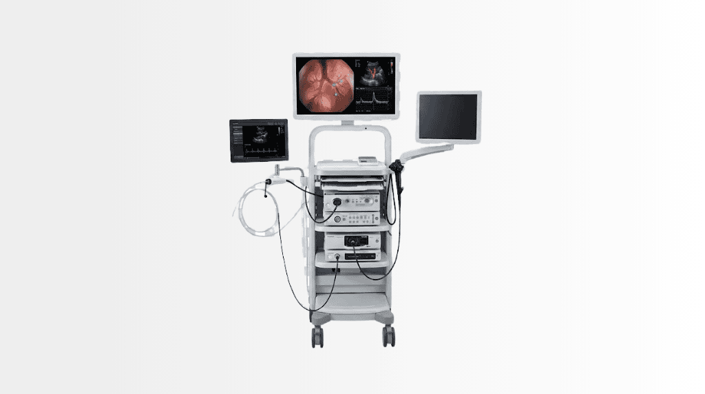
Modern ultrasound machine used for medical imaging at Acibadem Hospitals.

Quick answer

Endobronchial ultrasound (EBUS) is a minimally invasive bronchoscopic technology that uses ultrasound to assess lymph nodes and lesions near the airways and guide needle biopsies. At Acibadem in Turkey, EBUS is performed by respiratory specialists to support the diagnosis and staging of lung and chest conditions, often reducing the need for surgical diagnostic procedures.

EBUS: Endobronchial Ultrasound for Precise Lung and Lymph Node Diagnosis

Endobronchial ultrasound, commonly called EBUS, is an advanced diagnostic procedure that helps specialists examine areas in and around the airways without open surgery. It combines bronchoscopy, using a thin flexible camera passed through the mouth into the airways, with ultrasound imaging to view lymph nodes and selected lesions within the chest.

EBUS is particularly valuable when doctors need accurate information about enlarged chest lymph nodes, suspected lung cancer, inflammation, infection or other changes seen on imaging. During the procedure, ultrasound can guide a fine needle to collect tissue samples from areas that may otherwise require a surgical diagnostic procedure.

At Acibadem International, Pulmonology specialists use EBUS as part of a coordinated diagnostic pathway. The aim is to obtain clear, reliable information that supports timely decisions and a personalised treatment plan, while avoiding unnecessary invasive steps whenever clinically appropriate.

What Is EBUS in Plain Language?

EBUS is a type of bronchoscopy with an ultrasound probe at the tip of the scope. A bronchoscope is a narrow, flexible tube equipped with a camera. It is introduced through the mouth and gently guided into the breathing passages while the patient is sedated or under anaesthesia, according to the individual treatment plan.

Standard bronchoscopy allows the specialist to see the inside of the airways. EBUS adds ultrasound imaging, allowing the team to look beyond the airway wall and assess structures nearby. These may include lymph nodes in the centre of the chest, certain masses or lesions close to the airways, and surrounding tissues.

When a tissue sample is needed, EBUS can guide a small needle through the airway wall and into the target area. This technique is called EBUS-guided transbronchial needle aspiration, or EBUS-TBNA. It enables the specialist to collect cells or tissue for laboratory analysis without making an incision in the chest.

How EBUS Works

EBUS brings together real-time ultrasound images and precise bronchoscopic access. This combination helps the specialist identify the most appropriate area to examine or sample during the procedure.

Real-Time Ultrasound Guidance

The ultrasound probe produces images using sound waves rather than ionising radiation. These sound waves create a picture of tissues and lymph nodes located around the airways. The specialist can assess their location, size, shape and other features that may help guide the next diagnostic step.

Real-time imaging is important because it allows the needle to be directed accurately while the target area is visible. This helps improve sample quality and may reduce the need for repeat testing in suitable cases.

Targeted Tissue Sampling

If sampling is recommended, a fine needle is passed through the bronchoscope to obtain cells or small tissue samples. The samples are sent to a pathology laboratory, where specialists examine them for signs of cancer, infection, inflammatory disease or other conditions.

In some cases, the laboratory may perform additional tests on the specimen. These can include immunohistochemical, microbiological, molecular or genetic analyses, depending on the suspected condition and the amount of tissue available. Such results can play an important role in determining the most appropriate treatment approach.

A Collaborative Diagnostic Pathway

EBUS findings are considered alongside a patient’s symptoms, medical history, chest imaging and laboratory results. Depending on the case, Pulmonology specialists may work closely with radiologists, pathologists, thoracic surgeons, medical oncologists, radiation oncologists and infectious disease specialists.

This multidisciplinary approach is particularly important when EBUS is used to investigate suspected lung cancer or complex chest conditions. It helps ensure that diagnostic information is interpreted in the right clinical context before treatment recommendations are made.

Clinical Applications of EBUS

EBUS is most commonly used to evaluate lymph nodes and lesions in the chest that are close to the central airways. It may be recommended after a chest X-ray, CT scan, PET-CT scan or other investigation shows an area requiring closer assessment.

Suitability depends on the location of the finding, the patient’s general health, previous test results and the specific clinical question. Not every lung lesion or lymph node can be reached by EBUS, and some patients may need another diagnostic method. Your specialist will explain which approach is most appropriate for your case.

Clinical applications How EBUS may help What the team may assess
Assessment of enlarged chest lymph nodes Provides ultrasound-guided access to lymph nodes near the airways for detailed evaluation and tissue sampling. Possible infection, inflammation, sarcoidosis, lymphoma, metastatic disease or other causes of lymph node enlargement.
Lung cancer diagnosis May help obtain tissue from suspicious lymph nodes or selected lesions near the airways. Whether abnormal cells are present and which pathology tests may be needed to guide treatment planning.
Lung cancer staging Can assess lymph nodes in the chest to support accurate staging before treatment. Whether lymph nodes contain cancer cells, which can influence decisions about surgery, systemic treatment or radiotherapy.
Investigation of sarcoidosis May obtain samples from enlarged lymph nodes or related chest findings without surgical biopsy. Features consistent with granulomatous inflammation and the need for further clinical assessment.
Investigation of tuberculosis or other infections Can provide samples for pathology and microbiological testing when infection is suspected. Signs of infection and the potential need for culture, molecular testing or other laboratory analysis.
Evaluation of unexplained chest abnormalities Helps clarify findings on imaging when lymph nodes or lesions require a more direct assessment. The nature of the abnormality and whether monitoring, further testing or treatment is recommended.

Conditions EBUS May Help Investigate

EBUS is a diagnostic technology rather than a treatment for a specific disease. It may help the medical team investigate a range of conditions affecting the lungs, airways and lymph nodes in the chest.

  • Suspected or confirmed lung cancer requiring diagnosis or staging.
  • Enlarged mediastinal or hilar lymph nodes, including nodes located between the lungs or near the major airways.
  • Sarcoidosis and other inflammatory conditions that may affect chest lymph nodes.
  • Tuberculosis, fungal infections and selected other infections when tissue or microbiological samples are needed.
  • Possible spread of cancer to chest lymph nodes from the lung or another part of the body.
  • Abnormal findings on CT, PET-CT or chest X-ray that need further investigation.
  • Selected lung lesions located close enough to the airways to be safely accessed through bronchoscopy.

Pathology results are essential, but they are not interpreted in isolation. The treating specialist reviews the results alongside imaging findings and the patient’s overall health. In some cases, EBUS provides the necessary answer. In others, further imaging, follow-up or another biopsy method may be advised.

Benefits and Advantages for Patients

One of the main advantages of EBUS is that it can provide important diagnostic information through the natural airway route. For suitable patients, this may reduce the need for surgical procedures performed through the chest wall or under more extensive anaesthesia.

EBUS can also support earlier diagnosis and more accurate staging. This is especially important in cancer care, where treatment decisions are often based on the exact location and extent of disease. A well-targeted tissue sample can help the care team recommend the most appropriate next step with greater confidence.

Patient benefits at a glance What this can mean for you
Minimally invasive approach EBUS is performed through the mouth and airways, without an incision in the chest for many suitable diagnostic cases.
Real-time imaging Ultrasound helps the specialist see the target area while guiding the biopsy needle.
Targeted sampling Samples can be taken from specific lymph nodes or lesions, supporting accurate laboratory assessment.
Potentially fewer diagnostic procedures When EBUS provides an adequate sample, it may help avoid or delay the need for a more invasive surgical diagnostic procedure.
No ionising radiation from the ultrasound component The ultrasound images used during EBUS are produced with sound waves rather than X-ray radiation.
Support for personalised treatment planning Pathology and staging information can help the multidisciplinary team tailor recommendations to your diagnosis.
Usually short recovery period Many patients return home the same day, subject to their recovery, clinical status and travel arrangements.

What to Expect During an EBUS Procedure

Your EBUS journey begins with a detailed medical review. The Pulmonology specialist considers your symptoms, imaging scans, previous biopsy results, medicines, allergies and medical history. The team will confirm whether EBUS is suitable and explain why it is being recommended.

Before the Procedure

You may be asked to avoid food and drinks for a period before the procedure. The exact instructions depend on the planned sedation or anaesthesia and your individual health needs. It is important to tell the team about blood-thinning medicines, diabetes medication, respiratory conditions, previous reactions to anaesthesia and any allergies.

For international patients, our coordination team can help organise the review of medical records before travel. Please share recent scan reports, pathology reports, discharge summaries, medication lists and imaging files whenever available. This helps the specialist assess your case efficiently and plan the appropriate appointments.

Before EBUS, you will have the opportunity to discuss the procedure, expected benefits, possible alternatives and known risks. The team will ask for your informed consent after answering your questions.

During the Procedure

EBUS is usually performed in a dedicated bronchoscopy or procedure unit. Depending on your clinical needs and the hospital protocol, you may receive sedative medication, local anaesthetic to numb the throat, or general anaesthesia. Your breathing, heart rate, oxygen level and blood pressure are monitored throughout.

The specialist gently passes the flexible bronchoscope through the mouth and into the airways. The ultrasound probe is used to identify the target lymph node or lesion. If a biopsy is required, the specialist uses ultrasound guidance to take samples through a fine needle.

The procedure duration varies according to the number and location of areas being examined and sampled. Your specialist will give you an individual estimate before the procedure. You should not expect to feel pain from the needle sampling itself, although some patients may experience temporary throat discomfort or coughing afterwards.

After the Procedure

After EBUS, you will rest in a monitored recovery area until the effects of sedation or anaesthesia have worn off sufficiently. Your throat may feel numb for a short time, so the team will advise you when it is safe to drink and eat.

Mild sore throat, hoarseness, cough or small streaks of blood in the sputum can occur after bronchoscopy and biopsy. These symptoms are often temporary, but you should follow the discharge instructions carefully and contact the medical team if you have concerns.

If you received sedation or anaesthesia, you will need a responsible adult to accompany you after discharge. Driving, operating machinery, signing important documents and making major decisions should be avoided until the effects of medication have fully passed, as advised by your care team.

Understanding Results and Next Steps

Some initial findings may be discussed soon after the procedure, but final pathology results generally require laboratory processing. The time needed can vary depending on the type of analysis required. More complex testing, including molecular or microbiological studies, may take longer.

Once results are available, your specialist will explain what they mean and discuss the next steps. If cancer is diagnosed or suspected, your case may be reviewed by a multidisciplinary team to ensure that all relevant expertise is included in the treatment recommendation.

Possible next steps may include monitoring, medication, additional imaging, referral for surgery, radiotherapy, systemic treatment or further diagnostic testing. Every plan is individual and based on your diagnosis, overall health, preferences and treatment goals.

Safety, Quality and Accreditation

EBUS is routinely used in modern respiratory medicine and is performed by trained specialists in an appropriately equipped clinical setting. Like all medical procedures, it involves potential risks. Your team will explain the risks relevant to your individual case before treatment.

Possible complications can include temporary sore throat, cough, bleeding, infection, low oxygen levels, reactions to sedative or anaesthetic medicines, or pneumothorax in uncommon circumstances. The likelihood and significance of these risks depend on factors such as the location of the sampled area, your lung function, your medications and other health conditions.

At Acibadem International, EBUS procedures are delivered within JCI-accredited hospital environments, where patient safety, infection prevention, medication management and clinical quality processes are central to care. The procedure team follows established protocols for pre-procedure assessment, monitoring, recovery and follow-up.

Your safety assessment is personal. If EBUS is not the most appropriate option, the specialist may recommend another diagnostic pathway. The goal is always to balance diagnostic value with your comfort, safety and overall medical needs.

Why EBUS Matters for International Patients

Travelling for diagnosis can feel uncertain, especially when you are waiting for answers about a lung or chest condition. A coordinated EBUS pathway can help international patients access specialist assessment, imaging review, procedure planning and follow-up in an organised way.

Our international patient team supports communication before, during and after your visit. They can help coordinate appointments, arrange medical record review, assist with language support and keep you informed about the sequence of consultations, tests and expected recovery needs.

When planning travel, it is wise to allow sufficient time for the specialist consultation, the procedure, recovery and discussion of preliminary or final results. The appropriate length of stay depends on your individual case, the need for additional tests and your clinical condition. Our team can provide guidance after your records have been reviewed.

If you need to return home before all laboratory results are available, the care team can explain how results and medical reports will be shared and how follow-up recommendations can be coordinated. Where appropriate, your records can support continuity of care with your doctor at home.

Frequently Asked Questions About EBUS

Is EBUS safe?

EBUS is widely used by trained respiratory specialists and is generally considered a minimally invasive diagnostic procedure. However, no procedure is risk-free. Your medical team will review your health status, medications and imaging findings to assess whether it is appropriate for you and explain any case-specific considerations.

Is EBUS painful?

Patients receive sedation, local anaesthesia or general anaesthesia depending on the planned procedure and individual clinical needs. Most patients do not experience pain during EBUS, although throat irritation, coughing or mild discomfort can occur afterwards.

Will I need to stay overnight?

Many EBUS procedures are performed as day cases. However, observation or an overnight stay may be recommended if you have certain medical conditions, require additional procedures, experience a complication or need closer monitoring. Your specialist will advise you based on your individual situation.

Can EBUS replace surgery?

For many suitable patients, EBUS can obtain tissue from chest lymph nodes or selected lesions without a surgical diagnostic procedure. However, it does not replace surgery in every situation. If the target cannot be reached, if the sample is not sufficient or if a different type of tissue is needed, another approach may be recommended.

Will my insurance cover EBUS?

Insurance coverage depends on your policy, insurer, diagnosis and pre-authorisation requirements. You can share your insurance details with our international patient team, who can help clarify available options and explain the financial process before care proceeds.

How do I get started?

You can submit your medical reports, imaging findings and previous pathology results through a consultation request. A specialist will review the available information and advise whether EBUS may be appropriate within your diagnostic or treatment plan. The international patient team will then help coordinate the next steps in your preferred language.

Add Acıbadem on Google

Add us as a Preferred Source to see more of our trusted health content across Google Search, AI Overviews and Discover.

Share this page
Where it is used

Related Medical Units

Where to find it

Available at these Hospitals

Speak with our medical team

Find out how this technology could support your diagnosis or treatment.

We’re With You at Every Step

How can we help you today?

We value your privacy We use essential cookies to run this site and, with your consent, analytics cookies to understand how it is used and improve it. You can accept, reject, or choose what to allow. See our Cookie Policy.
















