

Quick answer
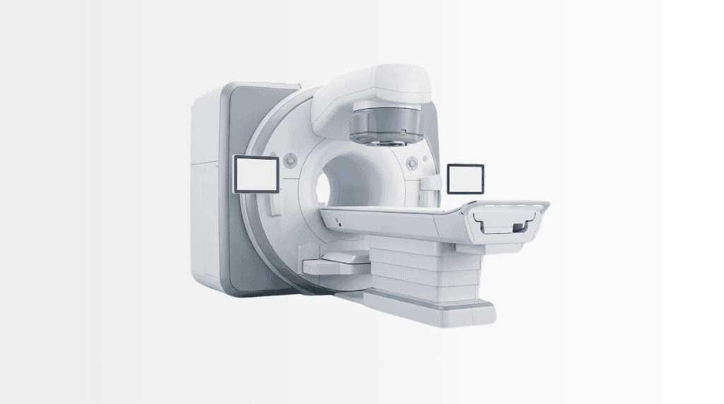
Harmony is an advanced MRI technology that produces detailed diagnostic images using magnetic fields and radio waves, without ionising radiation. At Acibadem in Turkey, Harmony MRI examinations are planned around the clinical question and individual safety needs, including assessment of implants and other relevant considerations.
Harmony MRI Technology
Harmony is an advanced magnetic resonance imaging (MRI) technology designed to provide detailed diagnostic images while keeping patient comfort at the centre of the experience. It supports radiology specialists in seeing internal structures clearly, helping them assess a wide range of conditions and plan the most appropriate next steps.
MRI is a non-invasive imaging method that uses strong magnetic fields and radio waves to create images of organs, soft tissues, bones, joints, blood vessels and other structures inside the body. Unlike X-ray-based imaging methods, MRI does not use ionising radiation. This can make it an important option when detailed soft-tissue imaging is needed or when repeat imaging may be clinically appropriate.
At Acibadem International, Harmony supports a patient-focused approach to diagnosis. Our radiology teams use advanced imaging to help identify changes earlier, measure findings accurately and provide the clinical information needed for personalised treatment planning. Every scan is planned around the patient’s medical history, symptoms and individual safety requirements.
What Is Harmony?
Harmony is an efficient MRI system developed to deliver high-quality imaging in a comfortable and well-coordinated clinical setting. It helps radiologists obtain detailed images that can support diagnosis, monitoring and treatment decisions across many medical specialties.
High-resolution imaging is especially valuable when doctors need to assess soft tissues that may not be as clearly visible with other imaging methods. This includes the brain, spine, muscles, ligaments, joints, abdominal organs, pelvic organs and certain blood vessels.
The technology is used as part of a wider diagnostic pathway. A Harmony MRI scan does not replace the expertise of the clinical team; rather, it provides precise information that specialists can interpret alongside your symptoms, examination findings, laboratory tests and previous medical reports.
How Harmony MRI Works
During an MRI scan, the body is placed within a magnetic field. Radiofrequency signals are then used to obtain information from tissues inside the body. A computer processes these signals and creates detailed cross-sectional images that can be reviewed from different angles.
Different tissues respond differently during the scan. This allows radiologists to distinguish between structures such as muscles, tendons, nerves, cartilage, fluid, fat and organs. In some cases, a contrast agent may be recommended to make specific tissues, blood vessels or areas of concern easier to assess. Your doctor will explain whether contrast is suitable and necessary for your scan.
Harmony is designed to support efficient imaging workflows and consistent image quality. The exact scan duration depends on the body area being examined, the clinical question and whether additional image sequences or contrast imaging are required.
Imaging Without Ionising Radiation
MRI uses magnetic fields and radio waves rather than ionising radiation. This is one of the key reasons MRI is frequently used for detailed evaluation of soft tissues and for follow-up imaging where clinically indicated.
However, MRI is not suitable for every patient or every clinical situation. Before the appointment, the team reviews your medical history and completes a safety assessment. This is particularly important if you have an implanted device, metal fragments, a previous operation, kidney disease, pregnancy, claustrophobia or any other condition that may affect the scan process.
Clinical Applications
Harmony MRI can support the work of radiology and multidisciplinary clinical teams across many areas of medicine. The scan protocol is selected according to the symptoms being investigated and the information your doctor needs.
| Clinical application | How MRI may help | Examples of conditions or concerns assessed |
|---|---|---|
| Brain and neurological imaging | Provides detailed views of the brain, spinal cord and surrounding structures. | Headaches, dizziness, seizures, stroke assessment, suspected brain lesions, multiple sclerosis evaluation and neurological follow-up. |
| Spine and nerve imaging | Helps assess discs, vertebrae, spinal nerves and soft tissues around the spine. | Back or neck pain, disc herniation, spinal stenosis, nerve compression, sciatica and spinal injury. |
| Musculoskeletal imaging | Shows muscles, tendons, ligaments, cartilage, joints and bone marrow in high detail. | Sports injuries, joint pain, ligament tears, meniscus injuries, shoulder conditions, hip disorders and unexplained musculoskeletal symptoms. |
| Abdominal imaging | Supports detailed assessment of abdominal organs and selected blood vessels. | Liver findings, pancreatic conditions, kidney concerns, adrenal lesions, bowel-related assessment and follow-up of known abnormalities. |
| Pelvic imaging | Helps evaluate pelvic organs and surrounding soft tissues. | Urological conditions, gynaecological concerns, prostate assessment, pelvic pain and selected cancer staging or monitoring needs. |
| Cardiovascular imaging | May provide information about the heart, major vessels and blood flow in suitable patients. | Selected heart muscle conditions, congenital heart concerns, vascular abnormalities and follow-up after specialist assessment. |
| Oncology support | Can help identify, characterise, stage or monitor certain tumours and treatment response. | Suspected soft-tissue masses, organ lesions, cancer staging, surveillance and treatment planning when MRI is clinically appropriate. |
The clinical use of MRI varies from person to person. Your specialist may recommend Harmony MRI as a first diagnostic test, as a follow-up examination after another scan, or as part of preparation for surgery, oncology treatment, rehabilitation or long-term monitoring.
Benefits and Advantages for Patients
For many patients, the main value of Harmony MRI is the combination of detailed imaging and a non-invasive approach. Clear images can help the clinical team make more confident decisions, avoid unnecessary procedures where possible and design a treatment plan that reflects your individual diagnosis.
Patient comfort is also an important part of the MRI experience. The care team explains each stage of the appointment, helps you prepare safely and remains available throughout the scan. If you are concerned about enclosed spaces, loud scanner sounds, contrast material or your ability to remain still, please discuss this before the examination so the team can guide you appropriately.
Non-invasive examinationMost scans do not require surgery or instrument insertion, helping make the diagnostic process more straightforward for many patients.
| Patient benefit | What it can mean for your care |
|---|---|
| Detailed image quality | Supports careful assessment of soft tissues, organs, joints, nerves and other structures that may require high-resolution imaging. |
| No ionising radiation | MRI uses magnetic fields and radio waves, rather than X-ray radiation, while still providing valuable diagnostic information. |
| Personalised scan planning | The examination is selected and tailored according to the body area, symptoms, medical history and clinical question. |
| Support for earlier decisions | Clear imaging may help specialists recognise clinically important changes and recommend the next appropriate step without unnecessary delay. |
| Multidisciplinary care support | Images can be reviewed by radiologists and relevant specialists to assist coordinated treatment planning. |
| International patient coordination | Patients travelling from abroad can receive help with medical record review, appointment planning, language support and follow-up arrangements. |
What to Expect During a Harmony MRI Procedure
Your experience begins before you arrive for the scan. For international patients, Acibadem International teams can help organise the process around your travel plans and medical needs. You may be asked to share previous reports, scan images, laboratory results, referral notes and information about any implants or prior procedures in advance.
Before Your Scan
A specialist reviews your history and confirms whether MRI is appropriate for your case. You will complete an MRI safety questionnaire and should mention any metal implants, pacemakers, neurostimulators, hearing devices, joint replacements, vascular clips, dental work, retained metal fragments or previous operations.
Some implants are MRI-compatible under specific conditions, while others may require special precautions or make MRI unsuitable. It is important not to assume that an implant is safe for MRI without a formal review. If you have documentation about an implant, please bring it or share it with the team beforehand.
You may also be asked about kidney function, allergies, pregnancy or breastfeeding if contrast material could be considered. The team will explain any preparation requirements. Depending on the planned scan, you may be asked to avoid food for a short period beforehand, although many MRI examinations require no special dietary preparation.
During the Scan
You will be asked to remove metal objects such as jewellery, watches, hair accessories, removable dental appliances, hearing aids, bank cards and other items that may be affected by the magnetic field. You may change into a gown if needed.
During imaging, you lie on a padded examination table that moves into the MRI scanner. The team positions the relevant body area carefully to obtain the required images. It is important to stay as still as possible during each image sequence, as movement can reduce image clarity.
The scanner produces tapping, knocking or humming sounds while it is working. Hearing protection is provided. You can communicate with the MRI team through the system during the examination, and staff monitor you throughout the scan.
If contrast is required, it may be given through a small intravenous line. Contrast-enhanced MRI can provide additional information in selected situations, but it is not needed for every examination. Your radiologist and referring doctor will consider the expected benefit and your individual medical history before recommending it.
After the Scan
Most patients can return to normal daily activities after a non-contrast MRI. If you have received contrast, the team may provide additional instructions based on your health status and the type of examination performed.
A radiologist reviews the images and prepares a report. Your treating specialist then discusses the findings in the context of your overall health and treatment goals. For patients returning home after medical travel, the international patient team can help coordinate access to reports, images and follow-up communication where appropriate.
Safety, Clinical Standards and Accreditation
MRI is routinely used worldwide and is considered a well-established diagnostic imaging method when appropriate safety screening is completed. At Acibadem International, Harmony MRI examinations are performed by trained radiology professionals who follow established imaging, infection prevention and patient safety procedures.
Acibadem hospitals serving international patients include JCI-accredited facilities. JCI accreditation reflects a commitment to recognised standards for quality improvement, patient safety and coordinated care. Safety processes include verifying patient identity, reviewing medical history, checking MRI suitability and ensuring that all relevant clinical information is available before scanning.
Although MRI does not use ionising radiation, the magnetic environment requires careful preparation. The MRI safety screening process is essential. Always tell your care team about implanted devices, metallic foreign bodies, prior surgery, possible pregnancy, kidney disease, anxiety in enclosed environments and any previous reaction to contrast materials.
If MRI is not the most suitable examination for you, your doctor may recommend another diagnostic option. The aim is always to choose the imaging method that provides the most useful information with appropriate attention to safety, comfort and clinical need.
Why Harmony Matters for International Patients
When travelling abroad for medical care, patients often need clear answers quickly. They may arrive with a complex medical history, reports in another language or images obtained at different hospitals. Harmony MRI can be integrated into a coordinated diagnostic pathway that helps specialists review your condition and determine the next steps efficiently.
Acibadem International supports patients before, during and after their visit. Our teams can assist with appointment coordination, medical document review, interpreter support and communication between relevant specialists. This can be particularly helpful when several consultations, imaging examinations or treatment planning sessions need to be arranged within a limited travel period.
Where clinically appropriate, MRI findings can be discussed in a multidisciplinary setting. For example, a radiologist may work alongside neurologists, orthopaedic specialists, oncologists, surgeons, cardiologists or other physicians involved in your care. This shared review helps ensure that imaging results are considered as part of the full clinical picture.
After your scan, you can receive guidance on the recommended next step, whether that involves further tests, treatment planning, a specialist consultation or follow-up with your doctor at home. The goal is to make the diagnostic journey understandable, well organised and centred on your individual needs.
Frequently Asked Questions
Is Harmony MRI safe?
Harmony MRI is used by trained specialists following strict safety procedures. MRI does not use ionising radiation, but it is not suitable for every person without assessment. Your team will review implants, metal exposure, medical conditions and any other factors that may affect safety.
Will the scan be uncomfortable?
MRI is generally painless, but some people find it challenging to remain still or feel anxious in an enclosed space. The scan also produces loud sounds. The team will explain what to expect, provide hearing protection and support you throughout the examination. Tell us in advance if you have claustrophobia, pain or difficulty lying flat.
Do I need contrast material?
Not every MRI examination requires contrast. Contrast may be recommended when it can provide additional information for your diagnosis or treatment planning. Your doctor will explain why it is being considered and review your medical history before proceeding.
Will my insurance cover Harmony MRI?
Coverage depends on your individual insurance policy, clinical indication and insurer requirements. You can share your insurance details with our international patient team, who can help clarify available options and explain the planned care pathway before your visit.
How do I get started?
You can submit a consultation request and share your available medical reports, previous imaging and clinical history. A specialist will review the information and advise whether Harmony MRI may be appropriate as part of your personalised assessment and treatment plan.
Related Medical Units
Available at these Hospitals


















Related Treatments
Speak with our medical team
Find out how this technology could support your diagnosis or treatment.