Oral Pathology
Oral pathology evaluates ulcers, lumps, color changes, cysts, and other mouth or jaw lesions to identify benign, infectious, inflammatory, or cancer-related conditions.
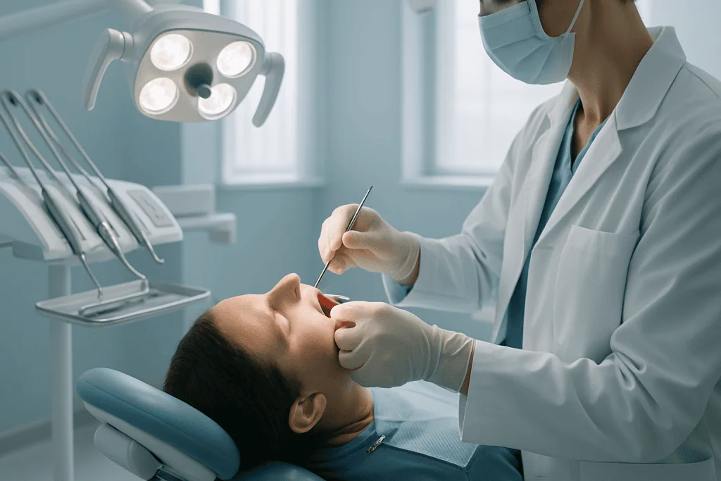
Quick answer
Oral pathology is the specialty that diagnoses diseases of the mouth, jaws, salivary glands, lips, tongue and gums. Evaluation typically involves a clinical examination, imaging where needed, and often a biopsy — removal of a small tissue sample for microscopic analysis. The findings determine whether a lesion is benign, infectious, inflammatory, precancerous or malignant, and guide treatment ranging from simple monitoring to surgery.
What Is Oral Pathology?
Oral pathology is the specialty that identifies and diagnoses diseases of the mouth, jaws, salivary glands, lips, tongue, palate, gums and related structures. It brings together clinical examination, imaging, laboratory testing and microscopic analysis of tissue to answer one essential question: what is this lesion, and what should be done next? Anyone with a persistent sore, lump, patch, swelling or cyst in the mouth or jaw region may need an oral pathology assessment, whether the finding turns out to be minor or serious.
A sore that does not heal, a lump in the cheek, a white or red patch on the tongue, swelling in the jaw or a cyst found on an X-ray can create understandable concern. Many oral changes are benign and treatable. Some require careful evaluation to rule out infection, inflammatory disease, precancerous change or oral cancer. The difficulty for you as a patient is that these possibilities often look alike at first. A harmless irritation, a fungal infection, an autoimmune disorder, a cyst, a benign tumour and an early cancer may each begin as a persistent spot, ulcer, swelling or change in colour. Appearance alone rarely settles the question.
That is why accurate diagnosis sits at the centre of oral pathology. It prevents two opposite mistakes: overtreatment of a harmless lesion and dangerous delay for a serious one. It also determines the right next step, whether that is medication, planned observation, minor surgical removal, further imaging or referral to a multidisciplinary cancer board. Without a confirmed diagnosis, treatment is guesswork. With one, care can be matched to the actual level of risk.
At Acibadem, oral pathology evaluation is approached as a structured diagnostic pathway rather than a single appointment. Depending on the finding, care may involve oral and maxillofacial surgery, dentistry, ear, nose and throat specialists, pathology, radiology, oncology, dermatology, infectious disease or other departments. This coordination consolidates evaluation, diagnosis and treatment planning within one organised medical setting rather than a series of disconnected visits.
What is oral pathology in dentistry?
In dentistry, oral pathology is the branch concerned with recognising, investigating and diagnosing disease of the oral cavity and its supporting structures — the mucosa, gums, jawbones, teeth-related tissues and minor salivary glands. Dentists are often the first clinicians to notice an abnormality, because many lesions are discovered during a routine check-up or on dental imaging rather than because of symptoms. Dental training therefore includes learning to distinguish normal anatomical variation from findings that need investigation. When a lesion cannot be confidently identified in the chair, the dentist refers the patient onward for specialist assessment and, where necessary, biopsy. In this sense oral pathology is the diagnostic backbone of dental and oral health care: it converts an uncertain finding into a named condition with a defined management plan.
Oral Pathology, Oral Medicine and Related Disciplines
Several overlapping specialties deal with disease of the mouth, and the terminology can be confusing when you are researching your own condition. The distinctions below matter less for the patient than for the referral pathway, but understanding them helps you see who does what.
What is oral and maxillofacial pathology?
Oral and maxillofacial pathology is the formal name of the specialty that diagnoses diseases affecting the mouth, jaws and adjacent facial structures. The “maxillofacial” part widens the scope beyond the oral cavity to include the jawbones, the temporomandibular region and related soft tissues of the face. In practice the terms oral pathology and oral and maxillofacial pathology describe the same discipline; the longer name simply reflects the full anatomical territory. The specialty spans both the clinical side — examining lesions, planning biopsies — and the laboratory side, where tissue samples are examined under the microscope.
What does an oral pathologist do?
An oral pathologist examines tissue samples from the mouth and jaws under a microscope and issues the diagnostic report that names the disease. This is laboratory medicine, not chairside dentistry: the oral pathologist processes the biopsy, studies thin stained sections of it, applies special stains or additional studies when the routine picture is ambiguous, and correlates what the tissue shows with the clinical description and imaging. Because oral lesions can be microscopically subtle — dysplasia grading, salivary gland tumours and rare jaw lesions are recognised areas of difficulty — the pathologist’s interpretation often determines whether a patient is monitored, treated with medication or scheduled for surgery. When findings do not match the clinical picture, a responsible pathologist recommends further investigation rather than forcing a conclusion.
How is oral medicine different from oral pathology?
Oral medicine is the clinical discipline that manages chronic, non-surgical diseases of the mouth — conditions that need diagnosis and long-term medical care rather than an operation. Where oral pathology answers “what is this lesion?”, oral medicine answers “how do we manage this condition over time?”. The two work together constantly: a biopsy diagnosed by the pathologist may be treated and followed by the oral medicine clinician. Typical oral medicine territory includes lichen planus, recurrent ulcers, immune-mediated blistering disorders, burning mouth symptoms, dry mouth and oral complications of systemic disease or its treatment.
When might you see an oral medicine specialist?
An oral medicine specialist is usually involved when an oral condition is chronic, recurrent or linked to a wider medical problem rather than to a single removable cause. Examples include ulcers that keep returning despite treatment, persistent burning or altered sensation without a visible lesion, painful inflammatory conditions of the mucosa, dry mouth related to autoimmune disease or medication, and oral changes accompanying dermatological or gastrointestinal disorders. These patients often need medication adjustment decisions, coordination with other physicians and structured follow-up — clinical management rather than a one-off procedure.
What does a center for oral pathology provide?
A center for oral pathology brings the whole diagnostic chain — clinical examination, imaging, biopsy and microscopic diagnosis — into one coordinated service instead of leaving the patient to arrange each step separately. This matters because oral diagnosis is iterative: the clinician’s findings shape what the pathologist looks for, and the pathology report shapes what the clinician does next. When those steps happen within one institution, information moves quickly, questions between clinician and laboratory are resolved directly, and the interval between first visit and confirmed diagnosis shortens. For lesions where cancer is a possibility, that compression of time has real clinical value.
Why would a dentist refer you to a pathologist?
A dentist refers you toward pathology when a lesion cannot be confidently identified by inspection alone and tissue diagnosis is needed. Common triggers for referral include an ulcer or patch that persists after obvious causes have been removed, a lump or swelling of unclear origin, a jaw lesion found on imaging, a lesion with suspicious features such as firmness or irregular borders, or a condition that has not responded to initial treatment as expected. Referral does not mean the dentist suspects cancer; most referred lesions prove benign. It means the finding has crossed the threshold where guessing is no longer acceptable and confirmation is the safer course.
Who May Need an Oral Pathology Evaluation
You may need an oral pathology evaluation if you have a mouth or jaw abnormality that persists, recurs, grows, changes appearance, causes unexplained symptoms or has no clear cause. A widely used rule in clinical practice is that any oral ulcer, patch, swelling or lump that has not improved within about two weeks of removing any obvious irritation should be assessed by a qualified clinician. Persistence past that point does not mean the lesion is serious. It means it should no longer be explained away without examination.
Common findings that bring patients to evaluation include:
- a sore or ulcerated area that does not heal;
- a lump inside the cheek, on the gum or under the tongue;
- a white patch that cannot be wiped away;
- a red or mixed red-and-white area of the mucosa;
- swelling of the jaw, or a cyst discovered on dental imaging;
- persistent pain, burning or altered taste;
- unexplained oral bleeding, or teeth loosening without a clear dental cause;
- numbness of the lip, chin, tongue or face.
Note that some significant lesions are painless. A painless lump or a painless white patch is easy to overlook precisely because it does not demand attention, which is one reason routine dental examinations remain an important detection route.
Diagnosis begins with a detailed medical and dental history. Your clinician will want to know when the lesion appeared, whether it has changed, whether it hurts, and whether trauma from a tooth edge, filling or dental appliance could be involved. You will be asked about tobacco in any form, alcohol, current medications, immune conditions and any history of cancer or radiation therapy, because these change the probability of particular diagnoses. Systemic symptoms matter too: fever, weight loss, skin lesions, dry mouth, dry eyes or joint pain can point toward an infection or a systemic disease expressing itself in the mouth.
The examination itself is systematic. The clinician inspects the lips, cheeks, gums, tongue, floor of the mouth, palate, throat region and jaw, gently palpates the lesion and surrounding tissue, assesses the salivary gland openings, checks the lymph nodes of the neck and reviews any dental structures that may be causing chronic irritation. Photographs are often taken — not for cosmetic reasons but because objective comparison at follow-up is far more reliable than memory.
Where the lesion involves the jawbone, teeth or deeper tissues, imaging is added to distinguish between dental infection, cyst, benign bone lesion, an impacted-tooth-related finding, a salivary gland problem or a tumour. Blood tests are useful in selected cases, particularly when an autoimmune disorder, nutritional deficiency, viral infection, blood disorder or systemic inflammatory disease is on the list of possibilities.
A biopsy is recommended when the diagnosis remains uncertain, when a lesion carries suspicious features, or when tissue confirmation is required before treatment can responsibly begin. Finally, some patients arrive not with a new lesion but with an old problem: an unclear previous diagnosis, an inconclusive biopsy, symptoms persisting despite treatment, or a recommendation for major surgery they want examined again. For these second-opinion situations, review of prior imaging, biopsy slides or pathology reports and clinical photographs can clarify the diagnosis and prevent both unnecessary surgery and unnecessary delay.
Conditions Assessed Through Oral Pathology
Oral pathology covers a broad territory: soft tissue disease, hard tissue disease and disorders of the salivary glands. Some conditions are minor and self-limited. Others need long-term management or coordinated multi-specialty care. The unifying purpose is classification — placing the lesion accurately so that treatment is proportionate to the risk it actually carries.
Orolabial lesions of the lips and mouth
Orolabial lesions are changes affecting the lips and the tissue around the mouth as well as the oral cavity itself, and they form a large part of everyday oral pathology practice. The lips are exposed to sun, trauma, infection and habit-related irritation, so lesions here range from viral blisters and inflammatory cracking at the corners of the mouth to mucous cysts of the minor salivary glands and, on the outer lip, sun-related precancerous change. Because lip lesions are visible and easily photographed, they are often noticed early — but visibility does not make them self-diagnosing, and persistent lip changes deserve the same structured assessment as any lesion inside the mouth.
Ulcers that do not heal
Ulcerative conditions are among the most common reasons for referral. Mouth ulcers may be traumatic, aphthous, viral, immune-mediated, drug-related, associated with inflammatory bowel disease or blood disorders, or — least commonly but most importantly — malignant. Features that raise concern include raised or rolled edges, firmness on palpation, bleeding, persistent pain and, above all, the absence of any healing trend. An ulcer that behaves like an ulcer should heal like one; an ulcer that does not is a diagnostic question, not a nuisance.
White, red and pigmented patches
Colour changes of the oral mucosa cover a wide diagnostic spectrum. White patches may be caused by friction, fungal infection, lichen planus, leukoplakia or other disorders. Red areas may reflect inflammation, vascular change, infection, erythroplakia or dysplasia. Brown, blue, black or purple areas may relate to normal pigmentation, vascular lesions, an amalgam tattoo, medication effects, systemic disease or, rarely, melanoma. The practical point is that colour alone does not determine seriousness. Clinical context, duration, texture and — when needed — tissue diagnosis do.
Cysts and lesions of the jaw
Jaw cysts and bone lesions are frequently found incidentally on dental imaging, or discovered after swelling, discomfort, tooth displacement, drainage or infection. Many jaw cysts are benign, but benign does not mean harmless: an expanding cyst can weaken bone, displace teeth and approach nerves. Certain benign jaw tumours behave in a locally aggressive way despite never spreading, which makes early and precise diagnosis essential for planning surgery of the right extent — enough to control the lesion, no more than the anatomy requires.
Salivary gland conditions
Salivary gland problems present as swelling, dry mouth, pain with meals, mucous cysts, infections, stones, autoimmune disease or tumours. Minor salivary gland lesions appear on the lips, palate, cheeks or floor of the mouth; the major glands near the jaw and under the tongue may need ultrasound, cross-sectional imaging or needle-based sampling depending on the presentation. Salivary gland tumours are a recognised area of microscopic difficulty, which is one context where specialist pathology review earns its place in the pathway.
Potentially malignant disorders and oral cancer
Oral pathology also addresses potentially malignant disorders and oral cancer itself: lesions showing dysplasia, leukoplakia, erythroplakia, certain chronic inflammatory conditions, and cancers of the tongue, floor of mouth, gums, palate, lips and other oral structures. Early oral cancer can be subtle — a small, sometimes painless patch or ulcer — which is exactly why biopsy of persistent suspicious lesions is essential rather than optional. When cancer is diagnosed or strongly suspected, the case moves into a multidisciplinary process involving surgical, medical, radiation, radiology, pathology and supportive care specialists, with dental preparation and maxillofacial prosthetic or reconstructive planning included where treatment will affect oral function.
Oral signs of systemic disease
The mouth can be the first place a systemic disease shows itself. Diabetes, anaemia, vitamin deficiency, autoimmune disease, dermatological disease, immune suppression, medication side effects and gastrointestinal disorders can all produce oral signs. In these cases an oral pathology evaluation opens the door to broader medical assessment, and the connection between the mouth and the rest of the body — explored further in oral health and systemic diseases — becomes central to the treatment plan rather than incidental to it.
What is the most common oral pathology?
The most frequently seen oral lesions in everyday practice are benign: traumatic ulcers, frictional white patches, mucous cysts, fibromas from chronic irritation, fungal infections such as oral candidiasis, aphthous ulcers and inflammatory gum conditions. Geographic tongue and lichen planus are also regular findings. This distribution is reassuring, but it carries a caveat. Common benign lesions and uncommon serious ones share the same early appearances, so frequency is never a reason to skip evaluation of a lesion that persists or changes. The value of specialist assessment is precisely that it identifies which lesions can safely be observed, which need medication and which require biopsy or prompt treatment.
How Oral Pathology Evaluation and Treatment Are Performed
Oral pathology care begins before any procedure, with careful information gathering. Prior dental records, medical reports, imaging files, laboratory results, medication lists and any existing biopsy or pathology reports all help the clinical team understand the history of a lesion, and clear photographs allow objective comparison with what is seen at examination. The more complete this background, the more efficiently the diagnostic sequence can be planned and the fewer steps need repeating.
The first consultation
At the first visit, your specialist reviews your history and performs a focused oral, dental, jaw and neck examination. The aim is to determine whether the lesion is most consistent with irritation, infection, inflammation, cystic disease, benign tumour, precancerous change or malignancy. The clinician documents size, colour, texture, firmness, borders, location, tenderness and the lesion’s relationship to teeth or dental appliances. If the finding appears related to an obvious irritant — a sharp tooth edge, a rough filling, a poorly fitting denture — the first step may simply be to remove the irritant and reassess after a defined period. A lesion that heals once its cause is gone has, in effect, diagnosed itself. Suspicious, persistent, enlarging or unexplained lesions are investigated more directly.
Imaging: choosing the right study
Imaging is selected according to the clinical question, not applied by default. Intraoral or panoramic dental imaging shows tooth-related infection, bone loss, impacted teeth, cystic borders and jaw involvement. Cone-beam computed tomography maps a lesion’s three-dimensional relationship to roots, nerves, sinuses and cortical bone — information that directly shapes surgical planning. Ultrasound suits salivary gland swelling and superficial soft tissue masses. Computed tomography or magnetic resonance imaging is reserved for deeper lesions, complex jaw involvement, soft tissue extension or cancer staging. Each study answers a specific question; together they establish depth, boundaries and extent before anyone decides what to remove.
How an oral biopsy is performed
When biopsy is needed, preparation is usually straightforward. Your clinician reviews medications, allergies, bleeding history and medical conditions, including whether you take blood thinners or immune-suppressing drugs; any decision about adjusting medication before a procedure belongs to the prescribing physician, and the team coordinates rather than improvises. You may be asked not to eat or drink beforehand if sedation or general anaesthesia is planned, while many local-anaesthetic biopsies need no extensive fasting. A typical biopsy under local anaesthesia proceeds as follows:
- The area is numbed with local anaesthetic.
- The sampling approach is confirmed: excisional biopsy removes the entire lesion when this can be done safely without compromising any later treatment; incisional biopsy removes a representative portion of a larger or more complex lesion.
- The clinician samples the most diagnostically useful area, deliberately avoiding surface debris or non-representative tissue when a deeper process is suspected. In selected cases, multiple samples are taken from different parts of a lesion.
- Bleeding is controlled, and the site is closed with sutures where needed — some dissolve on their own, others are removed at follow-up.
- The tissue is placed in an appropriate specimen container, labelled and sent to the laboratory with the clinical details the pathologist needs for interpretation.
Deeper lesions, jaw cysts and complex soft tissue procedures may instead require an operating room setting under sedation or general anaesthesia, sometimes as day-care surgery within the oral and maxillofacial surgery service.
What happens in the laboratory
In the pathology laboratory, the sample is processed into thin tissue sections, stained and examined under the microscope. Where the routine picture is ambiguous, the pathologist applies special stains or additional studies to identify fungal organisms, immune patterns, tumour type, degree of dysplasia, margin status or other diagnostic features. For suspected cancer, the pathology result is integrated with imaging and clinical findings to guide staging and treatment planning. Reporting time varies with the complexity of the case and any additional studies required; your clinician will tell you what to expect for your specific sample.
What are common treatments in oral pathology?
Treatment follows the diagnosis, and the range is wide. Removal of a chronic irritant may be all a traumatic lesion needs. Fungal and bacterial infections are treated with appropriate medication. Inflammatory and immune-mediated conditions are managed medically, often with topical treatment and structured follow-up. Mucous cysts, fibromas and similar benign lesions are removed surgically, frequently in the same minor procedure that confirms the diagnosis. Jaw cysts and benign jaw tumours are treated surgically at an extent matched to how the specific lesion behaves. Dysplastic lesions may be removed or kept under scheduled surveillance depending on grade and location. Confirmed cancer is managed through multidisciplinary planning that may combine surgery, radiation therapy, systemic therapy, reconstruction, dental rehabilitation and speech or swallowing support.
Procedure time, aftercare and results
Duration varies with complexity. A clinical examination fits within a standard outpatient visit. A minor biopsy under local anaesthesia is itself brief, though the full visit includes preparation, consent, anaesthesia, haemostasis and aftercare instructions. Most biopsy patients go home the same day. Mild soreness, slight swelling and small amounts of bleeding can occur early on; you will receive specific guidance on oral hygiene, rinsing, diet and pain control. Soft foods, careful brushing and protecting the site from trauma are the usual advice while healing begins.
Once the pathology report is available, your clinician explains the diagnosis and next steps. A benign lesion that was completely removed may need nothing beyond follow-up. An infection or inflammatory condition leads to medication or management of the underlying trigger. Dysplasia or cancer moves the case to a multidisciplinary board that defines the treatment strategy and the surveillance plan that follows it. Throughout, technology serves one purpose: high-quality imaging defines anatomy and extent, digital records and photography support comparison over time, and microscopic techniques refine the diagnosis — all of it to reduce uncertainty, not to accumulate data.
Why Acting Early Matters
Early evaluation matters because visible mouth changes span such a wide spectrum. Some conditions persist harmlessly only because their irritant has never been corrected. Infections can worsen or spread without treatment. Cysts and jaw lesions can enlarge silently, affecting teeth, bone strength, facial structure and nearby nerves. Chronic inflammatory and immune-mediated conditions can cause ongoing pain, scarring and difficulty eating when they are treated blindly instead of being properly diagnosed.
The strongest reason not to delay is the possibility of precancerous change or oral cancer. Early-stage oral cancer is generally more treatable than advanced disease, and treatment may be less extensive when disease is found before deep invasion or lymph node spread. A small persistent ulcer or patch is easier to biopsy and treat than a lesion that has grown for months. Most mouth changes are not cancer — but the consequences of missing the exception are severe enough that persistent or suspicious findings warrant prompt evaluation rather than watchful hoping.
Delay also muddies diagnosis itself. Repeated courses of medication without a confirmed diagnosis can suppress inflammation temporarily while leaving the underlying process untouched. Trauma from teeth or appliances can alter a lesion’s appearance. Infection can coexist with cystic or tumour-related disease and mask it. A structured oral pathology assessment replaces this cycle with evidence: examine, image, sample, confirm, then treat.
Early action also simplifies the practical side of care. When imaging, biopsy, pathology review and possibly multidisciplinary consultation will each be needed, starting early lets the care team sequence those steps deliberately — the right imaging before the biopsy, the biopsy before any treatment decision — rather than compressing them into a rushed timeline once a lesion has already grown or begun causing complications.
Benefits of Oral Pathology Evaluation and Treatment
The central benefits are clarity, timely diagnosis and a treatment plan matched to the true nature of the lesion — nothing more, nothing less.
| Benefit | What It Means for You |
|---|---|
| Accurate diagnosis | Clinical examination, imaging and biopsy when needed distinguish benign, infectious, inflammatory, precancerous and cancer-related conditions. |
| Appropriate treatment planning | Care is tailored to the diagnosis, avoiding unnecessary procedures while ensuring serious conditions are treated without avoidable delay. |
| Early cancer detection when present | Suspicious lesions are sampled and reviewed by pathology, allowing earlier referral to specialist boards and coordinated cancer care if needed. |
| Relief of symptoms | Identifying the cause of pain, burning, ulcers, swelling or bleeding guides medication, minor procedures, dental adjustments or other therapy. |
| Long-term monitoring | Conditions that can recur or progress — particularly dysplasia and chronic inflammatory disease — receive structured follow-up rather than being closed prematurely. |
Recovery After an Oral Biopsy or Minor Oral Pathology Procedure
Recovery depends on the type and location of the lesion, the extent of the biopsy or removal and your general health, but most minor oral procedures heal within a short outpatient recovery period. The timeline below describes a typical course; your own instructions take precedence.
| Time Period | What Patients Can Expect |
|---|---|
| Day 1 | Mild soreness, numbness while the anaesthetic wears off, slight swelling and minor oozing may occur. Soft foods and leaving the site undisturbed are the usual advice. |
| First week | Discomfort generally settles. Hygiene instructions, rinses and diet modifications protect the biopsy site. Sutures begin dissolving or are scheduled for removal. |
| First month | The site continues to mature and strengthen. The pathology report is reviewed with you, and any further treatment, monitoring or referral is planned. |
| Longer term | Benign, completely removed lesions may need only routine review. Chronic inflammatory, precancerous, cystic or cancer-related conditions follow a defined surveillance or treatment plan. |
Factors That Influence Outcomes
A good outcome in oral pathology begins with an accurate diagnosis, and the quality of that diagnosis is built at every step: the thoroughness of the clinical examination, the choice of imaging study, the timing of biopsy and the selection of the biopsy site. Lesions sampled too superficially or from an unrepresentative area may need repeat biopsy, particularly when clinical suspicion remains despite a reassuring first report. Good technique at the start saves procedures later.
The nature of the condition itself shapes what a good result looks like. A traumatic fibroma removed completely has a different outlook from a chronic immune-mediated disorder that needs management over years. A small jaw cyst has different treatment needs from a locally aggressive benign tumour. A lesion with mild dysplastic change requires a different plan from one with high-grade dysplasia or invasive cancer. Clear classification is what makes expectations realistic rather than optimistic.
Timing matters throughout. Lesions evaluated early tend to be smaller, easier to sample and less likely to have caused structural damage. In cancer-related diagnoses, earlier assessment can preserve treatment options and may reduce the need for extensive surgery or combined therapies. In cystic jaw disease, earlier treatment improves the chances of preserving teeth, nerves and bone where the anatomy allows.
Patient-related factors influence both disease and healing. Tobacco in any form, alcohol exposure, diabetes, immune suppression, dry mouth, nutritional deficiency, oral hygiene, chronic dental trauma and medication effects can each contribute to the development of oral lesions and slow recovery after procedures. When your clinician raises these, it is not a general lifestyle lecture — these factors act directly on the tissue being treated. Older patients with dentures or complex dental histories bring additional considerations, addressed within gerontological oral health care.
Pathology expertise is central and worth naming plainly. Microscopic interpretation of oral lesions can be genuinely difficult — inflammatory conditions, salivary gland tumours, dysplasia grading and rare jaw lesions are established problem areas. Responsible practice includes additional stains, second pathology review or correlation with radiology and clinical findings when the first answer does not fit the clinical picture. Accepting uncertainty is never the endpoint of good diagnostics.
Finally, follow-up completes the result. Benign lesions can recur if their cause remains. Inflammatory diseases fluctuate. Potentially malignant disorders need periodic monitoring even after treatment, and cancer diagnoses require structured surveillance afterwards. You should leave every stage of care knowing what to watch for, when to return and which changes should prompt an earlier review with your treating team.
How Care Is Coordinated When a Diagnosis Needs More Than One Specialist
Oral lesions often need more than one specialist, and that is the practical reason complex oral diagnosis suits a hospital environment. A mouth or jaw abnormality can involve dentistry, oral and maxillofacial surgery, pathology, radiology, ear, nose and throat medicine, oncology, dermatology, infectious disease or internal medicine. Acibadem’s hospital structure supports this collaboration, so a patient can move from evaluation to diagnosis to treatment planning within one coordinated system instead of carrying reports between unconnected providers.
Clear communication matters more in oral pathology than in many fields, because the decisions are nuanced. You need to understand why a biopsy is recommended, what each candidate diagnosis would mean, how long results take, what follow-up involves and what the next step would be if abnormal cells are found. Good coordination also works behind the scenes: the clinician who examined you, the radiologist who read your imaging and the pathologist who studied your tissue can resolve questions between themselves directly, instead of leaving contradictions in the record for you to reconcile.
When cancer or a potentially malignant disorder is suspected, cases are reviewed through multidisciplinary boards that bring the relevant physicians together over the imaging, pathology and clinical findings. The purpose is an evidence-based plan built from several perspectives rather than one. For oral and jaw cancers this can include surgical options, reconstruction, radiation therapy, systemic therapy, dental preparation, nutrition, speech and swallowing support and the follow-up strategy that ties it together.
Coordination also extends past diagnosis. When a lesion needs long-term surveillance, the follow-up schedule, the changes to watch for and the findings that prompted it are documented so that any dentist or physician involved in later care can see exactly what was found, what was done and what should happen next. Continuity of records is part of the diagnosis’s lasting value: a lesion that recurs years later is interpreted far more accurately when the original pathology report, images and photographs are available for comparison.
Moving From Uncertainty to a Diagnosis
Finding an unexplained lesion in your mouth or jaw is unsettling, and online reading rarely helps — it presents the harmless and the serious side by side without a way to tell them apart in your own case. Oral pathology exists to close that gap methodically: examine the lesion, identify the likely causes, confirm the diagnosis with tissue when needed, and set a plan proportionate to what is actually found. Most findings prove benign. The ones that are not benefit most from having been investigated early, precisely and without shortcuts. Either way, a confirmed diagnosis replaces worry with a defined next step — which is what this specialty is for.
Preparation
- Your dentist or oral specialist reviews your medical history, medications, allergies, and symptoms. Avoid eating or drinking shortly before the visit if a biopsy or minor procedure is planned. Bring previous dental X-rays, pathology reports, and a list of current medications.
Aftercare
- Most patients return to daily activities the same day. If a biopsy is performed, follow instructions for oral hygiene, soft foods, and prescribed pain relief or mouth rinses. Contact the clinic if bleeding, fever, increasing swelling, or severe pain occurs.
Turkey vs UK, Germany & USA
Oral pathology consultations help investigate mouth or jaw changes such as ulcers, lumps, cysts, color changes, and suspicious lesions. Costs and patient experience vary by diagnostic needs, biopsy requirements, pathology review, hospital setting, and follow-up planning.
For international patients, the main differences between destinations usually relate to access, diagnostic coordination, hospital standards, language support, and what is included in the care pathway.
| Factor | Turkey | UK | Germany | USA |
|---|---|---|---|---|
| Cost structure | Often offered as coordinated self-pay care with consultation, imaging, biopsy, pathology, and treatment planning arranged through the hospital. | Private care costs vary by clinic, consultant, biopsy, pathology, and imaging; public pathways may not be available to international patients. | Costs vary by specialist setting, diagnostics, pathology laboratory, and hospital fees; billing may be itemized. | Costs can vary widely by provider, facility, pathology laboratory, imaging, anesthesia, and insurance status. |
| Specialist and hospital factors | Care may involve oral and maxillofacial surgeons, oral medicine specialists, dentists, radiologists, and pathology teams within hospital networks such as JCI-accredited facilities. | Care may be delivered in dental hospitals, private oral surgery clinics, or maxillofacial units, depending on referral route. | Care may involve university hospitals, specialist clinics, or private practices with access to pathology and imaging. | Care may involve oral surgeons, dentists, oral medicine specialists, pathology laboratories, and hospital-based teams. |
| Waiting and scheduling | Private appointments and coordinated diagnostics may be scheduled around travel plans, subject to clinical urgency and availability. | Private scheduling may be flexible; public referral pathways can involve variable waiting depending on urgency. | Scheduling is generally based on specialist availability, referral needs, and diagnostic complexity. | Scheduling depends on provider access, insurance authorization, facility availability, and diagnostic requirements. |
| Accreditation and quality | International patients may choose hospitals with international accreditation such as JCI and established pathology pathways. | Quality is supported by national regulation, professional standards, and specialist training frameworks. | Quality is supported by national medical regulation, specialist certification, and established hospital standards. | Quality varies by provider and facility; many centers follow recognized accreditation and specialty standards. |
| Travel and language logistics | International patient departments may assist with appointments, translation, travel coordination, and medical document preparation. | English-language care is standard; international patients may still need help with referral documents and private billing. | Interpreter support may be needed; medical records and pathology reports may require translation. | English-language care is standard; travel distance, accommodation, and insurance administration may affect planning. |
| Typical package inclusions | May include specialist consultation, diagnostic planning, imaging coordination, biopsy or minor procedure, pathology review, report, and follow-up guidance. | Private packages may include consultation and selected diagnostics, while pathology, imaging, and procedures may be billed separately. | Packages are often tailored, with consultation, imaging, biopsy, pathology, and follow-up itemized according to need. | Services may be separated across provider, facility, laboratory, imaging, and anesthesia billing. |
- What affects your final cost
- Type, size, location, and suspected cause of the lesion.
- Need for imaging, biopsy, culture, blood tests, or specialist pathology review.
- Whether the procedure is performed under local anesthesia, sedation, or operating room conditions.
- Complexity of cyst, jaw, salivary gland, or soft tissue involvement.
- Need for multidisciplinary review, cancer pathway referral, surgery, medication, or surveillance.
- Hospital category, specialist experience, pathology laboratory fees, translation, travel, and follow-up arrangements.
Compare your options
Oral pathology may involve several diagnostic and treatment options. Suitability is decided by a specialist after examination, medical history review, and assessment of the lesion.
| Option | What it is | Typical use | Key considerations |
|---|---|---|---|
| Clinical oral examination | A detailed assessment of the mouth, jaw, tongue, gums, palate, lips, and lymph nodes. | Initial evaluation of ulcers, color changes, swellings, pain, bleeding, or non-healing areas. | Often guides whether observation, testing, biopsy, or referral is needed. |
| Imaging assessment | Dental radiography, panoramic imaging, ultrasound, or advanced jaw imaging when indicated. | Evaluation of cysts, jaw lesions, impacted teeth, bone changes, salivary concerns, or deep swelling. | Choice of imaging depends on the suspected condition and the area involved. |
| Laboratory and infection testing | Targeted tests such as swabs, cultures, blood tests, or immune-related investigations. | Used when infection, inflammatory disease, autoimmune disease, or systemic illness may be involved. | Results must be interpreted with clinical findings and medication history. |
| Biopsy and histopathology | Removal of a small tissue sample or the entire small lesion for microscope examination by a pathology specialist. | Used for persistent ulcers, unexplained lumps, white or red patches, cysts, and suspicious lesions. | Important for distinguishing benign, inflammatory, infectious, precancerous, and cancer-related changes. |
| Medical management | Medication or supportive care such as topical treatment, antimicrobial therapy, anti-inflammatory care, or management of dry mouth and irritation factors. | Used for confirmed inflammatory, infectious, immune-related, or trauma-associated conditions. | Follow-up is needed to confirm response and adjust treatment if the lesion does not improve. |
| Surgical removal or referral | Excision, cyst management, drainage, or referral to oral and maxillofacial surgery, oncology, dermatology, or another specialty. | Used for cysts, growths, recurrent lesions, jaw involvement, or suspected cancer-related disease. | The plan depends on diagnosis, lesion extent, pathology findings, and overall health. |
General information only — not medical or financial advice. Final costs depend on the factors above and your individual case; request a free, personalised quote.
Frequently Asked Questions
What affects the cost of oral pathology evaluation?
The final cost depends on the type and location of the lesion, the need for imaging, biopsy, pathology review, laboratory testing, anesthesia, specialist involvement, and follow-up care. A personalised quote can be prepared after review of your medical information and images.
How can I get a personalised quote from Acibadem?
You can request a free consultation and share photos of the lesion, dental or medical reports, imaging if available, medication history, and a description of symptoms. The medical team can then advise which assessments may be needed and prepare a tailored estimate.
Is biopsy always required?
No. Some lesions can be monitored or treated medically, while others need biopsy to confirm the diagnosis. The decision is made by a specialist based on the appearance, duration, symptoms, risk factors, and imaging findings.
What is usually included in an oral pathology care plan?
A care plan may include specialist examination, diagnostic imaging, biopsy if indicated, pathology reporting, medication or surgical planning, and follow-up recommendations. In international patient pathways, translation and appointment coordination may also be arranged.
Can oral pathology be planned as part of a short medical trip?
Many evaluations can be coordinated for international patients, but timing depends on clinical urgency, the need for biopsy, pathology processing, and follow-up. The team can advise on a realistic travel plan after reviewing your case.
Is this information medical or financial advice?
No. This is general educational information. Diagnosis, treatment suitability, and final cost can only be confirmed after specialist assessment, so a free consultation is recommended for personalised guidance.
Medically reviewed by the Acıbadem International Medical Board — September 1, 2026
See our medical review board →
Update history
- PublishedJune 8, 2026
- Medical review approvedSeptember 1, 2026
- Last content updateAugust 31, 2026
References1
- Oral Cancer — cancer.gov
Trusted care for international patients
Doctors Performing This Treatment

Assoc. Prof. Dr. Ferit Bayram
Oral & Dental Health
Dr. Ezgi Gülüm
Oral Dental & Maxillofacial Surgery
Dr. Emre Çengelli
Oral Dental & Maxillofacial Surgery
Dr. Arzu Morçiçek
Oral & Dental HealthDr. Deniz Turgut
Oral & Dental Health
Dr. Ceyda Sabancı
Oral & Dental Health
Dr. Çağla Su Doğangün Ayduk
Oral & Dental Health
Dr. Begüm Öykü Kesim
Oral & Dental Health
Dr. Eylül Türsen
Oral & Dental Health
Dr. Zeynep Ezgi Akan
Oral & Dental Health
Dr. Metin Kınacı
Oral & Dental Health
Dr. Duygu Yavuzer Karadeniz
PeriodontolgyDr. Ali Riza Özdurmuş
Oral & Dental Health
Dr. Bedii Ender Topçu
Oral & Dental Health
Dr. Sebiha Nihal Yılmaz
Oral Dental & Maxillofacial SurgeryDr. Seda Saygılı Özaydın
Oral & Dental Health
Dr. Zeynep Ekin Kılınç
Oral & Dental Health
Dr. Uğur Önder
Oral & Dental Health
Dr. Helin Kuşsever Topçu
Oral & Dental Health
Dr. Pelin Açık
Oral & Dental Health
Dr. İpek Saygılı
Oral & Dental Health
Dr. Halime Bayram
Oral & Dental Health
Dr. Havva Gölalan
Oral & Dental Health
Dr. Merve Ağartıoğlu
Oral & Dental HealthMedical Units
Available at These Hospitals