Knee Anatomy: A Complete Medical Overview

The knee is a complex hinge-type joint formed mainly by the femur, tibia, and patella. Cartilage, menisci, ligaments, and tendons help the knee move smoothly and stay stable.
Key Takeaways
- The knee is a complex hinge-type joint formed mainly by the femur, tibia, and patella.
- Cartilage, menisci, ligaments, and tendons help the knee move smoothly and stay stable.
- Pain, swelling, locking, or giving way can point to injury or wear affecting specific knee structures.
- Doctors assess knee anatomy through physical examination and, when needed, imaging such as X-rays or MRI.
- Protecting knee health involves strength, flexibility, weight management, proper technique, and timely medical evaluation.
Knee anatomy refers to the structures that make up the knee joint, including bones, cartilage, ligaments, tendons, muscles, and supporting nerves and blood vessels. Together, these parts allow the knee to bend, straighten, absorb force, and help the body stand, walk, run, and change direction.
Overview: what knee anatomy means
Knee anatomy is the study of the parts that form the knee joint and how they work together. In simple terms, the knee is where the thigh bone meets the shin bone, with the kneecap sitting in front. It is built to be strong enough to support body weight but flexible enough to allow bending, straightening, walking, climbing, and turning.
Although many people think of the knee as a simple hinge, it is more complex than that. The joint combines gliding, rolling, and rotating movements. This is why knee function depends not only on bone shape, but also on cartilage, ligaments, tendons, muscles, and the soft tissues around the joint.
Understanding knee anatomy can help explain why certain injuries cause instability, why arthritis leads to stiffness and pain, or why swelling can limit motion. It also helps patients understand what doctors are looking for during an exam and why different imaging tests may be recommended for bone, cartilage, or ligament problems.
Bones and joint surfaces of the knee
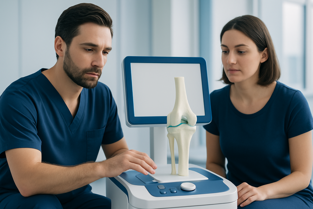
The main bones in the knee are the femur, the tibia, and the patella. The femur is the thigh bone, and its lower end forms the top part of the knee joint. The tibia is the larger bone in the lower leg and forms the lower part of the joint. The patella, or kneecap, sits at the front of the knee and helps the thigh muscles extend the leg more efficiently.
A smaller lower-leg bone called the fibula is close to the knee but does not directly form the main weight-bearing knee joint. It still matters because important ligaments, muscles, and nerves are nearby. The surfaces of the femur, tibia, and the back of the patella are covered with smooth articular cartilage, which reduces friction and helps the joint move with less wear.
The knee is often described as having two related joints: the tibiofemoral joint between the femur and tibia, and the patellofemoral joint between the patella and femur. These work together during daily activities such as standing from a chair, climbing stairs, or squatting. Problems in either area can cause pain, clicking, or difficulty with movement.
Cartilage and menisci: the knee's shock absorbers
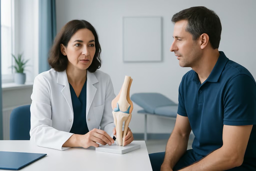
Two important forms of cartilage support the knee. The first is articular cartilage, the smooth coating on the ends of the bones. It allows surfaces to glide with minimal friction and helps spread force across the joint. Damage to this cartilage can lead to pain, stiffness, and, over time, knee osteoarthritis.
The second are the menisci, two crescent-shaped pads of fibrocartilage called the medial meniscus and lateral meniscus. They sit between the femur and tibia and act as shock absorbers. The menisci also help distribute body weight more evenly, improve joint stability, and support smooth knee motion.
Meniscal tissue can be injured by a twisting movement or worn down gradually with age. When this happens, a person may notice pain along the joint line, swelling, catching, or a feeling that the knee does not move normally. In some cases, evaluation for a meniscus repair or related surgery may be discussed if symptoms are persistent and function is limited.
Ligaments, tendons, and muscles that stabilize the knee
The ligaments are strong bands of tissue that connect bone to bone and keep the knee stable. Inside the joint are the anterior cruciate ligament (ACL) and posterior cruciate ligament (PCL), which help control forward, backward, and rotational movement. On the sides are the medial collateral ligament (MCL) and lateral collateral ligament (LCL), which resist side-to-side stress.
The tendons connect muscle to bone. The quadriceps tendon links the front thigh muscles to the patella, and the patellar tendon continues from the patella to the tibia. Together, these structures help straighten the knee. The hamstring muscles at the back of the thigh help bend the knee and also contribute to stability, especially during walking, running, and sudden direction changes.
Muscles around the hip and calf also influence knee mechanics. Weakness or poor movement patterns in these areas can increase stress on the knee even when the joint itself is structurally normal. Injuries to major stabilizers such as the ACL may cause a popping sensation, swelling, and instability, and some patients may be evaluated for ACL reconstruction depending on the injury pattern, activity level, and overall goals.
Nerves, blood supply, and other supporting structures
Knee anatomy includes more than the visible joint parts. Nerves around the knee carry signals for pain, touch, and muscle control. The area is closely related to branches of the femoral, tibial, common peroneal, and saphenous nerves. This is one reason some knee conditions cause pain patterns that radiate or feel different from simple soreness.
Blood vessels around the knee nourish the bones, ligaments, tendons, and other tissues. A network of arteries helps maintain circulation even when the knee bends deeply. Healthy blood supply is important for healing after injury, although some structures, such as parts of the meniscus, have more limited blood flow and may heal more slowly.
Other supporting structures include the joint capsule, synovial membrane, synovial fluid, and bursae. The capsule encloses the joint, while the synovial membrane produces lubricating fluid. Bursae are small fluid-filled sacs that reduce friction between tissues. When these structures become irritated, they can contribute to swelling, stiffness, or pain around the front, side, or back of the knee.
How knee anatomy relates to symptoms and common conditions
Different parts of the knee tend to produce different symptoms when they are injured or affected by wear. Problems involving articular cartilage or arthritis often cause stiffness, pain with weight-bearing, and reduced range of motion. Meniscal problems may lead to joint-line pain, swelling, catching, or locking. Ligament injuries are more likely to cause instability or a sense that the knee may give way.
Pain at the front of the knee can come from the patellofemoral joint, the patellar tendon, or surrounding soft tissues. Swelling after an injury may reflect bleeding or inflammation within the joint. Clicking can be harmless in some people, but when it comes with pain, locking, or swelling, it deserves assessment.
Common conditions linked to knee anatomy include ACL tears, meniscal injuries, tendon overuse, bursitis, and degenerative joint disease. In advanced cases of arthritis, where pain and function are significantly affected despite non-surgical care, doctors may discuss options such as knee replacement surgery.
- Pain with stairs may suggest patellofemoral involvement.
- Sudden swelling after twisting can suggest ligament or meniscus injury.
- Locking or catching may point to meniscal or cartilage problems.
- Giving way may indicate instability from ligament injury or muscle weakness.
How doctors evaluate the knee
Doctors usually begin with a medical history and physical examination. They ask when symptoms began, whether there was an injury, where the pain is felt, and what movements make it better or worse. During the exam, they assess swelling, alignment, tenderness, range of motion, walking pattern, and how stable the knee feels with specific maneuvers.
Imaging tests are chosen based on the suspected problem. X-rays are useful for bone alignment, fractures, and signs of arthritis. MRI is especially helpful for soft tissues such as ligaments, menisci, tendons, cartilage, and bone bruising. Ultrasound may be used for some tendon, bursa, or fluid-related issues, while CT can help in selected bone and surgical planning situations.
Not every painful knee needs advanced imaging immediately. In many cases, a careful clinical assessment provides the first and most important information. Test results are most useful when interpreted together with symptoms, physical findings, age, activity level, and overall health.
Prevention, self-care, and when to seek medical care
Protecting the knee starts with balanced strength, flexibility, and movement control. Strong quadriceps, hamstrings, gluteal muscles, and calf muscles can reduce stress on the joint. Warm-up before exercise, gradual training progression, supportive footwear, and good technique during sports or lifting can also help lower the risk of injury.
Self-care for mild knee symptoms often includes short-term activity modification, relative rest, ice, and guidance from a healthcare professional or physiotherapist. Weight management may reduce load across the knee, especially in people with degenerative changes. However, prolonged complete rest is usually not ideal, because gentle, appropriate movement often supports recovery and joint function.
Medical care should be sought if knee pain follows a significant injury, if the joint looks deformed, if weight-bearing becomes difficult, or if there is marked swelling, locking, repeated giving way, fever, or redness with warmth. Ongoing pain that does not improve, nighttime pain, or loss of function also deserves evaluation. Near the end of the care pathway, patients who need specialist assessment may be seen by multidisciplinary teams; Acibadem International’s JCI-accredited hospitals diagnose and treat knee conditions for international patients.
Frequently asked questions
What are the main parts of knee anatomy?
The main parts of knee anatomy are the femur, tibia, and patella, along with articular cartilage, menisci, ligaments, tendons, muscles, nerves, and blood vessels. All of these structures work together to support body weight and allow controlled movement.
Is the knee a hinge joint?
The knee is often described as a hinge joint because it mainly bends and straightens. However, it is more complex than a simple hinge because it also allows a small amount of rotation and gliding during normal movement.
What do the menisci do in the knee?
The menisci are two fibrocartilage pads between the femur and tibia. They help absorb shock, spread force across the joint, improve stability, and support smoother movement.
Which ligaments are most important in the knee?
The ACL and PCL help control front-to-back and rotational stability, while the MCL and LCL help stabilize the inner and outer sides of the knee. Each ligament has a distinct role, and injury to one can change how stable the knee feels.
Why does knee anatomy matter when a person has pain?
Knowing knee anatomy helps explain which structure may be causing symptoms. For example, meniscal problems often cause catching or locking, while ligament injuries may cause instability and cartilage wear may cause stiffness and pain with activity.
When should someone see a doctor for knee symptoms?
A doctor should assess knee symptoms after a major injury, if swelling is significant, if the knee gives way, or if walking becomes difficult. Persistent pain, locking, fever, or visible deformity also need medical evaluation.
References
- American Academy of Orthopaedic Surgeons
- National Institute of Arthritis and Musculoskeletal and Skin Diseases
- MedlinePlus
- OrthoInfo
- Mayo Clinic
This article is for general information only and is not a substitute for professional medical advice. Please consult a qualified doctor about your individual situation.
Joint and spine care in Turkey — expert assessment & treatment
JCI-accredited · board-certified surgeons · reply within 24h
Add us as a Preferred Source to see more of our trusted health content across Google Search, AI Overviews and Discover.

More from the Health Library



Related Specialists

Dr. Ömer Hersek
Internal Medicine
Dyt. Gizem Katkıcı Taşkın
Nutrition & Diet
Dr. Beyhan Levent Erdoğan
Pediatrics
Prof. Dr. Mahir Gülşen
Orthopedic Surgery & Traumatology