Thoracentesis: An Evidence-Based Guide for Patients

Thoracentesis removes excess fluid from the pleural space around the lungs. It may be done to relieve symptoms, diagnose the cause of pleural effusion, or both.
Key Takeaways
- Thoracentesis removes excess fluid from the pleural space around the lungs.
- It may be done to relieve symptoms, diagnose the cause of pleural effusion, or both.
- Most procedures are performed with imaging guidance and local anesthetic.
- Common side effects are mild soreness and temporary cough, while serious complications are uncommon.
- Follow-up care and testing depend on the amount of fluid removed and the underlying cause.
Thoracentesis is a common medical procedure used to remove fluid from the space between the lungs and chest wall. It can ease shortness of breath and chest pressure while also helping doctors identify why the fluid collected.
What thoracentesis is and why it is done
Thoracentesis is a procedure in which a doctor uses a thin needle or small catheter to remove fluid from the pleural space, the area between the lungs and the chest wall. When too much fluid collects there, it is called pleural effusion. Removing that fluid can make breathing easier and can provide a sample for laboratory testing.
This procedure may be performed for two main reasons. The first is therapeutic, meaning it is done to reduce symptoms such as shortness of breath, chest heaviness, or discomfort. The second is diagnostic, meaning the fluid is examined to help determine why it built up in the first place.
Thoracentesis does not treat every cause by itself. Instead, it often forms one part of a larger care plan. For example, a person may also need treatment for infection, heart failure, cancer, kidney disease, liver disease, or another condition linked to pleural effusion.
How fluid around the lungs can affect the body
The lungs need space to expand with each breath. When fluid fills the pleural space, that space becomes smaller, and the lungs may not expand as fully. As a result, a person may notice breathlessness during activity or even at rest if the effusion is large.
Symptoms vary depending on how much fluid is present, how quickly it accumulates, and what is causing it. Some people have only mild symptoms, while others feel marked chest pressure or fatigue. Occasionally, a pleural effusion is found on an imaging test before it causes any symptoms at all.
Common symptoms that may lead to thoracentesis include:
- Shortness of breath
- Chest pressure or heaviness
- Pain with deep breathing
- Cough
- Reduced exercise tolerance
These symptoms are not specific to pleural effusion, so doctors usually combine the physical examination with chest imaging and, when needed, fluid testing to make the diagnosis clear.
Why pleural effusion happens

Thoracentesis is usually recommended only after doctors identify fluid around the lungs and decide that removing some or all of it would be helpful. Pleural effusion itself is not a disease but a sign that another medical issue is present. The next steps depend on that underlying cause.
Common causes include heart failure, pneumonia, inflammatory diseases, cancer, kidney disease, liver cirrhosis, and blood clots in the lungs. In some cases, the cause is obvious from a person’s medical history. In others, laboratory analysis of the fluid is necessary to guide care.
Doctors often classify pleural fluid as a transudate or an exudate. This distinction helps narrow the cause. Transudative effusions are more often related to pressure or fluid balance problems, such as heart failure or liver disease. Exudative effusions are more often linked to inflammation, infection, or malignancy.
If there is concern for a complex chest condition, the evaluation may include advanced imaging, respiratory assessment, and consultation with specialists in pulmonology or interventional radiology.
What to expect before, during, and after thoracentesis
Before thoracentesis, the medical team reviews symptoms, medications, imaging results, and any factors that could increase bleeding risk. The patient is usually asked about blood thinners, allergies, lung disease, and previous procedures. In many cases, ultrasound is used to confirm the fluid location and choose the safest entry point.
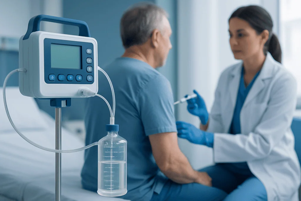
During the procedure, the person commonly sits upright and leans slightly forward, although another position may be used if needed. The skin is cleaned carefully, and local anesthetic numbs the area. A needle or small catheter is then guided into the pleural space to withdraw fluid. Many patients feel pressure rather than sharp pain.
The amount of fluid removed depends on why the procedure is being done and how the patient tolerates it. If thoracentesis is diagnostic, only a sample may be needed. If it is therapeutic, more fluid may be removed to improve breathing. The fluid is often sent for tests such as cell count, protein, glucose, culture, and cytology.
Afterward, the patient is monitored for symptoms such as cough, dizziness, chest discomfort, or shortness of breath. Some people go home the same day, while others remain in hospital because of the underlying illness rather than the procedure itself. Recovery is usually brief, and mild soreness at the needle site can occur for a day or two.
Benefits, risks, and possible complications
The main benefit of thoracentesis is that it can relieve symptoms relatively quickly when fluid around the lungs is making breathing difficult. It also gives doctors valuable information that may lead to faster diagnosis and more tailored treatment. For many patients, both goals are achieved in a single procedure.
Like any medical procedure, thoracentesis carries some risks, but serious complications are uncommon when it is performed by trained clinicians using proper technique and imaging guidance. Potential complications include pain, bruising, bleeding, infection, temporary cough, low blood pressure, or re-expansion discomfort after large-volume drainage.
A recognized but uncommon complication is pneumothorax, which means air enters the space around the lung and causes partial lung collapse. This may require observation or, less often, placement of a chest tube. Doctors reduce this risk by selecting the safest site, using ultrasound, and monitoring symptoms after the procedure.
Patients who have recurrent fluid buildup may need further treatment rather than repeated temporary drainage alone. Depending on the cause, options can include chest surgery or other targeted therapies planned by a multidisciplinary team.
How doctors use the results
The fluid obtained during thoracentesis can provide important diagnostic clues. Laboratory analysis may look for signs of infection, inflammation, heart-related fluid overload, cancer cells, blood, or abnormal chemistry values. These results are interpreted together with symptoms, imaging, and other blood tests.
If infection is suspected, the fluid may be sent for microbiology testing. If cancer is a concern, cytology may look for malignant cells, and additional imaging or tissue sampling may be considered. If the effusion appears linked to heart or kidney problems, treatment may focus more on the underlying condition than on repeated procedures.
Sometimes the answer is straightforward after one thoracentesis. In other cases, further evaluation is needed. This may include repeat imaging, bronchoscopy, biopsy, or ongoing care through oncology or other specialty services, depending on the clinical picture.
Self-care, follow-up, and when to seek medical care
After thoracentesis, most patients can resume light activity based on their doctor’s instructions. Keeping the bandage clean and dry for the recommended period and watching for new symptoms are simple but important steps. It also helps to attend follow-up appointments so the medical team can review test results and decide whether the fluid is likely to return.
Self-care mainly centers on the underlying condition. For example, a person with heart failure may need medication adjustment and fluid management, while someone with infection may need antibiotics. Thoracentesis works best when paired with treatment of the cause, not just the symptoms.
Medical care should be sought promptly if there is worsening shortness of breath, new chest pain, fever, chills, coughing up blood, fainting, or increasing redness or drainage at the procedure site. These symptoms do not always mean a serious complication, but they should be assessed without delay.
For patients who need coordinated evaluation or ongoing treatment, Acibadem International’s multidisciplinary specialists and JCI-accredited hospitals diagnose and treat pleural conditions for international patients in a structured, evidence-based setting.
Frequently asked questions
Is thoracentesis painful?
Thoracentesis is usually done with local anesthetic, so the area is numbed before the needle is inserted. Many people feel pressure or a brief stinging sensation from the anesthetic rather than significant pain. Mild soreness afterward is common and usually short-lived.
How long does a thoracentesis take?
The procedure itself often takes less than 30 minutes, although preparation and monitoring can add more time. The exact length depends on how much fluid is removed, whether imaging guidance is used, and the patient’s overall condition.
Do patients need to stay in the hospital after thoracentesis?
Not always. Some people have thoracentesis as an outpatient procedure and go home the same day. Others stay in hospital because of the condition causing the pleural effusion or because they need further testing and monitoring.
Can fluid come back after thoracentesis?
Yes, pleural fluid can return if the underlying cause is still active. Whether this happens depends on the reason for the effusion, such as heart failure, infection, or cancer. Follow-up care focuses on treating the cause and deciding whether more drainage or another procedure is needed.
Why is the fluid tested after thoracentesis?
Testing the fluid helps doctors understand why it collected around the lungs. The analysis may show signs of infection, inflammation, heart-related fluid imbalance, or cancer. This information helps guide the next steps in treatment.
What are the warning signs after thoracentesis?
Patients should contact a doctor promptly if they develop worsening shortness of breath, chest pain, fever, chills, fainting, or increasing redness and drainage at the needle site. These symptoms may be related to a complication or to the underlying illness and should be assessed by a clinician.
References
- American Thoracic Society
- National Heart, Lung, and Blood Institute
- Merck Manual Professional Edition
- British Thoracic Society
- Cleveland Clinic
This article is for general information only and is not a substitute for professional medical advice. Please consult a qualified doctor about your individual situation.
Explore treatments in Turkey — costs, top hospitals & a free quote
JCI-accredited · board-certified surgeons · reply within 24h
Add us as a Preferred Source to see more of our trusted health content across Google Search, AI Overviews and Discover.

More from the Health Library

Pacu nurse: Symptoms, Causes, and Treatment — Complete Patient Guide

Subcutaneous: A Complete Medical Overview

Pelvic Exam: A Complete Medical Overview

Stamina What Does It Mean — Explained by Medical Evidence, Not Myths

Tick Bite Pictures — Explained by Medical Evidence, Not Myths